In 3-D computer graphics, ray tracing is a technique for modeling light transport for use in a wide variety of rendering algorithms for generating digital images.

X-ray is a high-energy electromagnetic radiation. In many languages, it is referred to as Röntgen radiation, after the German scientist Wilhelm Conrad Röntgen, who discovered it in 1895 and named it X-radiation to signify an unknown type of radiation.

A computed tomography scan is a medical imaging technique used to obtain detailed internal images of the body. The personnel that perform CT scans are called radiographers or radiology technologists.

Radiography is an imaging technique using X-rays, gamma rays, or similar ionizing radiation and non-ionizing radiation to view the internal form of an object. Applications of radiography include medical and industrial radiography. Similar techniques are used in airport security,. To create an image in conventional radiography, a beam of X-rays is produced by an X-ray generator and it is projected towards the object. A certain amount of the X-rays or other radiation are absorbed by the object, dependent on the object's density and structural composition. The X-rays that pass through the object are captured behind the object by a detector. The generation of flat two-dimensional images by this technique is called projectional radiography. In computed tomography, an X-ray source and its associated detectors rotate around the subject, which itself moves through the conical X-ray beam produced. Any given point within the subject is crossed from many directions by many different beams at different times. Information regarding the attenuation of these beams is collated and subjected to computation to generate two-dimensional images on three planes which can be further processed to produce a three-dimensional image.

Angiography or arteriography is a medical imaging technique used to visualize the inside, or lumen, of blood vessels and organs of the body, with particular interest in the arteries, veins, and the heart chambers. Modern angiography is performed by injecting a radio-opaque contrast agent into the blood vessel and imaging using X-ray based techniques such as fluoroscopy.
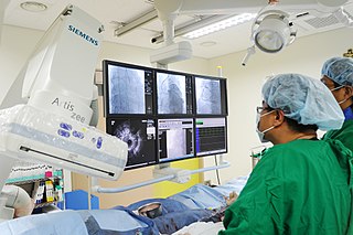
A coronary catheterization is a minimally invasive procedure to access the coronary circulation and blood filled chambers of the heart using a catheter. It is performed for both diagnostic and interventional (treatment) purposes.
Mechanism design is a field in economics and game theory that takes an objectives-first approach to designing economic mechanisms or incentives, toward desired objectives, in strategic settings, where players act rationally. Because it starts at the end of the game, then goes backwards, it is also called reverse game theory. It has broad applications, from economics and politics in fields such as market design, auction theory and social choice theory to networked-systems.
Radiocontrast agents are substances used to enhance the visibility of internal structures in X-ray-based imaging techniques such as computed tomography, projectional radiography, and fluoroscopy. Radiocontrast agents are typically iodine, or more rarely barium sulfate. The contrast agents absorb external X-rays, resulting in decreased exposure on the X-ray detector. This is different from radiopharmaceuticals used in nuclear medicine which emit radiation.
A contrast agent is a substance used to increase the contrast of structures or fluids within the body in medical imaging. Contrast agents absorb or alter external electromagnetism or ultrasound, which is different from radiopharmaceuticals, which emit radiation themselves. In x-ray imaging, contrast agents enhance the radiodensity in a target tissue or structure. In magnetic resonance imaging, contrast agents shorten the relaxation times of nuclei within body tissues in order to alter the contrast in the image.
Peter O’Fallon is an American television director.
Ytterbium(III) fluoride is an inorganic chemical compound that is insoluble in water. Like other Ytterbium compounds, it is a rather unremarkable white substance. Ytterbium fluoride has found a niche usage as a radio-opaque agent in the dental industry to aid in the identification of fillings under X-ray examination.

The autopsy of John F. Kennedy, the 35th president of the United States, was performed at the Bethesda Naval Hospital in Bethesda, Maryland. The autopsy began at about 8 p.m. Eastern Standard Time (EST) on November 22, 1963—the day of Kennedy's assassination—and ended in the early morning of November 23, 1963. The choice of autopsy hospital in the Washington, D.C. area was made by his widow, First Lady Jacqueline Kennedy, who chose the Bethesda as President Kennedy had been a naval officer during World War II.

Ioxilan is a diagnostic contrast agent. It is injected intravenously before taking X-ray images to increase arterial contrast in the final image. It was marketed in the US under the trade name Oxilan by Guerbet, L.L.C., but has been discontinued in 2017.
Secret Agent X-9 (1937) is a Universal film serial based on the comic strip Secret Agent X-9 by Dashiell Hammett and Alex Raymond.

Projectional radiography, also known as conventional radiography, is a form of radiography and medical imaging that produces two-dimensional images by X-ray radiation. The image acquisition is generally performed by radiographers, and the images are often examined by radiologists. Both the procedure and any resultant images are often simply called 'X-ray'. Plain radiography or roentgenography generally refers to projectional radiography. Plain radiography can also refer to radiography without a radiocontrast agent or radiography that generates single static images, as contrasted to fluoroscopy, which are technically also projectional.
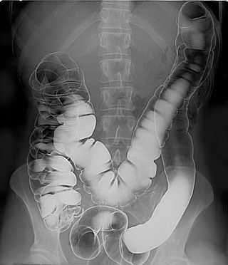
A double-contrast barium enema is a form of contrast radiography in which x-rays of the colon and rectum are taken using two forms of contrast to make the structures easier to see. A liquid containing barium is put into the rectum. Barium outlines the colon and rectum on an x-ray and helps show abnormalities. Air is also put into the rectum and colon to further enhance the x-ray.

Iomeprol is a pharmaceutical drug used as a radiocontrast agent in X-ray imaging. It is sold under the trade names Imeron and Iomeron.
Adipiodone is a pharmaceutical drug used as a radiocontrast agent in X-ray imaging. It was introduced in the 1950s.

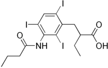
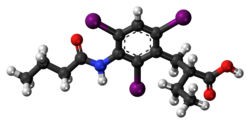
Iobenzamic acid is a pharmaceutical drug used as an X-ray contrast agent.
Spectral imaging is an umbrella term for energy-resolved X-ray imaging in medicine. The technique makes use of the energy dependence of X-ray attenuation to either increase the contrast-to-noise ratio, or to provide quantitative image data and reduce image artefacts by so-called material decomposition. Dual-energy imaging, i.e. imaging at two energy levels, is a special case of spectral imaging and is still the most widely used terminology, but the terms "spectral imaging" and "spectral CT" have been coined to acknowledge the fact that photon-counting detectors have the potential for measurements at a larger number of energy levels.