
A microscope is a laboratory instrument used to examine objects that are too small to be seen by the naked eye. Microscopy is the science of investigating small objects and structures using a microscope. Microscopic means being invisible to the eye unless aided by a microscope.

A scanning electron microscope (SEM) is a type of electron microscope that produces images of a sample by scanning the surface with a focused beam of electrons. The electrons interact with atoms in the sample, producing various signals that contain information about the surface topography and composition of the sample. The electron beam is scanned in a raster scan pattern, and the position of the beam is combined with the intensity of the detected signal to produce an image. In the most common SEM mode, secondary electrons emitted by atoms excited by the electron beam are detected using a secondary electron detector. The number of secondary electrons that can be detected, and thus the signal intensity, depends, among other things, on specimen topography. Some SEMs can achieve resolutions better than 1 nanometer.

The optical microscope, also referred to as a light microscope, is a type of microscope that commonly uses visible light and a system of lenses to generate magnified images of small objects. Optical microscopes are the oldest design of microscope and were possibly invented in their present compound form in the 17th century. Basic optical microscopes can be very simple, although many complex designs aim to improve resolution and sample contrast.

A monocular is a compact refracting telescope used to magnify images of distant objects, typically using an optical prism to ensure an erect image, instead of using relay lenses like most telescopic sights. The volume and weight of a monocular are typically less than half of a pair of binoculars with similar optical properties, making it more portable and also less expensive. This is because binoculars are essentially a pair of monoculars packed together — one for each eye. As a result, monoculars only produce two-dimensional images, while binoculars can use two parallaxed images to produce binocular vision, which allows stereopsis and depth perception.
The microscopic scale is the scale of objects and events smaller than those that can easily be seen by the naked eye, requiring a lens or microscope to see them clearly. In physics, the microscopic scale is sometimes regarded as the scale between the macroscopic scale and the quantum scale. Microscopic units and measurements are used to classify and describe very small objects. One common microscopic length scale unit is the micrometre, which is one millionth of a metre.

In optical engineering, an objective is an optical element that gathers light from an object being observed and focuses the light rays from it to produce a real image of the object. Objectives can be a single lens or mirror, or combinations of several optical elements. They are used in microscopes, binoculars, telescopes, cameras, slide projectors, CD players and many other optical instruments. Objectives are also called object lenses, object glasses, or objective glasses.
Magnification is the process of enlarging the apparent size, not physical size, of something. This enlargement is quantified by a size ratio called optical magnification. When this number is less than one, it refers to a reduction in size, sometimes called de-magnification.

A borescope is an optical instrument designed to assist visual inspection of narrow, difficult-to-reach cavities, consisting of a rigid or flexible tube with an eyepiece or display on one end, an objective lens or camera on the other, linked together by an optical or electrical system in between. The optical system in some instances is accompanied by illumination to enhance brightness and contrast. An internal image of the illuminated object is formed by the objective lens and magnified by the eyepiece which presents it to the viewer's eye.

An eyepiece, or ocular lens, is a type of lens that is attached to a variety of optical devices such as telescopes and microscopes. It is named because it is usually the lens that is closest to the eye when someone looks through an optical device to observe an object or sample. The objective lens or mirror collects light from an object or sample and brings it to focus creating an image of the object. The eyepiece is placed near the focal point of the objective to magnify this image to the eyes. The amount of magnification depends on the focal length of the eyepiece.

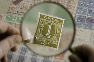
Macro photography is extreme close-up photography, usually of very small subjects and living organisms like insects, in which the size of the subject in the photograph is greater than life-size . By the original definition, a macro photograph is one in which the size of the subject on the negative or image sensor is life-size or greater. In some senses, however, it refers to a finished photograph of a subject that is greater than life-size.

In optics, the exit pupil is a virtual aperture in an optical system. Only rays which pass through this virtual aperture can exit the system. The exit pupil is the image of the aperture stop in the optics that follow it. In a telescope or compound microscope, this image is the image of the objective element(s) as produced by the eyepiece. The size and shape of this disc is crucial to the instrument's performance, because the observer's eye can see light only if it passes through the aperture. The term exit pupil is also sometimes used to refer to the diameter of the virtual aperture. Older literature on optics sometimes refers to the exit pupil as the Ramsden disc, named after English instrument-maker Jesse Ramsden.
The eye relief of an optical instrument is the distance from the last surface of an eyepiece within which the user's eye can obtain the full viewing angle. If a viewer's eye is outside this distance, a reduced field of view will be obtained. The calculation of eye relief is complex, though generally, the higher the magnification and the larger the intended field of view, the shorter the eye relief.

A micrograph is an image, captured photographically or digitally, taken through a microscope or similar device to show a magnified image of an object. This is opposed to a macrograph or photomacrograph, an image which is also taken on a microscope but is only slightly magnified, usually less than 10 times. Micrography is the practice or art of using microscopes to make photographs. A photographic micrograph is a photomicrograph, and one taken with an electron microscope is an electron micrograph.
A parfocal lens is a lens that stays in focus when magnification/focal length is changed. There is inevitably some amount of focus error, but too small to be considered significant.
A light field camera, also known as a plenoptic camera, is a camera that captures information about the light field emanating from a scene; that is, the intensity of light in a scene, and also the precise direction that the light rays are traveling in space. This contrasts with conventional cameras, which record only light intensity at various wavelengths.

A digital microscope is a variation of a traditional optical microscope that uses optics and a digital camera to output an image to a monitor, sometimes by means of software running on a computer. A digital microscope often has its own in-built LED light source, and differs from an optical microscope in that there is no provision to observe the sample directly through an eyepiece. Since the image is focused on the digital circuit, the entire system is designed for the monitor image. The optics for the human eye are omitted.

The stereo, stereoscopic or dissecting microscope is an optical microscope variant designed for low magnification observation of a sample, typically using light reflected from the surface of an object rather than transmitted through it. The instrument uses two separate optical paths with two objectives and eyepieces to provide slightly different viewing angles to the left and right eyes. This arrangement produces a three-dimensional visualization of the sample being examined. Stereomicroscopy overlaps macrophotography for recording and examining solid samples with complex surface topography, where a three-dimensional view is needed for analyzing the detail.
Hirox (ハイロックス) is a lens company in Tokyo, Japan that created the first digital microscope in 1985. This company is now known as Hirox Co Ltd. Hirox's main industry is digital microscopes, but still makes the lenses for a variety of items including rangefinders.

Afocal photography, also called afocal imaging or afocal projection is a method of photography where the camera with its lens attached is mounted over the eyepiece of another image forming system such as an optical telescope or optical microscope, with the camera lens taking the place of the human eye.
In optics, a relay lens is a lens or a group of lenses that receives the image from the objective lens and relays it to the eyepiece. Relay lenses are found in refracting telescopes, endoscopes, and periscopes to optically manipulate the light path, extend the length of the whole optical system, and usually serve the purpose of inverting the image. They may be made of one or more conventional lenses or achromatic doublets, or a long cylindrical gradient-index of refraction lens.


