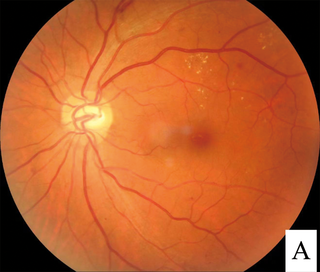
Diabetic retinopathy, is a medical condition in which damage occurs to the retina due to diabetes mellitus. It is a leading cause of blindness in developed countries.

Glaucoma is a group of eye diseases that lead to damage of the optic nerve, which transmits visual information from the eye to the brain. Glaucoma may cause vision loss if left untreated. It has been called the "silent thief of sight" because the loss of vision usually occurs slowly over a long period of time. A major risk factor for glaucoma is increased pressure within the eye, known as intraocular pressure (IOP). It is associated with old age, a family history of glaucoma, and certain medical conditions or medications. The word glaucoma comes from the Ancient Greek word γλαυκóς, meaning 'gleaming, blue-green, gray'.

Vitrectomy is a surgery to remove some or all of the vitreous humor from the eye.
The National Eye Institute (NEI) is part of the U.S. National Institutes of Health (NIH), an agency of the U.S. Department of Health and Human Services. The mission of NEI is "to eliminate vision loss and improve quality of life through vision research." NEI consists of two major branches for research: an extramural branch that funds studies outside NIH and an intramural branch that funds research on the NIH campus in Bethesda, Maryland. Most of the NEI budget funds extramural research.

Eye surgery, also known as ophthalmic surgery or ocular surgery, is surgery performed on the eye or its adnexa. Eye surgery is part of ophthalmology and is performed by an ophthalmologist or eye surgeon. The eye is a fragile organ, and requires due care before, during, and after a surgical procedure to minimize or prevent further damage. An eye surgeon is responsible for selecting the appropriate surgical procedure for the patient, and for taking the necessary safety precautions. Mentions of eye surgery can be found in several ancient texts dating back as early as 1800 BC, with cataract treatment starting in the fifth century BC. It continues to be a widely practiced class of surgery, with various techniques having been developed for treating eye problems.

Phacoemulsification is a cataract surgery method in which the internal lens of the eye which has developed a cataract is emulsified with the tip of an ultrasonic handpiece and aspirated from the eye. Aspirated fluids are replaced with irrigation of balanced salt solution to maintain the volume of the anterior chamber during the procedure. This procedure minimises the incision size and reduces the recovery time and risk of surgery induced astigmatism.

Retinal detachment is a disorder of the eye in which the retina peels away from its underlying layer of support tissue. Initial detachment may be localized, but without rapid treatment the entire retina may detach, leading to vision loss and blindness. It is a surgical emergency.

Intraocular pressure (IOP) is the fluid pressure inside the eye. Tonometry is the method eye care professionals use to determine this. IOP is an important aspect in the evaluation of patients at risk of glaucoma. Most tonometers are calibrated to measure pressure in millimeters of mercury (mmHg).

Cataract surgery, also called lens replacement surgery, is the removal of the natural lens of the eye that has developed a cataract, an opaque or cloudy area. The eye's natural lens is usually replaced with an artificial intraocular lens (IOL) implant.

In the anatomy of the eye, the retinal nerve fiber layer (RNFL) or nerve fiber layer, stratum opticum, is formed by the expansion of the fibers of the optic nerve; it is thickest near the optic disc, gradually diminishing toward the ora serrata.

Dilated fundus examination (DFE) is a diagnostic procedure that uses mydriatic eye drops to dilate or enlarge the pupil in order to obtain a better view of the fundus of the eye. Once the pupil is dilated, examiners use ophthalmoscopy to view the eye's interior, which makes it easier to assess the retina, optic nerve head, blood vessels, and other important features. DFE has been found to be a more effective method for evaluating eye health when compared to non-dilated examination, and is the best method of evaluating structures behind the iris. It is frequently performed by ophthalmologists and optometrists as part of an eye examination.

Blurred vision is an ocular symptom where vision becomes less precise and there is added difficulty to resolve fine details.
Pseudoexfoliation syndrome, often abbreviated as PEX and sometimes as PES or PXS, is an aging-related systemic disease manifesting itself primarily in the eyes which is characterized by the accumulation of microscopic granular amyloid-like protein fibers. Its cause is unknown, although there is speculation that there may be a genetic basis. It is more prevalent in women than men, and in persons past the age of seventy. Its prevalence in different human populations varies; for example, it is prevalent in Scandinavia. The buildup of protein clumps can block normal drainage of the eye fluid called the aqueous humor and can cause, in turn, a buildup of pressure leading to glaucoma and loss of vision. As worldwide populations become older because of shifts in demography, PEX may become a matter of greater concern.

Laser coagulation or laser photocoagulation surgery is used to treat a number of eye diseases and has become widely used in recent decades. During the procedure, a laser is used to finely cauterize ocular blood vessels to attempt to bring about various therapeutic benefits.
The Himalayan Cataract Project (HCP) was created in 1995 by Dr. Geoffrey Tabin and Dr. Sanduk Ruit with a goal of establishing a sustainable eye care infrastructure in the Himalaya. HCP empowers local doctors to provide ophthalmic care through skills-transfer and education. From its beginning, HCP responds to a pressing need for eye care in the Himalayan region. With programs in Nepal, Ethiopia, Ghana, Bhutan and India they have been able to restore sight to over 1.4 million people since 1995.

Josef Flammer is a Swiss ophthalmologist and long-time director of the Eye Clinic at Basel University Hospital. Flammer is a glaucoma specialist who developed a new pathogenetic concept of glaucomatous damage according to which unstable blood supply leads to oxidative stress, which in turn plays a major role in apoptosis of cells in the optic nerve and retina in glaucoma patients.

Yog Raj Sharma is an Indian ophthalmologist and ex-chief of Dr. Rajendra Prasad Centre for Ophthalmic Sciences of the All India Institute of Medical Sciences (AIIMS), New Delhi, the apex body of the National Programme for the Control of Blindness, a Government of India initiative to reduce the prevalence of blindness in India. He is the Chairman of the Task Force on Prevention and Control of Diabetic Retinopathy Group and the Co-Chairman of the National Task Force on Prevention of Blindness from Retinopathy of Prematurity under the Ministry of Health and Family Welfare of the Government of India. An advisor to the Ministry of Health and Family Welfare, India. Sharma was honored by the Government of India in 2015 with Padma Shri, the fourth highest Indian civilian award. In 2005, Yog Raj Sharma's published article on "Pars plana vitrectomy vs scleral buckling in rhegmatogenous retinal detachment" in Acta Ophthalmologica Scandinavica and in November 2021, American society of retina specialists cited it as top 100 publications on retinal detachment management in the last ~121 years. Of these top hundred publications, only nineteen countries contributed, three of the contributing countries were Asian and from India this study was the sole contribution. Dr Sharma called it 'the singular biggest achievement of his career" in an article published in Daily Excelsior, Jammu in December 2021.
Joan Whitten Miller is a Canadian-American ophthalmologist and scientist who has made notable contributions to the treatment and understanding of eye disorders. She is credited for developing photodynamic therapy (PDT) with verteporfin (Visudyne), the first pharmacologic therapy for retinal disease. She also co-discovered the role of vascular endothelial growth factor (VEGF) in eye disease and demonstrated the therapeutic potential of VEGF inhibitors, forming the scientific basis of anti-VEGF therapy for age-related macular degeneration (AMD), diabetic retinopathy, and related conditions.

John Gustaf Lindberg was a Finnish ophthalmologist who was the first to describe exfoliation syndrome, an age-related degenerative disease of the eye that often complicates glaucoma and cataract surgery, in his doctoral thesis that he wrote in St. Petersburg and defended in March 1917 in Helsinki, Finland, at that time a Grand Duchy in the Russian Empire.

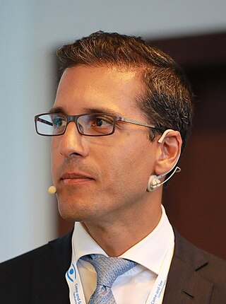
Kaweh Mansouri is an Iranian-Austrian ophthalmologist and researcher in glaucoma. He is an adjunct professor at the Department of Ophthalmology, University of Colorado School of Medicine and a consultant ophthalmologist at the Montchoisi Clinic, Lausanne. He serves as the Associate Executive Vice President of the World Glaucoma Association and is President of the Swiss Glaucoma Research Foundation.


