Uterine septum

Last updated
Uterine septum
Specialty Gynaecology   OOjs UI icon edit-ltr-progressive.svg

A uterine septum is a congenital uterine malformation where the uterine cavity is partitioned by a longitudinal septum; the outside of the uterus has a normal typical shape. The wedge-like partition may involve only the superior part of the cavity resulting in an incomplete septum or a subseptate uterus, or less frequently the total length of the cavity (complete septum) and the cervix resulting in a double cervix. The septation may also continue caudally into the vagina resulting in a "double vagina". [1]

Contents

Signs and symptoms

Four types of uterine malformations Uterine malformation.jpg
Four types of uterine malformations

The condition may not be known to the affected individual and not result in any reproductive problems; thus normal pregnancies may occur. In more serious cases have reported high infertility rates. [2] However, it is associated with a higher risk for miscarriage, premature birth, and malpresentation. According to the classical study by Buttram there is a 60% risk of a spontaneous abortion, this being more common in the second than in the first trimester. [3] However, there is no agreement on this number and other studies show a lower risk. Woelfer found that the miscarriage risk is more pronounced in the first trimester. [4]

The condition is also associated with abnormalities of the renal system. Further, skeletal abnormalities have been linked to the condition. [5]

Cause

The uterus is formed during embryogenesis by the fusion of the two Müllerian ducts. During this fusion a resorption process eliminates the partition between the two ducts to create a single cavity. This process begins caudally and advances cranially, thus a complete septum formation represents an earlier disturbance of this absorption than the incomplete form. Causes for incomplete absorption are not known.[ citation needed ]

Diagnosis

A pelvic examination may reveal a double vagina or double cervix that should be further investigated and may lead to the discovery of a uterine septum. In most patients, however, the pelvic examination is normal. Investigations are usually prompted on the basis of reproductive problems.[ citation needed ]

Helpful techniques to investigate a septum are transvaginal ultrasonography and sonohysterography, MRI, and hysteroscopy. More recently 3-D ultrasonography has been advocated as an excellent non-invasive method to delineate the condition. [4] Prior to modern imaging hysterosalpingography was used to help diagnose the uterine septum, however, a bicornuate uterus may deliver a similar image. An important category of septate uterus is the hybrid type a variant that may be misdiagnosed as bicornuate uterus when seen by laparoscopy [6] Professor El Saman From Egypt was the first to describe this anomaly and warned gynecologist about this common misdiagnosis, whenever there is a uterine fundus depression on laparoscopy gynecologists should compare the depth of this depression with the depth of the dividing internal interface. Hybrid septate uterus benefit from hysteroscopic metroplasty under laparoscopic control. [7]

Differential diagnosis

Other forms of uterine malformation need to be considered in the work-up for uterine septum. An arcuate uterus contains a residual cranial septum that is smaller than an incomplete septum but definitions between the two conditions are not standardized, - a cause for discrepancies in the literature. A bicornuate uterus is sometimes confused with a septate uterus as in each situation the cavity is partitioned, however, in the former case the uterine body is cranially doubled (two uterine horns) while in the latter a single uterine body is present. The former represents a malformation of incomplete fusion of the Müllerian systems, and the latter of incomplete absorption. A hysterosalpingogram may not be able to distinguish between the two conditions. The differentiation, however, is important as a septum can be corrected by hysteroscopy, while a bicornuate uterus would be corrected by a metroplasty via laparotomy if necessary.[ citation needed ]

Management

A septum can be resected with surgery. Hysteroscopic removal of a uterine septum is generally the preferred method, as the intervention is relatively minor and safe in experienced hands. A follow-up imaging study should demonstrate the removal of the septum. Tactile cold scissor metroplasty was described as a back technique for hysteroscopic challenges that interfere with proper visualization [8] or uterine distention.

It is not considered necessary to remove a septum that has not caused problems, especially in women who are not considering pregnancy. There is controversy over whether a septum should be removed prophylactically to reduce the risk of pregnancy loss prior to a pregnancy or infertility treatment. [9]

Epidemiology

Most studies are based on populations of women who have experienced a pregnancy loss and thus do not address the issue of the prevalence in the general population. A screening study by Woelfer et al. of women without a history of reproductive problems found that about 3% of women had a uterine septation; the most common anomaly in their study was an arcuate uterus (5%), while 0.5% were found to have a bicornuate uterus. [4] In contrast, in about 15% of patients with recurrent pregnancy loss anatomical problems are thought to be causative with the septate uterus as the most common finding. [10]

Related Research Articles

<span class="mw-page-title-main">Cervix</span> Lower part of the uterus in the human female reproductive system

The cervix or cervix uteri is the lower part of the uterus (womb) in the human female reproductive system. The cervix is usually 2 to 3 cm long and roughly cylindrical in shape, which changes during pregnancy. The narrow, central cervical canal runs along its entire length, connecting the uterine cavity and the lumen of the vagina. The opening into the uterus is called the internal os, and the opening into the vagina is called the external os. The lower part of the cervix, known as the vaginal portion of the cervix, bulges into the top of the vagina. The cervix has been documented anatomically since at least the time of Hippocrates, over 2,000 years ago.

<span class="mw-page-title-main">Uterus</span> Female sex organ in mammals

The uterus or womb is the organ in the reproductive system of most female mammals, including humans, that accommodates the embryonic and fetal development of one or more embryos until birth. The uterus is a hormone-responsive sex organ that contains glands in its lining that secrete uterine milk for embryonic nourishment.

<span class="mw-page-title-main">Paramesonephric duct</span> Paired ducts in the embryo in the primitive urogenital structures

The paramesonephric ducts are paired ducts of the embryo in the female reproductive system that run down the lateral sides of the genital ridge and terminate at the sinus tubercle in the primitive urogenital sinus. In the female, they will develop to form the fallopian tubes, uterus, cervix, and the upper one-third of the vagina.

Müllerian agenesis, also known as Müllerian aplasia, vaginal agenesis, or Mayer–Rokitansky–Küster–Hauser syndrome, is a congenital malformation characterized by a failure of the Müllerian ducts to develop, resulting in a missing uterus and variable degrees of vaginal hypoplasia of its upper portion. Müllerian agenesis is the cause in 15% of cases of primary amenorrhoea. Because most of the vagina does not develop from the Müllerian duct, instead developing from the urogenital sinus, along with the bladder and urethra, it is present even when the Müllerian duct is completely absent. Because ovaries do not develop from the Müllerian ducts, affected people might have normal secondary sexual characteristics but are infertile due to the lack of a functional uterus. However, biological motherhood is possible through uterus transplantation or use of gestational surrogates.

<span class="mw-page-title-main">Asherman's syndrome</span> Medical condition

Asherman's syndrome (AS) is an acquired uterine condition that occurs when scar tissue (adhesions) forms inside the uterus and/or the cervix. It is characterized by variable scarring inside the uterine cavity, where in many cases the front and back walls of the uterus stick to one another. AS can be the cause of menstrual disturbances, infertility, and placental abnormalities. Although the first case of intrauterine adhesion was published in 1894 by Heinrich Fritsch, it was only after 54 years that a full description of Asherman syndrome was carried out by Joseph Asherman. A number of other terms have been used to describe the condition and related conditions including: uterine/cervical atresia, traumatic uterine atrophy, sclerotic endometrium, and endometrial sclerosis.

<span class="mw-page-title-main">Hysteroscopy</span> Medical procedure

Hysteroscopy is the inspection of the uterine cavity by endoscopy with access through the cervix. It allows for the diagnosis of intrauterine pathology and serves as a method for surgical intervention.

<span class="mw-page-title-main">Gynecologic ultrasonography</span> Application of medical ultrasonography to the female pelvic organs

Gynecologic ultrasonography or gynecologic sonography refers to the application of medical ultrasonography to the female pelvic organs as well as the bladder, the adnexa, and the recto-uterine pouch. The procedure may lead to other medically relevant findings in the pelvis.This technique is useful to detect myomas or mullerian malformations.

<span class="mw-page-title-main">Uterine malformation</span> Medical condition

A uterine malformation is a type of female genital malformation resulting from an abnormal development of the Müllerian duct(s) during embryogenesis. Symptoms range from amenorrhea, infertility, recurrent pregnancy loss, and pain, to normal functioning depending on the nature of the defect.

<span class="mw-page-title-main">Uterine myomectomy</span> Surgical removal of uterine fibroid

Myomectomy, sometimes also called fibroidectomy, refers to the surgical removal of uterine leiomyomas, also known as fibroids. In contrast to a hysterectomy, the uterus remains preserved and the woman retains her reproductive potential. It still may impact hormonal regulation and the menstrual cycle.

Vaginal atresia is a condition in which the vagina is abnormally closed or absent. The main causes can either be complete vaginal hypoplasia, or a vaginal obstruction, often caused by an imperforate hymen or, less commonly, a transverse vaginal septum. It results in uterovaginal outflow tract obstruction. This condition does not usually occur by itself within an individual, but coupled with other developmental disorders within the female. The disorders that are usually coupled with a female who has vaginal atresia are Mayer-Rokitansky-Küster-Hauser syndrome, Bardet-Biedl syndrome, or Fraser syndrome. One out of every 5,000 women have this abnormality.

<span class="mw-page-title-main">Unicornuate uterus</span> Medical condition

A unicornuate uterus represents a uterine malformation where the uterus is formed from one only of the paired Müllerian ducts while the other Müllerian duct does not develop or only in a rudimentary fashion. The sometimes called hemi-uterus has a single horn linked to the ipsilateral fallopian tube that faces its ovary.

<span class="mw-page-title-main">Bicornuate uterus</span> Medical condition

A bicornuate uterus or bicornate uterus, is a type of müllerian anomaly in the human uterus, where there is a deep indentation at the fundus (top) of the uterus.

<span class="mw-page-title-main">Arcuate uterus</span> Medical condition

The arcuate uterus is a form of a uterine anomaly or variation where the uterine cavity displays a concave contour towards the fundus. Normally the uterine cavity is straight or convex towards the fundus on anterior-posterior imaging, but in the arcuate uterus the myometrium of the fundus dips into the cavity and may form a small septation. The distinction between an arcuate uterus and a septate uterus is not standardized.

<span class="mw-page-title-main">Uterus didelphys</span> Medical condition

Uterus didelphys represents a uterine malformation where the uterus is present as a paired organ when the embryogenetic fusion of the Müllerian ducts fails to occur. As a result, there is a double uterus with two separate cervices, and possibly a double vagina as well. Each uterus has a single horn linked to the ipsilateral fallopian tube that faces its ovary.

Hematocolpos is a medical condition in which the vagina is pooled with menstrual blood due to multiple factors leading to the blockage of menstrual blood flow. The medical definition of hematocolpos is 'an accumulation of blood within the vagina'. It is often caused by the combination of menstruation with an imperforate hymen. It is sometimes seen in Robinow syndrome, uterus didelphys, or other vaginal anomalies.

<span class="mw-page-title-main">Vaginal septum</span> Medical condition

A vaginal septum is a vaginal anomaly that is partition within the vagina; such a septum could be either longitudinal or transverse. In some affected women, the septum is partial or does not extend the length or width of the vagina. Pain during intercourse can be a symptom. A longitudinal vaginal septum develops during embryogenesis when there is an incomplete fusion of the lower parts of the two Müllerian ducts. As a result, there may appear to be two openings to the vagina. There may be associated duplications of the more cranial parts of the Müllerian derivatives, a double cervix, and either a uterine septum or uterus didelphys. A transverse septum forms during embryogenesis when the Müllerian ducts do not fuse to the urogenital sinus. A complete transverse septum can occur across the vagina at different levels. Menstrual flow can be blocked, and is a cause of primary amenorrhea. The accumulation of menstrual debris behind the septum is termed cryptomenorrhea. Some transverse septa are incomplete and may lead to dyspareunia or obstruction in labour.

<span class="mw-page-title-main">Cervical pregnancy</span> Medical condition

A cervical pregnancy is an ectopic pregnancy that has implanted in the uterine endocervix. Such a pregnancy typically aborts within the first trimester, however, if it is implanted closer to the uterine cavity – a so-called cervico-isthmic pregnancy – it may continue longer. Placental removal in a cervical pregnancy may result in major hemorrhage.

Müllerian duct anomalies are those structural anomalies caused by errors in Müllerian duct development during embryonic morphogenesis. Factors that precipitate include genetics, and maternal exposure to teratogens.
<span class="mw-page-title-main">T-shaped uterus</span> Medical condition

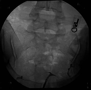
A t-shaped uterus is a type of uterine malformation wherein the uterus is shaped resembling the letter T. This is typically observed in DES-exposed women. It is recognised in the ESHRE/ESGE classification, and is associated with failed implantation, increased risk of ectopic pregnancy, miscarriage and preterm delivery. There is a surgical procedure to correct the malformation.

<span class="mw-page-title-main">Vaginal anomalies</span> Congenital defect; abnormal or absent vagina

Vaginal anomalies are abnormal structures that are formed during the prenatal development of the female reproductive system and are rare congenital defects that result in an abnormal or absent vagina.

References

  1. Heinonen PK (March 2006). "Complete septate uterus with longitudinal vaginal septum". Fertil. Steril. 85 (3): 700–5. doi: 10.1016/j.fertnstert.2005.08.039 . PMID   16500341.
  2. Creasy RK, Resnik R (1994). Maternal-Fetal Medicine: Principles and Practice. Philadelphia: W.B.Saunders. p.  447. ISBN   0-7216-6590-X.
  3. Buttram VC, Gibbons WE (July 1979). "Müllerian anomalies: a proposed classification. (An analysis of 144 cases)". Fertil. Steril. 32 (1): 40–6. doi:10.1016/s0015-0282(16)44114-2. PMID   456629.
  4. 1 2 3 Woelfer B, Salim R, Banerjee S, Elson J, Regan L, Jurkovic D (December 2001). "Reproductive outcomes in women with congenital uterine anomalies detected by three-dimensional ultrasound screening". Obstet Gynecol. 98 (6): 1099–103. doi:10.1016/S0029-7844(01)01599-X. PMID   11755560. S2CID   37650526.
  5. Li S, Qayyum A, Coakley FV, Hricak H (2000). "Association of renal agenesis and Müllerian duct anomalies". J Comput Assist Tomogr. 24 (6): 829–34. doi:10.1097/00004728-200011000-00001. PMID   11105695. S2CID   26387695.
  6. El Saman, AM; Nasr, A; Tawfik, RM; Saadeldeen, HS (Aug 2011). "Müllerian duct anomalies: successful endoscopic management of a hybrid bicornuate/septate variety". J Pediatr Adolesc Gynecol. 24 (4): e89–92. doi:10.1016/j.jpag.2011.02.013. PMID   21514191.
  7. El Saman, AM; Shahin, AY; Nasr, A; Tawfik, RM; Saadeldeen, HS; Othman, ER; Habib, DM; Abdel-Aleem, MA (Nov 2012). "Hybrid septate uterus, coexistence of bicornuate and septate varieties: a genuine report". Journal of Obstetrics and Gynaecology Research. 38 (11): 1308–14. doi:10.1111/j.1447-0756.2012.01866.x. PMID   22612567. S2CID   6177612.
  8. El Saman, AM; Darwish, AM; Zakherah, MS; Hamed, HO; Bedaiwy, MA; Nasr, AM (Aug 2010). "Tactile cold scissor metroplasty as a novel backup method for hysteroscopic metroplasty". Fertil Steril. 94 (3): 1086–9. doi:10.1016/j.fertnstert.2009.05.018. PMID   19591982.
  9. Ozgur K, Isikoglu M, Donmez L, Oehninger S (March 2007). "Is hysteroscopic correction of an incomplete uterine septum justified prior to IVF?". Reprod Biomed Online. 14 (3): 335–40. doi: 10.1016/S1472-6483(10)60876-0 . PMID   17359587.
  10. Propst AM, Hill JA (2000). "Anatomic factors associated with recurrent pregnancy loss". Semin. Reprod. Med. 18 (4): 341–50. doi:10.1055/s-2000-13723. PMID   11355792.