For a modern description of the substance referred to as 'vagusstoff', see Acetylcholine.
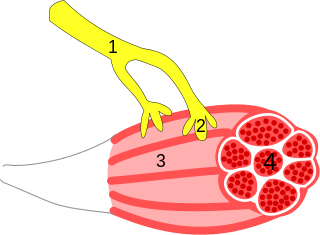
Figure 1. Diagram of the frog heart preparation used by Loewi. Vagus nerve stimulation slows heart rate while accelerator (sympathetic) nerve stimulation speeds up heart rate.
By the time Loewi began his experiments there was much discussion among scientists whether communication between nerves and muscles was chemical or electrical by nature. Experiments by Luigi Galvani in the 18th century had demonstrated that electrical stimulation of the frog sciatic nerve resulted in twitching of the leg muscles, and from this he developed the concept of bioelectricity. This led to the idea that direct electrical contact between nerves and muscles mediated transmission of excitation. However, work by John Newport Langley had suggested that in the autonomic nervous system communication in the ciliary ganglion was chemical. Loewi's experiments, published in 1921 [permanent dead link], finally settled the issue, proving that synaptic transmission was chemical.
Figure 2. Loewi's experiment proving that neurotransmision was chemical, rather than electrical.
Loewi performed a very simple yet elegant experiment. Using an isolated frog heart he had previously found that stimulation of the vagus nerve resulted in a slowing of the heart rate, while stimulation of the sympathetic nerve caused the heart rate to speed up (Figure 1). He reasoned that stimulation of either the vagus or sympathetic nerve would cause the nerve terminal to release a substance which would either slow or accelerate the heart rate. To prove this, he took a frog heart, which had been cannulated in order to perfuse the fluid surrounding the heart, and electrically stimulated the vagus nerve until the heart rate slowed. He then collected the fluid surrounding the heart and added it to a second frog heart which had been stripped of its vagal and sympathetic nerves. By adding the fluid surrounding the first heart to the second heart, he caused the heart rate of the second heart to slow down. This proved that stimulation of the vagus nerve caused the release of a substance which acted upon the heart tissue and directly caused the heart rate to slow down. (Figure 2) This substance was called vagusstoff. Vagustoff was later confirmed to be acetylcholine and was found to be the principal neurotransmitter in the parasympathetic nervous system.
Original records from Loewi's experiment in 1921. Saline from the stimulated heart was added to the unstimulated heart whenever the number "2" is indicated in the graph.
In an interesting aside, Loewi apparently had the idea for his experiment in a dream. He wrote it down in the middle of the night but the next morning could not decipher his writing. He eventually had the same dream on another night, and decided to run to the laboratory to perform the experiment in the middle of the night. About this incident, Loewi writes:
On mature consideration, in the cold light of the morning, I would not have done it. After all, it was an unlikely enough assumption that the vagus should secrete an inhibitory substance; it was still more unlikely that a chemical substance that was supposed to be effective at very close range between nerve terminal and muscle be secreted in such large amounts that it would spill over and, after being diluted by the perfusion fluid, still be able to inhibit another heart. (Loewi 1921)
Loewi was fortunate in his choice of experimental preparation. In the species of frog used (Rana esculenta), the vagus contains both inhibitory and stimulatory fibers. In the winter, inhibitory fibers predominate, so Loewi was also fortunate to have performed his experiments in February or March. Additionally, acetylcholinesterase activity (the enzyme that degrades acetylcholine) is low, particularly in an unheated laboratory, allowing the neurotransmitter to remain long enough to be collected and applied to a second heart. Thanks to this confluence of events, Loewi was able to describe the existence of vagusstoff and prove the existence of chemical synaptic transmission.
Endocrinology is a branch of biology and medicine dealing with the endocrine system, its diseases, and its specific secretions known as hormones. It is also concerned with the integration of developmental events proliferation, growth, and differentiation, and the psychological or behavioral activities of metabolism, growth and development, tissue function, sleep, digestion, respiration, excretion, mood, stress, lactation, movement, reproduction, and sensory perception caused by hormones. Specializations include behavioral endocrinology and comparative endocrinology.
A neuron or nerve cell is an electrically excitable cell that communicates with other cells via specialized connections called synapses. It is the main component of nervous tissue all animals except sponges and placozoa. Plants and fungi do not have nerve cells. The spelling neurone has become uncommon.
Neurotransmitters are chemical messengers that transmit a message from a nerve cell across the synapse to a target cell. The target can be another nerve cell, or a muscle cell, or a gland cell. They are chemicals made by the nerve cell specifically to transmit the message.
In biology, the nervous system is a highly complex part of an animal that coordinates its actions and sensory information by transmitting signals to and from different parts of its body. The nervous system detects environmental changes that impact the body, then works in tandem with the endocrine system to respond to such events. Nervous tissue first arose in wormlike organisms about 550 to 600 million years ago. In vertebrates it consists of two main parts, the central nervous system (CNS) and the peripheral nervous system (PNS). The CNS consists of the brain and spinal cord. The PNS consists mainly of nerves, which are enclosed bundles of the long fibers or axons, that connect the CNS to every other part of the body. Nerves that transmit signals from the brain are called motor or efferent nerves, while those nerves that transmit information from the body to the CNS are called sensory or afferent. Spinal nerves serve both functions and are called mixed nerves. The PNS is divided into three separate subsystems, the somatic, autonomic, and enteric nervous systems. Somatic nerves mediate voluntary movement. The autonomic nervous system is further subdivided into the sympathetic and the parasympathetic nervous systems. The sympathetic nervous system is activated in cases of emergencies to mobilize energy, while the parasympathetic nervous system is activated when organisms are in a relaxed state. The enteric nervous system functions to control the gastrointestinal system. Both autonomic and enteric nervous systems function involuntarily. Nerves that exit from the cranium are called cranial nerves while those exiting from the spinal cord are called spinal nerves.
Chemical synapses are biological junctions through which neurons' signals can be sent to each other and to non-neuronal cells such as those in muscles or glands. Chemical synapses allow neurons to form circuits within the central nervous system. They are crucial to the biological computations that underlie perception and thought. They allow the nervous system to connect to and control other systems of the body.
The vagus nerve, historically cited as the pneumogastric nerve, is the tenth cranial nerve or CN X, and interfaces with the parasympathetic control of the heart, lungs, and digestive tract. The vagus nerves are normally referred to in the singular. It is the longest nerve of the autonomic nervous system in the human body and comprises sensory and motor fibers. The sensory fibers originate from neurons of the nodose ganglion, whereas the motor fibers come from neurons of the dorsal motor nucleus of the vagus and the nucleus ambiguus.
Acetylcholine (ACh) is an organic chemical that functions in the brain and body of many types of animals as a neurotransmitter—a chemical message released by nerve cells to send signals to other cells, such as neurons, muscle cells and gland cells. Its name is derived from its chemical structure: it is an ester of acetic acid and choline. Parts in the body that use or are affected by acetylcholine are referred to as cholinergic. Substances that increase or decrease the overall activity of the cholinergic system are called cholinergics and anticholinergics, respectively.
The autonomic nervous system (ANS), formerly the vegetative nervous system, is a division of the peripheral nervous system that supplies smooth muscle and glands, and thus influences the function of internal organs. The autonomic nervous system is a control system that acts largely unconsciously and regulates bodily functions, such as the heart rate, digestion, respiratory rate, pupillary response, urination, and sexual arousal. This system is the primary mechanism in control of the fight-or-flight response.
The parasympathetic nervous system (PSNS) is one of the two divisions, the other being the sympathetic, that together are called the autonomic nervous system, which is a division of another system called the peripheral nervous system (PNS)). The autonomic nervous system is responsible for regulating the body's unconscious actions. The parasympathetic system is responsible for stimulation of "rest-and-digest" or "feed and breed" activities that occur when the body is at rest, especially after eating, including sexual arousal, salivation, lacrimation (tears), urination, digestion and defecation. Its action is described as being complementary to that of the sympathetic nervous system, which is responsible for stimulating activities associated with the fight-or-flight response.
In neuroscience, an excitatory postsynaptic potential (EPSP) is a postsynaptic potential that makes the postsynaptic neuron more likely to fire an action potential. This temporary depolarization of postsynaptic membrane potential, caused by the flow of positively charged ions into the postsynaptic cell, is a result of opening ligand-gated ion channels. These are the opposite of inhibitory postsynaptic potentials (IPSPs), which usually result from the flow of negative ions into the cell or positive ions out of the cell. EPSPs can also result from a decrease in outgoing positive charges, while IPSPs are sometimes caused by an increase in positive charge outflow. The flow of ions that causes an EPSP is an excitatory postsynaptic current (EPSC).
Otto Loewi was a German-born pharmacologist and psychobiologist who discovered the role of acetylcholine as an endogenous neurotransmitter. For his discovery he was awarded the Nobel Prize in Physiology or Medicine in 1936, which he shared with Sir Henry Dale, who was a lifelong friend that helped to inspire the neurotransmitter experiment. Loewi met Dale in 1902 when spending some months in Ernest Starling's laboratory at University College, London.
An excitatory synapse is a synapse in which an action potential in a presynaptic neuron increases the probability of an action potential occurring in a postsynaptic cell. Neurons form networks through which nerve impulses travel, each neuron often making numerous connections with other cells. These electrical signals may be excitatory or inhibitory, and, if the total of excitatory influences exceeds that of the inhibitory influences, the neuron will generate a new action potential at its axon hillock, thus transmitting the information to yet another cell.
A neuroeffector junction is a site where a motor neuron releases a neurotransmitter to affect a target—non-neuronal—cell. This junction functions like a synapse. However, unlike most neurons, somatic efferent motor neurons innervate skeletal muscle, and are always excitatory. Visceral efferent neurons innervate smooth muscle, cardiac muscle, and glands, and have the ability to be either excitatory or inhibitory in function. Neuroeffector junctions are known as neuromuscular junctions when the target cell is a muscle fiber.
In physiology, a stimulus is a detectable change in the physical or chemical structure of an organism's internal or external environment. The ability of an organism or organ to detect external stimuli, so that an appropriate reaction can be made, is called sensitivity. Sensory receptors can receive information from outside the body, as in touch receptors found in the skin or light receptors in the eye, as well as from inside the body, as in chemoreceptors and mechanoreceptors. When a stimulus is detected by a sensory receptor, it can elicit a reflex via stimulus transduction. An internal stimulus is often the first component of a homeostatic control system. External stimuli are capable of producing systemic responses throughout the body, as in the fight-or-flight response. In order for a stimulus to be detected with high probability, its level of strength must exceed the absolute threshold; if a signal does reach threshold, the information is transmitted to the central nervous system (CNS), where it is integrated and a decision on how to react is made. Although stimuli commonly cause the body to respond, it is the CNS that finally determines whether a signal causes a reaction or not.
A neuromuscular junction is a chemical synapse between a motor neuron and a muscle fiber. It allows the motor neuron to transmit a signal to the muscle fiber, causing muscle contraction.
Muscarinic acetylcholine receptors, or mAChRs, are acetylcholine receptors that form G protein-coupled receptor complexes in the cell membranes of certain neurons and other cells. They play several roles, including acting as the main end-receptor stimulated by acetylcholine released from postganglionic fibers in the parasympathetic nervous system.
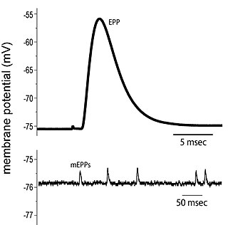
End plate potentials (EPPs) are the voltages which cause depolarization of skeletal muscle fibers caused by neurotransmitters binding to the postsynaptic membrane in the neuromuscular junction. They are called "end plates" because the postsynaptic terminals of muscle fibers have a large, saucer-like appearance. When an action potential reaches the axon terminal of a motor neuron, vesicles carrying neurotransmitters are exocytosed and the contents are released into the neuromuscular junction. These neurotransmitters bind to receptors on the postsynaptic membrane and lead to its depolarization. In the absence of an action potential, acetylcholine vesicles spontaneously leak into the neuromuscular junction and cause very small depolarizations in the postsynaptic membrane. This small response (~0.4mV) is called a miniature end plate potential (MEPP) and is generated by one acetylcholine-containing vesicle. It represents the smallest possible depolarization which can be induced in a muscle.
Neurotransmission is the process by which signaling molecules called neurotransmitters are released by the axon terminal of a neuron, and bind to and react with the receptors on the dendrites of another neuron a short distance away. A similar process occurs in retrograde neurotransmission, where the dendrites of the postsynaptic neuron release retrograde neurotransmitters that signal through receptors that are located on the axon terminal of the presynaptic neuron, mainly at GABAergic and glutamatergic synapses.
Neuromodulation is the physiological process by which a given neuron uses one or more chemicals to regulate diverse populations of neurons. Neuromodulators typically bind to metabotropic, G-protein coupled receptors (GPCRs) to initiate a second messenger signaling cascade that induces a broad, long-lasting signal. This modulation can last for hundreds of milliseconds to several minutes. Some of the effects of neuromodulators include: alter intrinsic firing activity, increase or decrease voltage-dependent currents, alter synaptic efficacy, increase bursting activity and reconfiguration of synaptic connectivity.
The catecholamines comprise the endogenous substances dopamine, noradrenaline (norepinephrine) and adrenaline (epinephrine) as well as numerous artificially synthesized compounds such as isoprenaline. Their investigation constitutes a prominent chapter in the history of physiology, biochemistry and pharmacology. Adrenaline was the first hormone extracted from its endocrine gland and obtained in pure form, before the word hormone was coined. It was also the first hormone the structure and biosynthesis of which were clarified. Apart from acetylcholine, adrenaline and noradrenaline were the first neurotransmitters to be discovered and the first intercellular biochemical signals to be found in intracellular vesicles. The β-adrenoceptor was the first G protein-coupled receptor the gene of which was cloned. Goal-directed catecholamine research began with the preparation by George Oliver and Edward Albert Sharpey-Schafer of a pharmacologically active extract from the adrenal glands.
This page is based on this Wikipedia article Text is available under the CC BY-SA 4.0 license; additional terms may apply. Images, videos and audio are available under their respective licenses.