In human anatomy, the thoracic duct is the larger of the two lymph ducts of the lymphatic system. The thoracic duct usually begins from the upper aspect of the cisterna chyli, passing out of the abdomen through the aortic hiatus into first the posterior mediastinum and then the superior mediastinum, extending as high up as the root of the neck before descending to drain into the systemic (blood) circulation at the venous angle.

In anatomy, the epidural space is the potential space between the dura mater and vertebrae (spine).

The occipital sinus is the smallest of the dural venous sinuses. It is usually unpaired, and is sometimes altogether absent. It is situated in the attached margin of the falx cerebelli. It commences near the foramen magnum, and ends by draining into the confluence of sinuses.

The vertebral vein is formed in the suboccipital triangle, from numerous small tributaries which spring from the internal vertebral venous plexuses and issue from the vertebral canal above the posterior arch of the atlas.

The occipital vein is a vein of the scalp. It originates from a plexus around the external occipital protuberance and superior nuchal line to the back part of the vertex of the skull. It usually drains into the internal jugular vein, but may also drain into the posterior auricular vein. It drains part of the scalp.
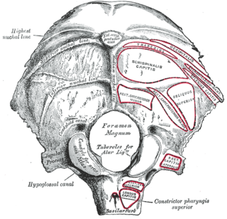
The condylar canal is a canal in the condyloid fossa of the lateral parts of occipital bone behind the occipital condyle. Resection of the rectus capitis posterior major and minor muscles reveals the bony recess leading to the condylar canal, which is situated posterior and lateral to the occipital condyle. It is immediately superior to the extradural vertebral artery, which makes a loop above the posterior C1 ring to enter the foramen magnum. The anteriomedial wall of the condylar canal thickens to join the foramen magnum rim and connect to the occipital condyle.

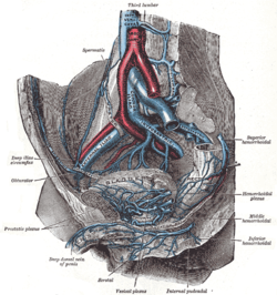
The rectal venous plexus surrounds the rectum, and communicates in front with the vesical venous plexus in the male, and the vaginal venous plexus in the female.
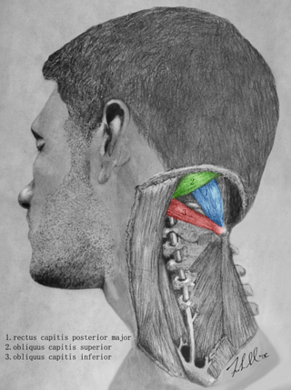
The suboccipital triangle is a region of the neck bounded by the following three muscles of the suboccipital group of muscles:

The basilar plexus consists of several interlacing venous channels between the layers of the dura mater over the basilar part of the occipital bone, and serves to connect the two inferior petrosal sinuses.

The external vertebral venous plexuses best marked in the cervical region, consist of anterior and posterior plexuses which anastomose freely with each other.
The internal vertebral venous plexuses lie within the vertebral canal in the epidural space, and receive tributaries from the bones and from the spinal cord.
The basivertebral veins are veins within the vertebral column. They are contained in large, tortuous channels in the substance of the bones, similar in every respect to those found in the diploë of the cranial bones.

The spinal veins are situated in the pia mater and form a minute, tortuous, venous plexus.
Vertebral venous plexuses may refer to:

The prostatic veins form a well-marked prostatic plexus which lies partly in the fascial sheath of the prostate and partly between the sheath and the prostatic capsule. It communicates with the pudendal and vesical plexuses.

The vaginal venous plexus is a group of veins draining blood from the vagina. It lies around the sides of the vagina. Its blood is eventually into the internal iliac veins.
The Batson venous plexus is a network of valveless veins in the human body that connect the deep pelvic veins and thoracic veins to the internal vertebral venous plexuses. Because of their location and lack of valves, they are believed to provide a route for the spread of cancer metastases. These metastases commonly arise from cancer of the pelvic organs such as the rectum and prostate and may spread to the vertebral column or brain. The plexus is named after anatomist Oscar Vivian Batson, who first described it in 1940. Batson's plexus is part of the Cerebrospinal venous system.
The suboccipital venous plexus drains deoxygenated blood from the back of the head.
Anterior spinal veins are veins that receive blood from the anterior spinal cord.
The cerebrospinal venous system (CSVS) consists of the interconnected venous systems of the brain and the spine.