Warfarin necrosis

Last updated
Warfarin necrosis
Other namesAnticoagulant-induced skin necrosis
Right leg affected by warfarin necrosis.png
Right leg affected by warfarin necrosis.

Warfarin-induced skin necrosis is a condition in which skin and subcutaneous tissue necrosis (tissue death) occurs due to acquired protein C deficiency following treatment with anti-vitamin K anticoagulants (4-hydroxycoumarins, such as warfarin). [1]

Contents

Warfarin necrosis is a rare but severe complication of treatment with warfarin or related anticoagulants. [2] The typical patient appears to be an obese, middle aged woman (median age 54 years, male to female ratio 1:3). [1] [3] :122–3 This drug eruption usually occurs between the third and tenth days of therapy with warfarin derivatives. [1] The first symptoms are pain and redness in the affected area. As they progress, lesions develop a sharp border and become petechial, then hard and purpuric. They may then resolve or progress to form large, irregular, bloody bullae with eventual necrosis and slow-healing eschar formation. Favored sites are breasts, thighs, buttocks and penis, [1] all areas with subcutaneous fat. [3] :122 In rare cases, the fascia and muscle are involved. [4]

Development of the syndrome is associated with the use of large loading doses at the start of treatment. [5]

Mechanism

The coagulation cascade. Coagulation full.svg
The coagulation cascade.

Warfarin necrosis usually occurs three to five days after drug therapy is begun, and a high initial dose increases the risk of its development. [3] :122 Warfarin-induced necrosis can develop both at sites of local injection and - when infused intravenously - in a widespread pattern. [3] :123

In warfarin's initial stages of action, inhibition of protein C and Factor VII is stronger than inhibition of the other vitamin K-dependent coagulation factors II, IX, and X. This results from the fact that these proteins have different half-lives: 1.5 to six hours for factor VII and eight hours for protein C, versus one day for factor IX, two days for factor X and two to five days for factor II. The larger the initial dose of vitamin K-antagonist, the more pronounced these differences are. This coagulation factor imbalance leads to paradoxical activation of coagulation, resulting in a hypercoagulable state and thrombosis. The blood clots interrupt the blood supply to the skin, causing necrosis. Protein C is an innate anticoagulant, and as warfarin further decreases protein C levels, it can lead to massive thrombosis with necrosis and gangrene of limbs.

Notably, the prothrombin time (or international normalized ratio, INR) used to test the effect of warfarin is highly dependent on factor VII, which explains why patients can have a therapeutic INR (indicating good anticoagulant effect) but still be in a hypercoagulable state. [1]

In one third of cases, warfarin necrosis occurs in patients with an underlying, innate and previously unknown deficiency of protein C. The condition is related to purpura fulminans, a complication in infants with sepsis which also involves skin necrosis. These infants often have protein C deficiency as well. There have also been cases in patients with other deficiency, including protein S deficiency, [6] [7] activated protein C resistance (Factor V Leiden) [8] and antithrombin III deficiency. [9]

Although the above hypothesis is the most commonly accepted, others believe that it is a hypersensitivity reaction or a direct toxic effect. [1]

Diagnosis

Differential diagnosis

Many conditions mimic or may be mistaken for warfarin necrosis, including pyoderma gangrenosum or necrotizing fasciitis. Warfarin necrosis is also different from another drug eruption associated with warfarin, purple toe syndrome, which usually occurs three to eight weeks after the start of anticoagulation therapy. No report has described this disorder in the immediate postpartum period in patients with protein S deficiency. [10]

Prevention

Vitamin K1 can be used to reverse the effects of warfarin, and heparin or its low molecular weight heparin (LMWH) can be used in an attempt to prevent further clotting. None of these suggested therapies have been studied in clinical trials.

Heparin and LMWH act by a different mechanism than warfarin, so these drugs can also be used to prevent clotting during the first few days of warfarin therapy and thus prevent warfarin necrosis (this is called 'bridging').

Treatment

The first element of treatment is usually to discontinue the offending drug, although there have been reports describing how the eruption evolved little after it had established in spite of continuing the medication. [11] [12]

Based on the assumption that low levels of protein C are involved in the underlying mechanism, common treatments in this setting include fresh frozen plasma or pure activated protein C. [13]

Since the clot-promoting effects of starting administration of 4-hydroxycoumarins are transitory, patients with protein C deficiency or previous warfarin necrosis can still be restarted on these drugs if appropriate measures are taken. [14] These include gradual increase starting from low doses and supplemental administration of protein C (pure or from fresh frozen plasma). [15]

The necrotic skin areas are treated as in other conditions, sometimes healing spontaneously with or without scarring, sometimes going on to require surgical debridement or skin grafting. [1]

History

While skin necrosis in patients had been previously described, Verhagen was the first to publish a paper on this relationship in the medical literature, in 1954. [12]

See also

Related Research Articles

<span class="mw-page-title-main">Anticoagulant</span> Class of drugs

Anticoagulants, commonly known as blood thinners, are chemical substances that prevent or reduce coagulation of blood, prolonging the clotting time. Some of them occur naturally in blood-eating animals such as leeches and mosquitoes, where they help keep the bite area unclotted long enough for the animal to obtain some blood. As a class of medications, anticoagulants are used in therapy for thrombotic disorders. Oral anticoagulants (OACs) are taken by many people in pill or tablet form, and various intravenous anticoagulant dosage forms are used in hospitals. Some anticoagulants are used in medical equipment, such as sample tubes, blood transfusion bags, heart–lung machines, and dialysis equipment. One of the first anticoagulants, warfarin, was initially approved as a rodenticide.

<span class="mw-page-title-main">Venous thrombosis</span> Blood clot (thrombus) that forms within a vein

Venous thrombosis is blockage of a vein caused by a thrombus. A common form of venous thrombosis is deep vein thrombosis (DVT), when a blood clot forms in the deep veins. If a thrombus breaks off (embolizes) and flows to the lungs to lodge there, it becomes a pulmonary embolism (PE), a blood clot in the lungs. The conditions of DVT only, DVT with PE, and PE only, are all captured by the term venous thromboembolism (VTE).

<span class="mw-page-title-main">Warfarin</span> Anticoagulant medication and rat poison

Warfarin is an anticoagulant used as a medication under several brand names including Coumadin, and as a poison for rats and other pests. While the drug is described as a "blood thinner", it does not reduce viscosity but inhibits coagulation, and is commonly used to prevent blood clots in the circulatory system such as deep vein thrombosis and pulmonary embolism, and to protect against stroke in people who have atrial fibrillation, valvular heart disease, or artificial heart valves. Less commonly, it is used following ST-segment elevation myocardial infarction and orthopedic surgery. It is usually taken by mouth, but may also be administered intravenously.

<span class="mw-page-title-main">Antiphospholipid syndrome</span> Medical condition

Antiphospholipid syndrome, or antiphospholipid antibody syndrome, is an autoimmune, hypercoagulable state caused by antiphospholipid antibodies. APS provokes blood clots (thrombosis) in both arteries and veins as well as pregnancy-related complications such as miscarriage, stillbirth, preterm delivery, and severe preeclampsia. Although the exact etiology of APS is still not clear, genetics is believed to play a key role in the development of the disease. The diagnostic criteria require one clinical event and two positive blood test results spaced at least three months apart that detect lupus anticoagulant, anti-apolipoprotein antibodies, or anti-cardiolipin antibodies.

Low-molecular-weight heparin (LMWH) is a class of anticoagulant medications. They are used in the prevention of blood clots and treatment of venous thromboembolism and in the treatment of myocardial infarction.

<span class="mw-page-title-main">Prothrombin time</span> Assay for evaluating the extrinsic pathway & common pathway of coagulation

The prothrombin time (PT) – along with its derived measures of prothrombin ratio (PR) and international normalized ratio (INR) – is an assay for evaluating the extrinsic pathway and common pathway of coagulation. This blood test is also called protime INR and PT/INR. They are used to determine the clotting tendency of blood, in such things as the measure of warfarin dosage, liver damage, and vitamin K status. PT measures the following coagulation factors: I (fibrinogen), II (prothrombin), V (proaccelerin), VII (proconvertin), and X.

<span class="mw-page-title-main">Heparin-induced thrombocytopenia</span> Low platelet count due to heparin, associated with a risk of thrombosis

Heparin-induced thrombocytopenia (HIT) is the development of thrombocytopenia, due to the administration of various forms of heparin, an anticoagulant. HIT predisposes to thrombosis because platelets release microparticles that activate thrombin, thereby leading to thrombosis. When thrombosis is identified the condition is called heparin-induced thrombocytopenia and thrombosis (HITT). HIT is caused by the formation of abnormal antibodies that activate platelets. If someone receiving heparin develops new or worsening thrombosis, or if the platelet count falls, HIT can be confirmed with specific blood tests.

<span class="mw-page-title-main">Thrombophilia</span> Abnormality of blood coagulation

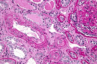
Thrombophilia is an abnormality of blood coagulation that increases the risk of thrombosis. Such abnormalities can be identified in 50% of people who have an episode of thrombosis that was not provoked by other causes. A significant proportion of the population has a detectable thrombophilic abnormality, but most of these develop thrombosis only in the presence of an additional risk factor.

<span class="mw-page-title-main">Argatroban</span> Pharmaceutical drug

Argatroban is an anticoagulant that is a small molecule direct thrombin inhibitor. In 2000, argatroban was licensed by the Food and Drug Administration (FDA) for prophylaxis or treatment of thrombosis in patients with heparin-induced thrombocytopenia (HIT). In 2002, it was approved for use during percutaneous coronary interventions in patients who have HIT or are at risk for developing it. In 2012, it was approved by the MHRA in the UK for anticoagulation in patients with heparin-induced thrombocytopenia Type II (HIT) who require parenteral antithrombotic therapy.

<span class="mw-page-title-main">Fresh frozen plasma</span>

Fresh frozen plasma (FFP) is a blood product made from the liquid portion of whole blood. It is used to treat conditions in which there are low blood clotting factors or low levels of other blood proteins. It may also be used as the replacement fluid in plasma exchange. Using ABO compatible plasma, while not required, may be recommended. Use as a volume expander is not recommended. It is given by slow injection into a vein.

<span class="mw-page-title-main">Hypoprothrombinemia</span> Medical condition

Hypoprothrombinemia is a rare blood disorder in which a deficiency in immunoreactive prothrombin, produced in the liver, results in an impaired blood clotting reaction, leading to an increased physiological risk for spontaneous bleeding. This condition can be observed in the gastrointestinal system, cranial vault, and superficial integumentary system, affecting both the male and female population. Prothrombin is a critical protein that is involved in the process of hemostasis, as well as illustrating procoagulant activities. This condition is characterized as an autosomal recessive inheritance congenital coagulation disorder affecting 1 per 2,000,000 of the population, worldwide, but is also attributed as acquired.

<span class="mw-page-title-main">Protein C deficiency</span> Medical condition

Protein C deficiency is a rare genetic trait that predisposes to thrombotic disease. It was first described in 1981. The disease belongs to a group of genetic disorders known as thrombophilias. Protein C deficiency is associated with an increased incidence of venous thromboembolism, whereas no association with arterial thrombotic disease has been found.

<span class="mw-page-title-main">Phenprocoumon</span> Drug

Phenprocoumon is a long-acting blood thinner drug to be taken by mouth, and a derivative of coumarin. It acts as a vitamin K antagonist and inhibits blood clotting (coagulation) by blocking synthesis of coagulation factors II, VII, IX and X. It is used for the prophylaxis and treatment of thromboembolic disorders such as heart attacks and pulmonary (lung) embolism. The most common adverse effect is bleeding. The drug interacts with a large number of other medications, including aspirin and St John's Wort. It is the standard coumarin used in Germany, Austria, and other European countries.

Prothrombin complex concentrate (PCC), also known as factor IX complex, is a medication made up of blood clotting factors II, IX, and X. Some versions also contain factor VII. It is used to treat and prevent bleeding in hemophilia B if pure factor IX is not available. It may also be used for reversal of warfarin therapy. It is given by slow injection into a vein.

Purpura fulminans is an acute, often fatal, thrombotic disorder which manifests as blood spots, bruising and discolouration of the skin resulting from coagulation in small blood vessels within the skin and rapidly leads to skin necrosis and disseminated intravascular coagulation.

Hypercoagulability in pregnancy is the propensity of pregnant women to develop thrombosis. Pregnancy itself is a factor of hypercoagulability, as a physiologically adaptive mechanism to prevent post partum bleeding. However, when combined with an additional underlying hypercoagulable states, the risk of thrombosis or embolism may become substantial.

Direct factor Xa inhibitors (xabans) are anticoagulants, used to both treat and prevent blood clots in veins, and prevent stroke and embolism in people with atrial fibrillation (AF).

<span class="mw-page-title-main">Vitamin K antagonist</span>

Vitamin K antagonists (VKA) are a group of substances that reduce blood clotting by reducing the action of vitamin K. The term "vitamin K antagonist" is technically a misnomer, as the drugs do not directly antagonise the action of vitamin K in the pharmacological sense, but rather the recycling of vitamin K.

Thromboelastometry (TEM), previously named rotational thromboelastography (ROTEG) or rotational thromboelastometry (ROTEM), is an established viscoelastic method for hemostasis testing in whole blood. It is a modification of traditional thromboelastography (TEG).

Direct thrombin inhibitors (DTIs) are a class of anticoagulant drugs that can be used to prevent and treat embolisms and blood clots caused by various diseases. They inhibit thrombin, a serine protease which affects the coagulation cascade in many ways. DTIs have undergone rapid development since the 90's. With technological advances in genetic engineering the production of recombinant hirudin was made possible which opened the door to this new group of drugs. Before the use of DTIs the therapy and prophylaxis for anticoagulation had stayed the same for over 50 years with the use of heparin derivatives and warfarin which have some well known disadvantages. DTIs are still under development, but the research focus has shifted towards factor Xa inhibitors, or even dual thrombin and fXa inhibitors that have a broader mechanism of action by both inhibiting factor IIa (thrombin) and Xa. A recent review of patents and literature on thrombin inhibitors has demonstrated that the development of allosteric and multi-mechanism inhibitors might lead the way to a safer anticoagulant.

References

  1. 1 2 3 4 5 6 7 McKnight JT, Maxwell AJ, Anderson RL (1992). "Warfarin necrosis". Arch Fam Med. 1 (1): 105–8. doi:10.1001/archfami.1.1.105. PMID   1341581.
  2. Rapini, Ronald P.; Bolognia, Jean L.; Jorizzo, Joseph L. (2007). Dermatology: 2-Volume Set. St. Louis: Mosby. pp. 331, 340. ISBN   978-1-4160-2999-1.
  3. 1 2 3 4 James, William; Berger, Timothy; Elston, Dirk (2005). Andrews' Diseases of the Skin: Clinical Dermatology. (10th ed.). Saunders. ISBN   0-7216-2921-0.
  4. Schleicher SM, Fricker MP (April 1980). "Coumadin necrosis". Arch Dermatol. 116 (4): 444–5. doi:10.1001/archderm.116.4.444. PMID   7369776.
  5. Chan YC, Valenti D, Mansfield AO, Stansby G (March 2000). "Warfarin induced skin necrosis". Br J Surg. 87 (3): 266–72. doi:10.1046/j.1365-2168.2000.01352.x. PMC   4928566 . PMID   10718793.
  6. Sallah S, Abdallah JM, Gagnon GA (1998). "Recurrent warfarin-induced skin necrosis in kindreds with protein S deficiency". Haemostasis. 28 (1): 25–30. doi:10.1159/000022380. PMID   9885367. S2CID   46762929.
  7. Grimaudo V, Gueissaz F, Hauert J, Sarraj A, Kruithof EK, Bachmann F (January 1989). "Necrosis of skin induced by coumadin in a patient deficient in protein S". BMJ. 298 (6668): 233–4. doi:10.1136/bmj.298.6668.233. PMC   1835547 . PMID   2522326.
  8. Makris M, Bardhan G, Preston FE (March 1996). "Warfarin induced skin necrosis associated with activated protein C resistance". Thromb. Haemost. 75 (3): 523–4. doi:10.1055/s-0038-1650312. PMID   8701423. S2CID   30576117.
  9. Kiehl R, Hellstern P, Wenzel E (January 1987). "Hereditary antithrombin III (AT III) deficiency and atypical localization of a coumadin necrosis". Thromb. Res. 45 (2): 191–3. doi:10.1016/0049-3848(87)90173-3. PMID   3563984.
  10. Cheng, A; Scheinfeld, NS; McDowell, B; Dokras, AA (1997). "Warfarin skin necrosis in a postpartum woman with protein S deficiency". Obstetrics and Gynecology. 90 (4 Pt 2): 671–2. doi:10.1016/S0029-7844(97)00393-1. PMID   11770590. S2CID   20764940.
  11. Nalbandian RM, Mader IJ, Barrett JL, Pearce JF, Rupp EC (May 1965). "Petechiae, ecchymoses, and necrosis of skin induced by coumadin congeners: rare, occasionally lethal complication of anticoagulant therapy". JAMA. 192: 603–8. doi:10.1001/jama.1965.03080200021006. PMID   14284863.
  12. 1 2 Verhagen H (1954). "Local haemorrhage and necrosis of the skin and underlying tissues, during anti-coagulant therapy with dicumarol or dicumacyl". Acta Med Scand. 148 (6): 453–67. doi:10.1111/j.0954-6820.1954.tb01741.x. PMID   13171021.
  13. Schramm W, Spannagl M, Bauer KA, et al. (June 1993). "Treatment of coumadin-induced skin necrosis with a monoclonal antibody purified protein C concentrate". Arch Dermatol. 129 (6): 753–6. doi:10.1001/archderm.129.6.753. PMID   8507079.
  14. Zauber NP, Stark MW (May 1986). "Successful warfarin anticoagulation despite protein C deficiency and a history of warfarin necrosis". Ann. Intern. Med. 104 (5): 659–60. doi:10.7326/0003-4819-104-5-659. PMID   3754407.
  15. De Stefano V, Mastrangelo S, Schwarz HP, et al. (August 1993). "Replacement therapy with a purified protein C concentrate during initiation of oral anticoagulation in severe protein C congenital deficiency". Thromb. Haemost. 70 (2): 247–9. doi:10.1055/s-0038-1649478. PMID   8236128. S2CID   28480921.