Well-woman examination

Last updated

A well-woman examination is an exam offered to women to review elements of their reproductive health. The exam includes a breast examination, a pelvic examination and a Pap smear but may include other procedures. Hospitals employ strict policies relating to the provision of consent by the patient, the availability of chaperones at the examination, and the absence of other parties. [1] [2]

Contents

Importance

Although women often undergo well-woman examinations on an annual basis, the interval for this visit and exam will vary depending on the needs of the patient. [3] The purpose of this exam in asymptomatic women is to screen for potential abnormalities, such as sexually transmitted infections, and malignancy. [4] [5]

Breast examination

Visual inspection

The breast examination begins with a visual inspection. With the patient in a supine or seated position, the medical professional will look at both breasts to check the color, symmetry, dimensions according to age, lean body mass, the physiological (pregnancy and lactation) and race, looking for abnormalities, such as bulges and shrinkage. [6] One of these abnormalities is changed in the areola or nipple. If it is flattened or retracted (umbilicated), it is necessary to consider the possibility of a cancerous lesion which has caused the malformation. [6] [7]

Palpation

Next, the breasts are palpated, again with the patient lying or sitting. The patient has to lift the arm and put one hand behind her head. With this position, the entire gland is palpated. It is also important to examine the armpits, because of masses that may be found there. [6] The test is executed pressing the gland with two or three fingers against the chest wall, making a radial route or by quadrants. The nipples are also squeezed to check for secretions, such as secretion of milk (galactorrhea), serous, blood or purulent secretions. If a node is detected, it is necessary to determine its place, size, shape, edges, consistency and sensitivity. [6]

Self-examination

Breast self-examination is not recommended as this practice is associated with increased false positive findings and no evidence of benefit. [8] Instead breast self-awareness is encouraged. Breast self-awareness includes being familiar with the normal contour of one's breast but does not involve monthly self-examinations. [9]

Further examination of the breasts

A mammogram is a special X-ray of the breasts. They are the procedure most likely to detect early breast cancer in asymptomatic women. Mammograms can show tumors long before they are large enough to palpate. They are recommended for women who have symptoms of breast cancer or who are at increased risk of developing the disease. They are performed with the patient standing, the breast pressed between two plastic plates, as the image is taken. The interpretation has to be performed by a specialist. [10]

Breast ultrasound is a complementary study of mammography. In many women the tissue that makes up the breast is very dense, representing fibrous tissue and glandular tissue, which produces milk during lactation. This limits the radiologist interpreting the study, so, in these cases, the ultrasound is helpful, since this is capable of distinguishing tumors in women with dense breast tissue, where identification is otherwise difficult. Additionally, it is advisable to follow up a mammogram that shows indications of tumors with an ultrasound, to confirm, before more invasive procedures are undertaken. [11]

Pelvic exam

The pelvic exam is part of the physical examination of the internal pelvic organs (uterus, cervix, ovaries), vagina, and vulva. This exam often includes three parts:

  1. Inspection of the vulva
  2. Bimanual examination
  3. Inspection of the cervix and vagina using a speculum. [12]

Inspection of vulva

The patient is placed in a supine position on a special examination table, which has two protrusions called "stirrups". With the feet in these stirrups, the legs are placed in a position such that the medical professional can access the pelvic area. The external genitalia is examined first, looking for abnormalities like lesions, ulcers, warts and color changes. The elements of this exam include the vulva, which contains the mons pubis, of which there are two longitudinal folds of skin forming the labia majora; then the labia minora and hair follicles. The clitoral hood is also checked. [12]

Bimanual exam

The purpose of this exam is to palpate or feel the pelvic organs. The index and middle finger are inserted into the vagina. This maneuver allows the doctor to palpate the vagina for any deep lacerations, masses, or nodularity. Next, the cervix is palpated to check position, size, consistency, and mobility. [13] The other hand is placed on the abdomen, compressing the uterus between both hands. This maneuver allows the clinician to assess the size, shape, consistency, tilt, and mobility of the uterus. With this technique, the ovaries may also be palpable. [13] This examination is useful for identifying clinical signs of medical conditions, such as infection, [14] presence of a mass, [15] or structural abnormality. [15] "Regular performance of this examination is crucial in early detection and management of gynecological conditions, which can significantly enhance treatment outcomes."

Speculum examination

The speculum is an instrument made of metal or plastic and is constructed with two flaps. Its purpose is to separate and widen the vaginal opening and keep it open. This allows direct observation by the physician into the vaginal canal with the help of a lamp or a mirror. [16] There are different types of speculums used within the different characteristics of each patient such as age, sex life, and other factors. The first step is to open the vaginal opening with two fingers at the vulvo-perineal angle, then separate the fingers slightly and press down, then insert the speculum arranging the width of the tip of the flaps in anteroposterior. Then the speculum is moved into the vagina at an angle of 45°, following the natural contour of the posterior vaginal wall. When the speculum is in place, the fingers are removed and the device is rotated so that the flaps are horizontal. The flaps are then separated and locked into place when the cervical neck is completely visible. [16] The speculum examination is not necessary for adolescents who are asymptomatic. [17]

Screening tests

Samples for screening tests may be collected during the pelvic exam. These screening tests include:

  1. Cervical cancer screening - A Pap smear and/or HPV testing may be performed as a screening test for cervical cancer. [18] The procedure begins by gently scraping or sampling the cells of the cervix using a special spatula, brush or swab. Some women experience temporary bleeding from this procedure. The scrapings are placed on a slide, covered with a fixative for later examination under a microscope to determine if they are normal or abnormal. [19] Depending on patient's age or Pap smear result, HPV testing may also be performed.
  2. Sexually transmitted infection screening - Depending on age and risk factors, clinicians may recommend gonorrhea or chlamydia testing at the time of the well-woman exam. This sample can be collected via a swab of the cervix or vagina. This swab can be collected by the clinician or the patient. Urine samples can also be used for this test. Additional screening tests include blood tests for hepatitis C, HIV, and syphilis. [20]

Related Research Articles

<span class="mw-page-title-main">Pap test</span> Cervical screening test to detect potential cancers

The Papanicolaou test is a method of cervical screening used to detect potentially precancerous and cancerous processes in the cervix or, more rarely, anus. Abnormal findings are often followed up by more sensitive diagnostic procedures and, if warranted, interventions that aim to prevent progression to cervical cancer. The test was independently invented in the 1920s by the Greek physician Georgios Papanikolaou and named after him. A simplified version of the test was introduced by the Canadian obstetrician Anna Marion Hilliard in 1957.

Obstetrics and gynaecology is the medical specialty that encompasses the two subspecialties of obstetrics and gynaecology. The specialization is an important part of care for women's health.

<span class="mw-page-title-main">Gynaecology</span> Science of the treatment of diseases of the female sexual organs and reproductive tract

Gynaecology or gynecology is the area of medicine that involves the treatment of women's diseases, especially those of the reproductive organs. It is often paired with the field of obstetrics, forming the combined area of obstetrics and gynaecology (OB-GYN).

<span class="mw-page-title-main">Colposcopy</span> Medical examination of the cervix

Colposcopy is a medical diagnostic procedure to visually examine the cervix as well as the vagina and vulva using a colposcope.

Dyspareunia is painful sexual intercourse due to medical or psychological causes. The term dyspareunia covers both female dyspareunia and male dyspareunia, but many discussions that use the term without further specification concern the female type, which is more common than the male type. In females, the pain can primarily be on the external surface of the genitalia, or deeper in the pelvis upon deep pressure against the cervix. Medically, dyspareunia is a pelvic floor dysfunction and is frequently underdiagnosed. It can affect a small portion of the vulva or vagina or be felt all over the surface. Understanding the duration, location, and nature of the pain is important in identifying the causes of the pain.

<span class="mw-page-title-main">Speculum (medicine)</span> Medical device for investigating body orifices by direct vision

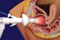
A speculum is a medical tool for investigating body orifices, with a form dependent on the orifice for which it is designed. In old texts, the speculum may also be referred to as a diopter or dioptra. Like an endoscope, a speculum allows a view inside the body; endoscopes, however, tend to have optics while a speculum is intended for direct vision.

<span class="mw-page-title-main">Cervicitis</span> Inflammation of the uterine cervix

Cervicitis is inflammation of the uterine cervix. Cervicitis in women has many features in common with urethritis in men and many cases are caused by sexually transmitted infections. Non-infectious causes of cervicitis can include intrauterine devices, contraceptive diaphragms, and allergic reactions to spermicides or latex condoms. Cervicitis affects over half of all women during their adult life.

<span class="mw-page-title-main">Vaginal bleeding</span> Medical condition

Vaginal bleeding is any expulsion of blood from the vagina. This bleeding may originate from the uterus, vaginal wall, or cervix. Generally, it is either part of a normal menstrual cycle or is caused by hormonal or other problems of the reproductive system, such as abnormal uterine bleeding.

<span class="mw-page-title-main">Vaginal discharge</span> Medical condition

Vaginal discharge is a mixture of liquid, cells, and bacteria that lubricate and protect the vagina. This mixture is constantly produced by the cells of the vagina and cervix, and it exits the body through the vaginal opening. The composition, amount, and quality of discharge varies between individuals and can vary throughout the menstrual cycle and throughout the stages of sexual and reproductive development. Normal vaginal discharge may have a thin, watery consistency or a thick, sticky consistency, and it may be clear or white in color. Normal vaginal discharge may be large in volume but typically does not have a strong odor, nor is it typically associated with itching or pain. While most discharge is considered physiologic or represents normal functioning of the body, some changes in discharge can reflect infection or other pathological processes. Infections that may cause changes in vaginal discharge include vaginal yeast infections, bacterial vaginosis, and sexually transmitted infections. The characteristics of abnormal vaginal discharge vary depending on the cause, but common features include a change in color, a foul odor, and associated symptoms such as itching, burning, pelvic pain, or pain during sexual intercourse.

Vaginal cancer is an extraordinarily rare form of cancer that develops in the tissue of the vagina. Primary vaginal cancer originates from the vaginal tissue – most frequently squamous cell carcinoma, but primary vaginal adenocarcinoma, sarcoma, and melanoma have also been reported – while secondary vaginal cancer involves the metastasis of a cancer that originated in a different part of the body. Secondary vaginal cancer is more common. Signs of vaginal cancer may include abnormal vaginal bleeding, dysuria, tenesmus, or pelvic pain, though as many as 20% of women diagnosed with vaginal cancer are asymptomatic at the time of diagnosis. Vaginal cancer occurs more frequently in women over age 50, and the mean age of diagnosis of vaginal cancer is 60 years. It often can be cured if found and treated in early stages. Surgery alone or surgery combined with pelvic radiation is typically used to treat vaginal cancer.

<span class="mw-page-title-main">Pelvic examination</span> Physical medical examination

A pelvic examination is the physical examination of the external and internal female pelvic organs. It is frequently used in gynecology for the evaluation of symptoms affecting the female reproductive and urinary tract, such as pain, bleeding, discharge, urinary incontinence, or trauma. It can also be used to assess a woman's anatomy in preparation for procedures. The exam can be done awake in the clinic and emergency department, or under anesthesia in the operating room. The most commonly performed components of the exam are 1) the external exam, to evaluate the vulva 2) the internal exam with palpation to examine the uterus, ovaries, and structures adjacent to the uterus (adnexae) and 3) the internal exam using a speculum to visualize the vaginal walls and cervix. During the pelvic exam, sample of cells and fluids may be collected to screen for sexually transmitted infections or cancer.

<span class="mw-page-title-main">Endometrial biopsy</span> Diagnostic medical procedure

The endometrial biopsy is a medical procedure that involves taking a tissue sample of the lining of the uterus. The tissue subsequently undergoes a histologic evaluation which aids the physician in forming a diagnosis.

<span class="mw-page-title-main">Hematometra</span> Medical condition

Hematometra is a medical condition involving collection or retention of blood in the uterus. It is most commonly caused by an imperforate hymen or a transverse vaginal septum.

A vaginal disease is a pathological condition that affects part or all of the vagina.

<span class="mw-page-title-main">Cervical cancer staging</span> Medical condition

Cervical cancer staging is the assessment of cervical cancer to determine the extent of the spread of cancer beyond the cervix. This is important for determining how serious the cancer is and to create the best treatment plan.

<span class="mw-page-title-main">Fertility testing</span>

Fertility testing is the process by which fertility is assessed, both generally and also to find the "fertile window" in the menstrual cycle. General health affects fertility, and STI testing is an important related field.

<span class="mw-page-title-main">Postcoital bleeding</span> Medical condition

Postcoital bleeding (PCB) is non-menstrual vaginal bleeding that occurs during or after sexual intercourse. Though some causes are with associated pain, it is typically painless and frequently associated with intermenstrual bleeding.

A rectovaginal examination is a type of gynecological examination used to supplement a pelvic examination. In the rectovaginal examination, a doctor or other health care provider places one finger in the vagina and another in the rectum to assess the rectovaginal septum. The examiner will look for any scarring or masses that may indicate cancer or another disease. Typically, a rectovaginal examination is performed to assess pelvic pain, rectal symptoms, or a pelvic mass. It can also provide a sample for fecal occult blood testing.

Pediatric gynaecology or pediatric gynecology is the medical practice dealing with the health of the vagina, vulva, uterus, and ovaries of infants, children, and adolescents. Its counterpart is pediatric andrology, which deals with medical issues specific to the penis and testes.

<span class="mw-page-title-main">Squamous cell carcinoma of the vagina</span> Medical condition

Squamous cell carcinoma of the vagina is a potentially invasive type of cancer that forms in the tissues of the vagina. Though uncommonly diagnosed, squamous cell cancer of the vagina (SCCV) is the most common type of vaginal cancer, accounting for 80-90% of cases as well as 2% of all gynecological cancers. SCCV forms in squamous cells, which are the thin, flat cells lining the vagina. SCCV initially spreads superficially within the vaginal wall and can slowly spread to invade other vaginal tissues. Because of its slow growth, this cancer may cause no symptoms, or it may present with signs like irregular bleeding, pain, or a vaginal mass. This carcinoma can metastasize to the lungs or less frequently to the liver, bone, or other sites. SCCV has many risk factors in common with cervical cancer and is similarly strongly associated with infection with oncogenic strains of human papillomavirus (HPV). Diagnosis of SCCV is done by pelvic exam and biopsy of the tissue. Treatment and prognosis will depend on the stage, location, and characteristics of the cancer.

References

  1. Kath Evans (July 2003). "INTIMATE EXAMINATION, PROCEDURE, CARE & CHAPERONING POLICY" (PDF). Whittington Hospital NHS Trust.{{cite journal}}: Cite journal requires |journal= (help)
  2. Helen Barratt (October 2001). "School pupil allowed to observe intimate examination". Student BMJ. 09. BMJ Publishing Group Ltd: 357–398.
  3. "ACOG Committee Opinion No. 755 Summary: Well-Woman Visit". Obstetrics & Gynecology. 132 (4): 1084–1085. October 2018. doi:10.1097/AOG.0000000000002898. ISSN   0029-7844. PMID   30247360. S2CID   52348080.
  4. "Reproductive Health Information - The Well Woman Exam". Baylor College of Medicine. Center for research on women with disabilities. Retrieved 2013-04-17.
  5. "Well Woman's Exam". trive. Campus Health Services. Retrieved 2013-04-17.
  6. 1 2 3 4 "Examen de las Mamas". Manual de Semiología. Retrieved 2013-04-19.
  7. "Manual de exploración Física de las Mamas" (PDF). Secretaría de Salud. Retrieved 2013-04-19.
  8. Fuller MS, Lee CI, Elmore JG (Oct 2016). "Breast Cancer Screening: An Evidence-Based Update". The Medical Clinics of North America. 99 (3). Med Clin North Am: 451–468. doi:10.1016/j.mcna.2015.01.002. PMC   5064844 . PMID   25841594.
  9. "Practice Bulletin Number 179: Breast Cancer Risk Assessment and Screening in Average-Risk Women". Obstetrics & Gynecology. 130 (1): e1–e16. July 2017. doi:10.1097/AOG.0000000000002158. ISSN   0029-7844. PMID   28644335.
  10. "Mammography". Medline Plus. U.S. Department of Health and Human Services National Institutes of Health. Retrieved 2013-04-22.
  11. "Ultrasonido mamario". FUCAM. Retrieved 2013-04-22.
  12. 1 2 Parrondo P, Pérez-Medina T, Álvarez-Heros J (April 2013). "Anatomía del aparato genital femenino" (PDF). Salud Mujer: 15–18.
  13. 1 2 "Examen ginecológico". Manual de Semiología. Retrieved 2013-04-19.
  14. "Pelvic Exam". Stanford Medicine 25. Retrieved 2020-03-31.
  15. 1 2 "UCSD's Practical Guide to Clinical Medicine". meded.ucsd.edu. Retrieved 2020-03-31.
  16. 1 2 "Técnica de colocación del eséculo". Omar Morera. Retrieved 2013-04-18.
  17. Cavanaugh, Robert M. (2007-09-01). "Screening Adolescent Gynecology in the Pediatrician's Office: Have a Listen, Take a Look". Pediatrics in Review. 28 (9): 332–342. doi:10.1542/pir.28-9-332. ISSN   0191-9601. PMID   17766592.
  18. "Screening Guidelines - ASCCP". www.asccp.org. Retrieved 2020-04-01.
  19. Klein S., Miller S., Thomson F002E (May 2000). "El examen pélvico" (PDF). Atención del embarazo, el parto y la salud de la mujer: 373–387.{{cite journal}}: CS1 maint: multiple names: authors list (link) CS1 maint: numeric names: authors list (link)
  20. "Recommendations for Women's Healthcare Coverage | Women's Preventive Services Initiative". www.womenspreventivehealth.org. 2018-10-01. Retrieved 2020-04-01.