
Neutron activation analysis (NAA) is a nuclear process used for determining the concentrations of elements in many materials. NAA allows discrete sampling of elements as it disregards the chemical form of a sample, and focuses solely on atomic nuclei. The method is based on neutron activation and thus requires a neutron source. The sample is bombarded with neutrons, causing its constituent elements to form radioactive isotopes. The radioactive emissions and radioactive decay paths for each element have long been studied and determined. Using this information, it is possible to study spectra of the emissions of the radioactive sample, and determine the concentrations of the various elements within it. A particular advantage of this technique is that it does not destroy the sample, and thus has been used for the analysis of works of art and historical artifacts. NAA can also be used to determine the activity of a radioactive sample.

X-ray fluorescence (XRF) is the emission of characteristic "secondary" X-rays from a material that has been excited by being bombarded with high-energy X-rays or gamma rays. The phenomenon is widely used for elemental analysis and chemical analysis, particularly in the investigation of metals, glass, ceramics and building materials, and for research in geochemistry, forensic science, archaeology and art objects such as paintings.

A scintillation counter is an instrument for detecting and measuring ionizing radiation by using the excitation effect of incident radiation on a scintillating material, and detecting the resultant light pulses.

Nuclear medicine, or nucleology, is a medical specialty involving the application of radioactive substances in the diagnosis and treatment of disease. Nuclear imaging is, in a sense, radiology done inside out, because it records radiation emitted from within the body rather than radiation that is transmitted through the body from external sources like X-ray generators. In addition, nuclear medicine scans differ from radiology, as the emphasis is not on imaging anatomy, but on the function. For such reason, it is called a physiological imaging modality. Single photon emission computed tomography (SPECT) and positron emission tomography (PET) scans are the two most common imaging modalities in nuclear medicine.

A scintillator is a material that exhibits scintillation, the property of luminescence, when excited by ionizing radiation. Luminescent materials, when struck by an incoming particle, absorb its energy and scintillate. Sometimes, the excited state is metastable, so the relaxation back down from the excited state to lower states is delayed. The process then corresponds to one of two phenomena: delayed fluorescence or phosphorescence. The correspondence depends on the type of transition and hence the wavelength of the emitted optical photon.

Radioactive contamination, also called radiological pollution, is the deposition of, or presence of radioactive substances on surfaces or within solids, liquids, or gases, where their presence is unintended or undesirable.
A semiconductor detector in ionizing radiation detection physics is a device that uses a semiconductor to measure the effect of incident charged particles or photons.

A gamma camera (γ-camera), also called a scintillation camera or Anger camera, is a device used to image gamma radiation emitting radioisotopes, a technique known as scintigraphy. The applications of scintigraphy include early drug development and nuclear medical imaging to view and analyse images of the human body or the distribution of medically injected, inhaled, or ingested radionuclides emitting gamma rays.

Scintigraphy, also known as a gamma scan, is a diagnostic test in nuclear medicine, where radioisotopes attached to drugs that travel to a specific organ or tissue (radiopharmaceuticals) are taken internally and the emitted gamma radiation is captured by gamma cameras, which are external detectors that form two-dimensional images in a process similar to the capture of x-ray images. In contrast, SPECT and positron emission tomography (PET) form 3-dimensional images and are therefore classified as separate techniques from scintigraphy, although they also use gamma cameras to detect internal radiation. Scintigraphy is unlike a diagnostic X-ray where external radiation is passed through the body to form an image.
Explorer 11 was a NASA satellite that carried the first space-borne gamma-ray telescope. This marked the beginning of space gamma-ray astronomy. Launched on 27 April 1961 by a Juno II, the satellite returned data until 17 November 1961, when power supply problems ended the science mission. During the spacecraft's seven-month lifespan it detected twenty-two events from gamma-rays and approximately 22,000 events from cosmic radiation.

Gamma-ray spectroscopy is the qualitative study of the energy spectra of gamma-ray sources, such as in the nuclear industry, geochemical investigation, and astrophysics. Gamma-ray spectrometry, on the other hand, is the method used to acquire a quantitative spectrum measurement.
A gamma counter is an instrument to measure gamma radiation emitted by a radionuclide. Unlike survey meters, gamma counters are designed to measure small samples of radioactive material, typically with automated measurement and movement of multiple samples.
Hal Oscar Anger was an American electrical engineer and biophysicist at Donner Laboratory, University of California, Berkeley, known for his invention of the gamma camera.
Neutron detection is the effective detection of neutrons entering a well-positioned detector. There are two key aspects to effective neutron detection: hardware and software. Detection hardware refers to the kind of neutron detector used and to the electronics used in the detection setup. Further, the hardware setup also defines key experimental parameters, such as source-detector distance, solid angle and detector shielding. Detection software consists of analysis tools that perform tasks such as graphical analysis to measure the number and energies of neutrons striking the detector.

A rectilinear scanner is an imaging device, used to capture emission from radiopharmaceuticals in nuclear medicine. The image is created by physically moving a radiation detector over the surface of a radioactive patient. It has become obsolete in medical imaging, largely replaced by the gamma camera since the late 1960s.

Radioactivity is generally used in life sciences for highly sensitive and direct measurements of biological phenomena, and for visualizing the location of biomolecules radiolabelled with a radioisotope.
The formation evaluation gamma ray log is a record of the variation with depth of the natural radioactivity of earth materials in a wellbore. Measurement of natural emission of gamma rays in oil and gas wells are useful because shales and sandstones typically have different gamma ray levels. Shales and clays are responsible for most natural radioactivity, so gamma ray log often is a good indicator of such rocks. In addition, the log is also used for correlation between wells, for depth correlation between open and cased holes, and for depth correlation between logging runs.
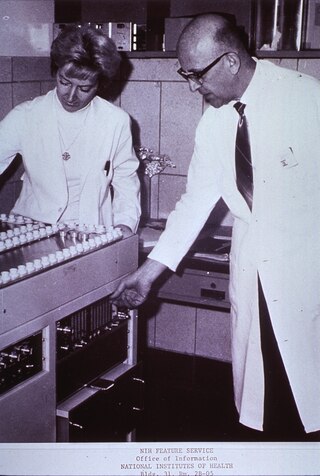
In health physics, whole-body counting refers to the measurement of radioactivity within the human body. The technique is primarily applicable to radioactive material that emits gamma rays. Alpha particle decays can also be detected indirectly by their coincident gamma radiation. In certain circumstances, beta emitters can be measured, but with degraded sensitivity. The instrument used is normally referred to as a whole body counter.
Nuclear medicine physicians, also called nuclear radiologists or simply nucleologists, are medical specialists that use tracers, usually radiopharmaceuticals, for diagnosis and therapy. Nuclear medicine procedures are the major clinical applications of molecular imaging and molecular therapy. In the United States, nuclear medicine physicians are certified by the American Board of Nuclear Medicine and the American Osteopathic Board of Nuclear Medicine.
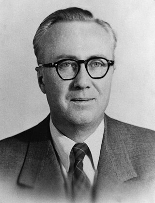
Leonidas D. Marinelli was the American radiological physicist who is best known for founding the field of Human Radiobiology and developing the Marinelli Beaker.
