Zona limitans intrathalamica

Last updated

The zona limitans intrathalamica (ZLI) is a lineage-restriction compartment and primary developmental boundary in the vertebrate forebrain (which is analogous to the human cerebrum) that serves as a signaling center and a restrictive border between the thalamus (also known as the dorsal thalamus) and the prethalamus (ventral thalamus).

Contents

Sonic hedgehog (shh) signaling from the ZLI is crucial in the development of the diencephalon , [1] which develops into the thalamus, the pretectum, and the anterior tegmental structures. [2] The ZLI together with the prethalamus and thalamus make up the mid-diencephalic territory (MDT). [3]

Forebrain structures in the fish at 3-4 days (with pretectum from midbrain for better depiction of boundaries), with shh expression in red; rostral extension between the prethalamus and thalamus depicts the ZLI. Labeled cichlid forebrain diagram with shh expression.jpg
Forebrain structures in the fish at 3-4 days (with pretectum from midbrain for better depiction of boundaries), with shh expression in red; rostral extension between the prethalamus and thalamus depicts the ZLI.

Discovery

Cell lineage restriction boundaries, across which replicating cells cannot migrate, were first discovered in invertebrates, where the expression of various Hox genes in each compartment confer the differentiation of observable segments in the adult body of Drosophila melanogaster. Analogous structures were discovered in the developing vertebrate brain. Rhombomeres, which extend down the embryo from the hindbrain, contain clear boundaries and each express various Hox genes that are necessary for differentiation of structures within the body. More anterior regions of the brain were examined in search of other cell lineage restriction boundaries, and multiple potential boundaries continue to be studied (see Developmental Boundaries). [4]

Layout of Hox genes in the fruitfly leading to differential development of segments Hoxgenesoffruitfly.svg
Layout of Hox genes in the fruitfly leading to differential development of segments

The importance of these compartments as local signaling centers, areas which chemically influence surrounding tissue, was elucidated by first observing differential expression of Hox genes in various compartments and second by observing mutant D. melanogaster and corresponding phenotypic (physical) changes. [4]

The ZLI was first discovered in the chick using explant and lineage-labeling experiments. In the explant experiments, cells in the region that will become the ZLI, the prethalamus, and the thalamus in the chick were removed and placed into separate cultures; the cells continued to grow and retained their identity (the ZLI began transcribing shh , whereas the prethalamus and thalamus did not). Necessity of the ZLI and its corresponding shh expression were evidenced by lack of thalamic and prethalamic genetic markers in culture (see Signaling). [5] These experiments confirmed the ZLI as a signaling center. In lineage-labeling experiments, cells were genetically marked, so that each time a labeled cell replicated, its progeny were marked as well. Cells that were marked in the developing ZLI and their progeny remained restricted to the zone. These experiments demonstrated the ZLI as a cell-lineage restriction boundary. [6]

Not only a boundary, the ZLI is also a compartment with separate cell lineage restriction boundaries both anterior and posterior of a section of shh expression. The importance of the ZLI was once more confirmed by ectopic expression of shh in other regions of the forebrain, known as the prosencephalon during development (both the telencephalon and diencephalon), inducing a ZLI-like region that induces thalamic fate. [7]

Developmental boundaries

During development in both vertebrates and invertebrates, cell lineage restriction boundaries and signaling centers are formed in order to ensure proper differentiation of the body. Chemical signals, like shh from the ZLI, are often released from these boundaries and compartments in a concentration gradient (the chemicals are in much higher concentrations closer to the source) and confer identity to flanking regions. Other genes differentially expressed in these flanking regions aid in ensuring proper differentiation (see Signaling).

Main structures of the developing brain: the prosencephalon (forebrain) consists of the telencephalon and diencephalon, the mesencephalon is the midbrain, and the rhombencephalon is the hindbrain. Four week embryo brain.svg
Main structures of the developing brain: the prosencephalon (forebrain) consists of the telencephalon and diencephalon, the mesencephalon is the midbrain, and the rhombencephalon is the hindbrain.

Many developmental boundaries have been studied: within the forebrain alone, the confirmed cell lineage restriction boundaries are the pallial-subpallial boundary (PSB) dividing the dorsal and ventral telencephalon, the diencephalon-midbrain boundary (DMB) posterior to the ZLI, and the ZLI. The ZLI, like each rhombomere, serves as an independent compartment that confers the identity of diencephalon in anterior and posterior regions. Other developmental boundaries serve as cell-lineage restriction boundaries but not signaling centers, while others are signaling centers to and from which cells can migrate. Despite discoveries of cell lineage restriction boundaries and compartments in the brain, many of the regions studied have been disproven as segmental boundaries. These areas have potential as signaling centers, which have influence over the development of neighboring tissues. [4]

These boundaries have great influence over other regions of the brain: the placement of the ZLI not only affects the size of adjacent regions but also the size of the telencephalon. A posterior shift in the ZLI allows more cells to be allocated to the telencephalon. The same is true for other developmental boundaries in the brain and throughout the body: shifts in boundaries responsible for allocating a certain amount of tissue to a certain function result in drastic changes in the adult structure. These boundaries are of crucial importance for proper differentiation.

Formation

Initial axis patterning

After gastrulation, the embryo is completely undifferentiated and requires many different cues to initiate proper differentiation of the body. The top (roof plate, on the dorsal side of the embryo) and bottom (floor plate, on the ventral side) play a crucial role in these first steps: each acts through global signaling (signaling throughout the entire embryo) for dorsoventral neural patterning. After completion of the development of the dorsoventral axis, more local signaling occurs in the developing brain: developmental boundaries such as the midbrain-hindbrain boundary (MHB), rhombomeres, and the ZLI aid in anteroposterior organization. [5]

Emergence of Shh expression

Shortly after the beginning of dorsoventral patterning, Shh is expressed along the basal plate (the bottom) of the embryo, which functions in "ventralization of the neural tube, promotion of growth and proliferation, and formation of the hypothalamus". [8] As the embryo continues to develop, Shh expression characteristic of the ZLI extends dorsally to form a wedge that eventually narrows to a strip at approximately 22 somites (the number of developed rhombomeres) or less than one day in zebrafish. Although Shh expression extends dorsally from the basal plate, the ZLI is capable of forming even without the basal plate or mesodermal tissue. Shh cooperates with dlx2 and fezl anteriorly and IRX3 and dbx1a posteriorly (genes expressed vary among different organisms), which are genes that are expressed in the prethalamus and thalamus, respectively. [3]

The ZLI is also characterized by a lack of lunatic fringe (lfng), which is observable even before Shh expression is observed in the forming ZLI. This indicates that cells are fated for ZLI formation before induction by Shh (n.b. Shh]' signals from the ZLI and is characteristic of the compartment during and after formation). Lfng is expressed very early (soon after gastrulation) in the region that will become the ZLI, but shortly thereafter expression recedes through migration of lfng-expressing cells to form the lfng-free wedge characteristic of the developing ZLI. Shh expression does not extend dorsally until a few hours later. [5]

Positioning

The factors influencing the formation and location of the ZLI are widely studied but still disputed. Differences between different animal models further complicate elucidation of the genetic pathways, specifically between chicks/ mammalian models and zebrafish.

Wnt (the family wingless) genes are crucial for the development of the ZLI both directly and indirectly in all animal systems. Along with the role of Wnt genes in patterning the anteroposterior axis through gradient polarization, Wnt8b is expressed within the ZLI itself and may help guide dorsal movement of Shh expression. [9] The Wnt polarization gradient has been linked to induction of ZLI-patterning genes IRX3 and SIX3 , which border the ZLI posteriorly and anteriorly, respectively. However, these genes have been shown to be non-essential for ZLI formation in zebrafish and have been reevaluated in other models. [1] [3]

Specification of the ZLI may also involve the ventral tissues of the embryo: the prechordal and epichordal plates, or neuroepithelial tissue (see figure). Interactions between these tissues may be responsible for the recession of expression of lfng which allows dorsal movement Shh expression. [10] The specific prechordal and epichordal plates, characterized by expression of SIX3 and IRX3, respectively, may influence positioning of the ZLI more so than the genes themselves. [5] The prechordal plate ventrally borders the telencephalon, with the epichordal plate posterior to it.

Studies of the formation of the ZLI performed in zebrafish have revealed the significance of OTX2 and IRX1 in ZLI positioning. OTX2 expression characterizes the developing optic tectum, which is responsible for sight processing. Expression extends anteriorly and ends sharply at the ZLI, with high expression along the line where Shh is expressed. [11] Before the ZLI is formed, OTX2] is expressed ubiquitously throughout the forebrain, and begins to recede to the position of the putative ZLI. Experiments where OTX2] expression was repressed showed no dorsal movement of Shh expression and no ZLI formation. [3] Irx1, which is analogous to IRX3 in birds and mammals, is expressed posterior to the ZLI. Studies have suggested that while OTX2 positively restricts the ZLI anteriorly ( Shh cannot be expressed where OTX2 is not), 'IRX1 negatively restricts the ZLI posteriorly. [3] Other genes crucial for differentiation in the brain, including Fgf genes responsible for patterning of the midbrain-hindbrain boundary (MHB), have been implicated in positioning of the ZLI. [9]

Studies on the role of Shh signaling in the ZLI were difficult to study for many years, because mutants lacking expression have many developmental deficits including lack of a diencephalon. [12] Explant and lineage-labeling experiments previously described aided in elucidation of the role of Shh and other genes in differentiation of these tissues. More recently, the mouse Shh;Gli3 double mutant was found to have an enlarged diencephalon with a ring of Fgf8 and Wnt in place of the ZLI, indicating a complex interaction between Shh and these genes at the ZLI. [13] This also indicates that other patterning cues are able to establish Fgf8 and Wnt signaling domains at the ZLI in the absence of Shh and Gli3.

Differentiation after ZLI degradation

After differentiation of the progenitor cells (at a precise stage yet to be fully determined), the ZLI and its lineage restriction disappears, allowing cells to migrate across the former boundary and the dorsal and ventral thalami to merge into one functional unit, as shown by replication-incompetent retroviral experiments that marked cells and showed their migration throughout the diencephalon. [9]

Signaling

After establishment of the ZLI, shh has been shown to induce expression of thalamic and prethalamic markers, gbx2 and dlx2/ nkx2.1, respectively. This differential induction most likely is due to the expression of genes such as IRX3 in the thalamus: ectopic expression experiments showed that if IRX3, which is normally expressed in the developing thalamus, is expressed anterior to the ZLI, then the developing prethalamus will change identity. [10] Note that these genes which help confer shh competence help to pattern the ZLI.

Signaling from the ZLI cooperating with thalamic and prethalamic markers ensures the migration of post-mitotic (neural progenitor) cells to the mantle zone where the cells assemble into nuclei characteristic of the thalamus. These nuclei are the mechanism of the relay of information from the thalamus to the cortex. The thalamus itself is highly diverse, with each nucleus having distinct morphologies and physiologies according to the region of the brain to which it is connected. These differences are thought to originate in differential gene expression in the thalamus and prethalamus, which allows for a single structure with multiple different and separate functions once the two have merged and completed growth and differentiation. [9] So a functional interplay between Shh from the ZLI and the bHLH factor Her6 (homolog to HES1) determines the neuronal identity within the thalamus: Her6 positive cells in the prethalamus and rostral thalamus differentiate into GABAergic inhibitory neurons, whereas her negative cells become glutamatergic relay neurons. Both cell types depend on Shh signal as trigger to initiate the developmental programme. [14]

Related Research Articles

<span class="mw-page-title-main">Central nervous system</span> Brain and spinal cord

The central nervous system (CNS) is the part of the nervous system consisting primarily of the brain and spinal cord. The CNS is so named because the brain integrates the received information and coordinates and influences the activity of all parts of the bodies of bilaterally symmetric and triploblastic animals—that is, all multicellular animals except sponges and diploblasts. It is a structure composed of nervous tissue positioned along the rostral to caudal axis of the body and may have an enlarged section at the rostral end which is a brain. Only arthropods, cephalopods and vertebrates have a true brain, though precursor structures exist in onychophorans, gastropods and lancelets.

<span class="mw-page-title-main">Thalamus</span> Structure within the brain

The thalamus is a large mass of gray matter located in the dorsal part of the diencephalon. Nerve fibers project out of the thalamus to the cerebral cortex in all directions, allowing hub-like exchanges of information. It has several functions, such as the relaying of sensory signals, including motor signals to the cerebral cortex and the regulation of consciousness, sleep, and alertness.

The development of the nervous system, or neural development (neurodevelopment), refers to the processes that generate, shape, and reshape the nervous system of animals, from the earliest stages of embryonic development to adulthood. The field of neural development draws on both neuroscience and developmental biology to describe and provide insight into the cellular and molecular mechanisms by which complex nervous systems develop, from nematodes and fruit flies to mammals.

<span class="mw-page-title-main">Neural tube</span> Developmental precursor to the central nervous system

In the developing chordate, the neural tube is the embryonic precursor to the central nervous system, which is made up of the brain and spinal cord. The neural groove gradually deepens as the neural fold become elevated, and ultimately the folds meet and coalesce in the middle line and convert the groove into the closed neural tube. In humans, neural tube closure usually occurs by the fourth week of pregnancy.

<span class="mw-page-title-main">Sonic hedgehog protein</span> Signaling molecule in animals

Sonic hedgehog protein(SHH) is encoded for by the SHH gene. The protein is named after the character Sonic the Hedgehog.

<span class="mw-page-title-main">Diencephalon</span> Division of the forebrain around the third ventricle

In the human brain, the diencephalon is a division of the forebrain. It is situated between the telencephalon and the midbrain. The diencephalon has also been known as the 'tweenbrain in older literature. It consists of structures that are on either side of the third ventricle, including the thalamus, the hypothalamus, the epithalamus and the subthalamus.

<span class="mw-page-title-main">Neurulation</span> Embryological process forming the neural tube

Neurulation refers to the folding process in vertebrate embryos, which includes the transformation of the neural plate into the neural tube. The embryo at this stage is termed the neurula.

Neuromeres are morphologically or molecularly defined transient segments of the early developing brain. Rhombomeres are such segments that make up the rhombencephalon or hindbrain. More controversially, some argue that there exist early developmental segments that give rise to structures of the midbrain (mesomeres) and forebrain (prosomeres).

<span class="mw-page-title-main">Metencephalon</span> Part of the embryonic brain

The metencephalon is the embryonic part of the hindbrain that differentiates into the pons and the cerebellum. It contains a portion of the fourth ventricle and the trigeminal nerve, abducens nerve, facial nerve, and a portion of the vestibulocochlear nerve.

The primitive node is the organizer for gastrulation in most amniote embryos. In birds it is known as Hensen's node, and in amphibians it is known as the Spemann-Mangold organizer. It is induced by the Nieuwkoop center in amphibians, or by the posterior marginal zone in amniotes including birds.

Compartments can be simply defined as separate, different, adjacent cell populations, which upon juxtaposition, create a lineage boundary. This boundary prevents cell movement from cells from different lineages across this barrier, restricting them to their compartment. Subdivisions are established by morphogen gradients and maintained by local cell-cell interactions, providing functional units with domains of different regulatory genes, which give rise to distinct fates. Compartment boundaries are found across species. In the hindbrain of vertebrate embryos, rhombomeres are compartments of common lineage outlined by expression of Hox genes. In invertebrates, the wing imaginal disc of Drosophila provides an excellent model for the study of compartments. Although other tissues, such as the abdomen, and even other imaginal discs are compartmentalized, much of our understanding of key concepts and molecular mechanisms involved in compartment boundaries has been derived from experimentation in the wing disc of the fruit fly.

<span class="mw-page-title-main">Apical ectodermal ridge</span>

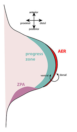
The apical ectodermal ridge (AER) is a structure that forms from the ectodermal cells at the distal end of each limb bud and acts as a major signaling center to ensure proper development of a limb. After the limb bud induces AER formation, the AER and limb mesenchyme—including the zone of polarizing activity (ZPA)—continue to communicate with each other to direct further limb development.

The limb bud is a structure formed early in vertebrate limb development. As a result of interactions between the ectoderm and underlying mesoderm, formation occurs roughly around the fourth week of development. In the development of the human embryo the upper limb bud appears in the third week and the lower limb bud appears four days later.

In the field of developmental biology, regional differentiation is the process by which different areas are identified in the development of the early embryo. The process by which the cells become specified differs between organisms.

The development of the nervous system in humans, or neural development or neurodevelopment involves the studies of embryology, developmental biology, and neuroscience to describe the cellular and molecular mechanisms by which the complex nervous system forms in humans, develops during prenatal development, and continues to develop postnatally.

<span class="mw-page-title-main">Orthodenticle homeobox 2</span> Protein-coding gene in the species Homo sapiens

Homeobox protein OTX2 is a protein that in humans is encoded by the OTX2 gene.

<span class="mw-page-title-main">Zone of polarizing activity</span>

The zone of polarizing activity (ZPA) is an area of mesenchyme that contains signals which instruct the developing limb bud to form along the anterior/posterior axis. Limb bud is undifferentiated mesenchyme enclosed by an ectoderm covering. Eventually, the limb bud develops into bones, tendons, muscles and joints. Limb bud development relies not only on the ZPA, but also many different genes, signals, and a unique region of ectoderm called the apical ectodermal ridge (AER). Research by Saunders and Gasseling in 1948 identified the AER and its subsequent involvement in proximal distal outgrowth. Twenty years later, the same group did transplantation studies in chick limb bud and identified the ZPA. It wasn't until 1993 that Todt and Fallon showed that the AER and ZPA are dependent on each other.

<span class="mw-page-title-main">Andrew Lumsden (scientist)</span>

Andrew Gino Lumsden is an English neurobiologist, Emeritus Professor of the University of London and founder in 2000 of the Medical Research Council Centre for Developmental Neurobiology at King's College London.

<span class="mw-page-title-main">Retinal homeobox protein Rx</span> Protein-coding gene in the species Homo sapiens

Retinal homeobox protein Rx also known as retina and anterior neural fold homeobox is a protein that in humans is encoded by the RAX gene. The RAX gene is located on chromosome 18 in humans, mice, and rats.

The isthmic organizer, or isthmus organizer, also known as the midbrain−hindbrain boundary (MHB), is a secondary organizer region that develops at the junction of the midbrain and metencephalon. The MHB expresses signaling molecules that regulate the differentiation and patterning of the adjacent neuroepithelium. This allows for the development of the midbrain and hindbrain as well as the specification of neuronal subtypes in these regions. The fact that the MHB is sufficient for the development of the mid and hindbrain was shown in an experiment where quail MHB cells transplanted into the forebrain of a chick were able to induce an ectopic midbrain and cerebellum.

References

  1. 1 2 Scholpp S, Lumsden A (Aug 2010). "Building a bridal chamber: development of the thalamus". Trends Neurosci. 33 (8): 373–380. doi:10.1016/j.tins.2010.05.003. PMC   2954313 . PMID   20541814.
  2. Garcia-Lopez R, Vieira C, Echevarria D, Martinez S (2004). "Fate map of the diencephalon and the zona limitans at 10-somites stage in chick embryos". Developmental Biology. 268 (2): 514–530. doi: 10.1016/j.ydbio.2003.12.038 . PMID   15063186.
  3. 1 2 3 4 5 Scholpp S, Foucher I, Staudt N, Peukert D, Lumsden A, Houart C (2007). "Otx1l, Otx2, and Irx1b establish and position the ZLI in the diencephalon". Development. 134 (17): 3167–3176. doi: 10.1242/dev.001461 . PMC   7116068 . PMID   17670791.
  4. 1 2 3 Kiecker C, Lumsden A (2005). "Compartments and their boundaries in vertebrate brain development". Nature Reviews Neuroscience. 6 (7): 553–564. doi:10.1038/nrn1702. PMID   15959467. S2CID   10869618.
  5. 1 2 3 4 Guinazu MF, Chambers D, Lumsden A, Kiecker C (2007). "Tissue interactions in the developing chick diencephalon". Neural Development. 2 (25): 1–15. doi: 10.1186/1749-8104-2-25 . PMC   2217525 . PMID   17999760.
  6. Zeltser LM, Larsen CW, Lumsden A (2001). "A new developmental compartment in the forebrain regulated by Lunatic fringe". Nature Neuroscience. 4 (7): 683–684. doi:10.1038/89455. PMID   11426219. S2CID   26205097.
  7. Puelles L, Rubenstein JL (1993). "Expression patterns of homeobox and other putative regulatory genes in the embryonic mouse forebrain suggests a neuromeric organization". Trends in Neurosciences. 16 (11): 472–479. doi:10.1016/0166-2236(93)90080-6. PMID   7507621. S2CID   3987564.
  8. Scholpp S, Wolf O, Brand M, Lumsden A (2005). "Hedgehog signaling from the zona limitans intrathalamica orchestrates patterning of the zebrafish diencephalon". Development. 133 (5): 855–864. doi: 10.1242/dev.02248 . PMID   16452095.
  9. 1 2 3 4 Lim Y, Golden JA (2007). "Patterning the developing diencephalon". Brain Research Reviews. 53 (1): 17–26. doi:10.1016/j.brainresrev.2006.06.004. PMID   16876871. S2CID   20491034.
  10. 1 2 Vieira C, Garda AL, Shimamura K, Martinez S (2005). "Thalamic development induced by Shh in the chick embryo". Developmental Biology. 284 (2): 351–363. doi: 10.1016/j.ydbio.2005.05.031 . PMID   16026780.
  11. Ba-Charvet KT, von Boxberg Y, Guazzi S, Boncinelli E, Godement P (1998). "A potential role for the OTX2 homeoprotein in creating early 'highways' for axon extension in the rostral brain". Development. 125 (21): 4273–4282. doi:10.1242/dev.125.21.4273. PMID   9753681.
  12. Hashimoto-Torii K, Motoyama J, Hui CC, Kuroiwa A, Nakafuku M, Shimamura K (2003). "Differential activities of Sonic hedgehog mediated by Gli transcription factors define distinct neuronal subtypes in the dorsal thalamus". Mechanisms of Development. 120 (10): 1097–1111. doi:10.1016/j.mod.2003.09.001. PMID   14568100.
  13. Rash, BG; Grove, EA (15 November 2011). "Shh and Gli3 regulate formation of the telencephalic-diencephalic junction and suppress an isthmus-like signaling source in the forebrain". Developmental Biology. 359 (2): 242–50. doi:10.1016/j.ydbio.2011.08.026. PMC   3213684 . PMID   21925158.
  14. Scholpp S, Delogu A, Gilthorpe J, Peukert D, Schindler S, Lumsden A. Her6 regulates the neurogenetic gradient and neuronal identity in the thalamus. Proc Natl Acad Sci U S A. 2009 Nov 24;106(47):19895-900