Anterior superior iliac spine

Last updated
Anterior superior iliac spine
Gray435.png
The obturator membrane (anterior superior iliac spine visible in upper right of illustration)
Gray abdomen front surface en.png
Anterior superior iliac spine labeled second to bottom, right
Details
Identifiers
Latin spina iliaca anterior superior
TA98 A02.5.01.111
TA2 1327
FMA 49465
Anatomical terms of bone

The anterior superior iliac spine (ASIS) is a bony projection of the iliac bone, and an important landmark of surface anatomy. It refers to the anterior extremity of the iliac crest of the pelvis. It provides attachment for the inguinal ligament, and the sartorius muscle. [1] The tensor fasciae latae muscle attaches to the lateral aspect of the superior anterior iliac spine, and also about 5  cm away at the iliac tubercle. [2] [3]

Contents

Structure

The anterior superior iliac spine refers to the anterior extremity of the iliac crest of the pelvis. This is a key surface landmark, and easily palpated. It provides attachment for the inguinal ligament, the sartorius muscle, [1] [4] and the tensor fasciae latae muscle. [2] [3]

A variety of structures lie close to the anterior superior iliac spine, including the subcostal nerve, [5] the femoral artery (which passes between it and the pubic symphysis), [4] and the iliohypogastric nerve. [6]

Clinical significance

The anterior superior iliac spine provides a clue in identifying some other clinical landmarks, including McBurney's point, Roser-Nélaton line, and true leg length. It is an important surface landmark for various surgical approaches, such as treatment of hernia. [7] The severity of symptoms of damage to the iliohypogastric nerve can show whether damage occurred above or below the anterior superior iliac spine. [6]

Bone may be harvested from the nearby iliac crest for use elsewhere in the body. [5] As the subcostal nerve lies close to the anterior superior iliac spine, this is put at risk of damage. [5]

The iliotibial tract may be irritated where it passes over the anterior superior iliac spine in iliotibial band syndrome. [3]

The line around anterior superior iliac spine is sometimes called the panty line or "bikini line". [8] It is considered to be a "discreet" location for concealing cosmetic surgery scars and ports. [9]

Additional images

See also

Related Research Articles

<span class="mw-page-title-main">Sartorius muscle</span> Longest muscle in the human body

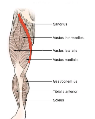
The sartorius muscle is the longest muscle in the human body. It is a long, thin, superficial muscle that runs down the length of the thigh in the anterior compartment.

<span class="mw-page-title-main">Quadriceps</span> Group of human leg muscle

The quadriceps femoris muscle is a large muscle group that includes the four prevailing muscles on the front of the thigh. It is the sole extensor muscle of the knee, forming a large fleshy mass which covers the front and sides of the femur. The name derives from Latin four-headed muscle of the femur.

<span class="mw-page-title-main">Obturator internus muscle</span> One of six small hip muscles in the lateral rotator group

The internal obturator muscle or obturator internus muscle originates on the medial surface of the obturator membrane, the ischium near the membrane, and the rim of the pubis.

<span class="mw-page-title-main">Serratus posterior inferior muscle</span> Muscle of the mid-low back

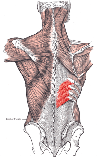
The serratus posterior inferior muscle, also known as the posterior serratus muscle, is a muscle of the human body.

<span class="mw-page-title-main">Iliopsoas</span> Joined psoas and the iliacus muscles

The iliopsoas muscle refers to the joined psoas major and the iliacus muscles. The two muscles are separate in the abdomen, but usually merge in the thigh. They are usually given the common name iliopsoas. The iliopsoas muscle joins to the femur at the lesser trochanter. It acts as the strongest flexor of the hip.

<span class="mw-page-title-main">Inferior epigastric artery</span> Blood vessel

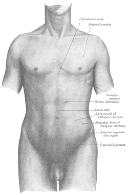
In human anatomy, the inferior epigastric artery is an artery that arises from the external iliac artery. It is accompanied by the inferior epigastric vein; inferiorly, these two inferior epigastric vessels together travel within the lateral umbilical fold The inferior epigastric artery then traverses the arcuate line of rectus sheath to enter the rectus sheath, then anastomoses with the superior epigastric artery within the rectus sheath.

<span class="mw-page-title-main">Femoral nerve</span> Long nerve down the thigh and inner leg

The femoral nerve is a nerve in the thigh that supplies skin on the upper thigh and inner leg, and the muscles that extend the knee. It is the largest branch of the lumbar plexus.

The gluteal tuberosity is the lateral one of the three upward prolongations of the linea aspera of the femur, extending to the base of the greater trochanter. It serves as the principal insertion site for the gluteus maximus muscle.

<span class="mw-page-title-main">Superior gluteal nerve</span> Nerve on the pelvis and leg

The superior gluteal nerve is a mixed nerve of the sacral plexus that originates in the pelvis. It provides motor innervation to the gluteus medius, gluteus minimus, tensor fasciae latae, and piriformis muscles; it also has a cutaneous branch.

<span class="mw-page-title-main">Iliolumbar artery</span>

The iliolumbar artery is the first branch of the posterior trunk of the internal iliac artery.

<span class="mw-page-title-main">Lateral sacral artery</span> Artery in the pelvis

The lateral sacral arteries is an artery in the pelvis that arises from the posterior division of the internal iliac artery. It later splits into two smaller branches, a superior and an inferior.

<span class="mw-page-title-main">Iliocostalis</span>

Iliocostalis muscle is the muscle immediately lateral to the longissimus that is the nearest to the furrow that separates the epaxial muscles from the hypaxial. It lies very deep to the fleshy portion of the serratus posterior muscle. It laterally flexes the vertebral column to the same side.

<span class="mw-page-title-main">Transversalis fascia</span> Aponeurosis between the transverse abdominal muscle and the extraperitoneal fat

The transversalis fascia is the fascial lining of the anterolateral abdominal wall situated between the inner surface of the transverse abdominal muscle, and the preperitoneal fascia. It is directly continuous with the iliac fascia, the internal spermatic fascia, and pelvic fascia.

<span class="mw-page-title-main">Greater sciatic foramen</span> Part of the pelvis

The greater sciatic foramen is an opening in the posterior human pelvis. It is formed by the sacrotuberous and sacrospinous ligaments. The piriformis muscle passes through the foramen and occupies most of its volume. The greater sciatic foramen is wider in women than in men.

<span class="mw-page-title-main">Long plantar ligament</span> Ligament that connects the calcaneus and cuboid bones in the human foot

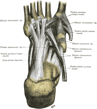
The long plantar ligament is a long ligament on the underside of the foot that connects the calcaneus with the 2nd to 5th metatarsal.

<span class="mw-page-title-main">Pubic tubercle</span> Bone

The pubic tubercle is a prominent tubercle on the superior ramus of the pubis bone of the pelvis.

<span class="mw-page-title-main">Ischial spine</span> Part of the posterior border of the body of the ischium bone of the pelvis

The ischial spine is part of the posterior border of the body of the ischium bone of the pelvis. It is a thin and pointed triangular eminence, more or less elongated in different subjects.

<span class="mw-page-title-main">Iliac crest</span> Top border of the hip

The crest of the ilium is the superior border of the wing of ilium and the superiolateral margin of the greater pelvis.

<span class="mw-page-title-main">Iliac fossa</span>

The iliac fossa is a large, smooth, concave surface on the internal surface of the ilium.

The soleal line, also known as the popliteal line, is a prominent ridge on the posterior surface of the tibia. It is the site of many muscle origins and insertions, such as those of popliteus muscle, soleus muscle, flexor digitorum longus muscle, and tibialis posterior muscle.

References

  1. 1 2 Chaitow, Leon; DeLany, Judith (2011-01-01), Chaitow, Leon; DeLany, Judith (eds.), "Chapter 12 - The hip", Clinical Application of Neuromuscular Techniques, Volume 2 (Second Edition), Oxford: Churchill Livingstone, pp. 391–445, doi:10.1016/b978-0-443-06815-7.00012-7, ISBN   978-0-443-06815-7 , retrieved 2020-12-15
  2. 1 2 Garten, Hans (2013), "M. tensor fasciae latae", The Muscle Test Handbook, Elsevier, pp. 236–237, doi:10.1016/b978-0-7020-3739-9.00091-2, ISBN   978-0-7020-3739-9 , retrieved 2020-12-15
  3. 1 2 3 Chaitow, Leon; DeLany, Judith (2011-01-01), Chaitow, Leon; DeLany, Judith (eds.), "Chapter 11 - The pelvis", Clinical Application of Neuromuscular Techniques, Volume 2 (Second Edition), Oxford: Churchill Livingstone, pp. 299–389, doi:10.1016/b978-0-443-06815-7.00011-5, ISBN   978-0-443-06815-7 , retrieved 2020-12-15
  4. 1 2 Jacob, S. (2008-01-01), Jacob, S. (ed.), "Chapter 6 - Lower limb", Human Anatomy, Churchill Livingstone, pp. 135–179, doi:10.1016/b978-0-443-10373-5.50009-9, ISBN   978-0-443-10373-5 , retrieved 2020-12-15
  5. 1 2 3 Rea, Paul (2015-01-01), Rea, Paul (ed.), "Chapter 3 - Lower Limb Nerve Supply", Essential Clinically Applied Anatomy of the Peripheral Nervous System in the Limbs, Academic Press, pp. 101–177, doi:10.1016/b978-0-12-803062-2.00003-6, ISBN   978-0-12-803062-2 , retrieved 2020-12-15
  6. 1 2 Mirjalili, S. Ali (2015-01-01), Tubbs, R. Shane; Rizk, Elias; Shoja, Mohammadali M.; Loukas, Marios (eds.), "Chapter 45 - Anatomy of the Lumbar Plexus", Nerves and Nerve Injuries, San Diego: Academic Press, pp. 609–617, doi:10.1016/b978-0-12-410390-0.00047-0, ISBN   978-0-12-410390-0 , retrieved 2020-12-15
  7. Molloy, Robert E. (2005-01-01), Benzon, Honorio T.; Raja, Srinivasa N.; Molloy, Robert E.; Liu, Spencer S. (eds.), "Chapter 75 - Truncal Blocks: Intercostal, Paravertebral, Interpleural, Suprascapular, Ilioinguinal, and Iliohypogastric Nerve Blocks", Essentials of Pain Medicine and Regional Anesthesia (Second Edition), Philadelphia: Churchill Livingstone, pp. 636–644, doi:10.1016/b978-0-443-06651-1.50079-4, ISBN   978-0-443-06651-1 , retrieved 2020-12-15
  8. Kim, Ji Hun (2013). "Robotic cholecystectomy with new port sites". World Journal of Gastroenterology. 19 (20): 3077. doi: 10.3748/wjg.v19.i20.3077 . ISSN   1007-9327. PMC   3662947 . PMID   23716987.
  9. Leggett, P.L.; Bissell, C.D.; Churchman-Winn, R. (2001). "Cosmetic minilaparoscopic cholecystectomy". Surgical Endoscopy. 15 (10): 1229–1231. doi:10.1007/s004640041018. ISSN   0930-2794.