Surface anatomy

Last updated
Surface projections of the major organs of the trunk, using the vertebral column and rib cage as main reference points of surface anatomy. Surface projections of the organs of the trunk.png
Surface projections of the major organs of the trunk, using the vertebral column and rib cage as main reference points of surface anatomy.

Surface anatomy (also called superficial anatomy and visual anatomy) is the study of the external features of the body of an animal. [1] In birds, this is termed topography. Surface anatomy deals with anatomical features that can be studied by sight, without dissection. As such, it is a branch of gross anatomy, along with endoscopic and radiological anatomy. [2] Surface anatomy is a descriptive science. [3] In particular, in the case of human surface anatomy, these are the form and proportions of the human body and the surface landmarks which correspond to deeper structures hidden from view, both in static pose and in motion.

Contents

In addition, the science of surface anatomy includes the theories and systems of body proportions and related artistic canons.[ citation needed ] The study of surface anatomy is the basis for depicting the human body in classical art.

Some pseudo-sciences such as physiognomy, phrenology and palmistry rely on surface anatomy.

Human surface anatomy

Surface anatomy of the thorax

Front of thorax, showing surface relations of bones, lungs (purple), pleura (blue), and heart (red outline). Heart valves are labeled with "B", "T", "A", and "P".

First heart sound: caused by atrioventricular valves - Bicuspid/Mitral (B) and Tricuspid (T).

Second heart sound caused by semilunar valves -- Aortic (A) and Pulmonary/Pulmonic (P). Gray1216.svg
Front of thorax, showing surface relations of bones, lungs (purple), pleura (blue), and heart (red outline). Heart valves are labeled with "B", "T", "A", and "P".

First heart sound: caused by atrioventricular valves - Bicuspid/Mitral (B) and Tricuspid (T).

Second heart sound caused by semilunar valves -- Aortic (A) and Pulmonary/Pulmonic (P).

Knowledge of the surface anatomy of the thorax (chest) is particularly important because it is one of the areas most frequently subjected to physical examination, like auscultation and percussion. [4] In cardiology, Erb's point refers to the third intercostal space on the left sternal border where S2 heart sound is best auscultated. [5] [6] Some sources include the fourth left interspace. [7]

Human female breasts are located on the chest wall, most frequently between the second and sixth rib. [4]

Anatomical landmarks

  1. On the trunk of the body in the thoracic area, the shoulder in general is the acromial, while the curve of the shoulder is the deltoid.
  2. The back as a general area is the dorsum or dorsal area, and the lower back as the limbus or lumbar region.
  3. The shoulderblades are the scapular area and the breastbone is the sternal region.
  4. The abdominal area is the region between the chest and the pelvis.
  5. The breast is called the mamma or mammary, the armpit as the axilla and axillary, and the navel as the umbilicus and umbilical.
  6. The pelvis is the lower torso, between the abdomen and the thighs.
  7. The groin, where the thigh joins the trunk, are the inguen and inguinal area.
  8. The entire arm is referred to as the brachium and brachial, the front of the elbow as the antecubitis and antecubital, the back of the elbow as the olecranon or olecranal, the forearm as the antebrachium and antebrachial, the wrist as the carpus and carpal area, the hand as the manus and manual, the palm as the palma and palmar, the thumb as the pollex, and the fingers as the digits, phalanges, and phalangeal.
  9. The buttocks are the gluteus or gluteal region and the pubic area is the pubis.
  10. Anatomists divide the lower limb into the thigh (the part of the limb between the hip and the knee) and the leg (which refers only to the area of the limb between the knee and the ankle).
  11. The thigh is the femur and the femoral region.
  12. The kneecap is the patella and patellar while the back of the knee is the popliteus and popliteal area.
  13. The leg (between the knee and the ankle) is the crus and crural area, the lateral aspect of the leg is the peroneal area, and the calf is the sura and sural region.
  14. The ankle is the tarsus and tarsal, and the heel is the calcaneus or calcaneal. The foot is the pes and pedal region, and the sole of the foot the planta and plantar.
  15. As with the fingers, the toes are also called the digits, phalanges, and phalangeal area.
  16. The big toe is referred to as the hallux.

List of features

Following are lists of surface anatomical features in humans and other animals. Sorted roughly from head to tail, cranial to caudal. Homologues share a bullet point and are separated by commas. Subcomponents are nested. Class in which component occurs in italic.

In humans

In other animals

See also

Notes

  1. Seeley (2003) chap.1 p.2
  2. Standring (2008) Introduction, Anatomical nomenclature, p.2
  3. "Journal of Morphology and Anatomy Surface Anatomy". Hilaris. Retrieved 1 June 2021.
  4. 1 2 Drake (2009) Ch.3 Thorax - Thorax surface anatomy, pp. 224-6 and Fig. 3.96 A
  5. "Week 4". Archived from the original on 2008-01-29. Retrieved 2007-10-11.
  6. Gavaghan, Mary (1998). "Cardiac anatomy and physiology: a review - includes examination questions AORN Journal - Find Articles". AORN Journal. Archived from the original on 2012-07-10. Retrieved 2007-10-11.
  7. Constant, Jules (1999). Bedside cardiology. Hagerstwon, MD: Lippincott Williams & Wilkins. p. 228. ISBN   0-7817-2168-7.

Related Research Articles

<span class="mw-page-title-main">Foot</span> Anatomical structure found in vertebrates

The foot is an anatomical structure found in many vertebrates. It is the terminal portion of a limb which bears weight and allows locomotion. In many animals with feet, the foot is a separate organ at the terminal part of the leg made up of one or more segments or bones, generally including claws and/or nails.

<span class="mw-page-title-main">Human leg</span> Lower extremity or limb of the human body (foot, lower leg, thigh and hip)

The leg is the entire lower limb of the human body, including the foot, thigh or sometimes even the hip or buttock region. The major bones of the leg are the femur, tibia, and adjacent fibula. The thigh is between the hip and knee, while the calf (rear) and shin (front) are between the knee and foot.

<span class="mw-page-title-main">Metatarsal bones</span> Five long bones in the foot

The metatarsal bones or metatarsus are a group of five long bones in the midfoot, located between the tarsal bones and the phalanges (toes). Lacking individual names, the metatarsal bones are numbered from the medial side : the first, second, third, fourth, and fifth metatarsal. The metatarsals are analogous to the metacarpal bones of the hand. The lengths of the metatarsal bones in humans are, in descending order, second, third, fourth, fifth, and first. A bovine hind leg has two metatarsals.

<span class="mw-page-title-main">Ankle</span> Region where the foot and the leg meet

The ankle, the talocrural region or the jumping bone (informal) is the area where the foot and the leg meet. The ankle includes three joints: the ankle joint proper or talocrural joint, the subtalar joint, and the inferior tibiofibular joint. The movements produced at this joint are dorsiflexion and plantarflexion of the foot. In common usage, the term ankle refers exclusively to the ankle region. In medical terminology, "ankle" can refer broadly to the region or specifically to the talocrural joint.

<span class="mw-page-title-main">Epiphysis</span> End of a long bone that ossifies from a secondary center

An epiphysis is one of the rounded ends or tips of a long bone that ossify from a secondary center of ossification. Between the epiphysis and diaphysis lies the metaphysis, including the epiphyseal plate. At the joint, the epiphysis is covered with articular cartilage; below that covering is a zone similar to the epiphyseal plate, known as subchondral bone. In evolution, reptiles do not have epiphyses and diaphyses, being restricted to mammals.

<span class="mw-page-title-main">Appendicular skeleton</span> Part of the skeletal system

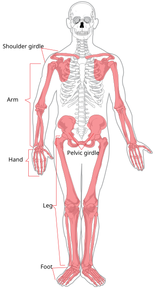
The appendicular skeleton is the portion of the vertebrate endoskeleton consisting of the bones and cartilages that support the paired appendages. In most terrestrial vertebrates, the appendicular skeleton and the associated skeletal muscles are the predominant locomotive structures.

<span class="mw-page-title-main">Strike (attack)</span> Directed physical attack

A strike is a directed, forceful physical attack with either a part of the human body or with a handheld object, intended to cause blunt or penetrating trauma upon an opponent.

<span class="mw-page-title-main">Deltoid muscle</span> Shoulder muscle

The deltoid muscle is the muscle forming the rounded contour of the human shoulder. It is also known as the 'common shoulder muscle', particularly in other animals such as the domestic cat. Anatomically, the deltoid muscle appears to be made up of three distinct sets of muscle fibers, namely the

  1. anterior or clavicular part
  2. posterior or scapular part
  3. intermediate or acromial part
<span class="mw-page-title-main">Digitigrade</span> Standing or walking on digits/toes; animals which do so

In terrestrial vertebrates, digitigrade locomotion is walking or running on the toes. A digitigrade animal is one that stands or walks with its toes (phalanges) on the ground, and the rest of its foot lifted. Digitigrades include birds, cats, dogs, and many other mammals, but not plantigrades or unguligrades. Digitigrades generally move more quickly than other animals.

<span class="mw-page-title-main">Piriformis muscle</span> One of six small hip muscles in the lateral rotator group

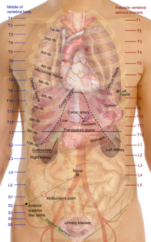
The piriformis muscle is a flat, pyramidally-shaped muscle in the gluteal region of the lower limbs. It is one of the six muscles in the lateral rotator group.

<span class="mw-page-title-main">Phalanx bone</span> Digital bone in the hands and feet of most vertebrates

The phalanges are digital bones in the hands and feet of most vertebrates. In primates, the thumbs and big toes have two phalanges while the other digits have three phalanges. The phalanges are classed as long bones.

<span class="mw-page-title-main">Anatomical terms of motion</span> Terms describing animal motion

Motion, the process of movement, is described using specific anatomical terms. Motion includes movement of organs, joints, limbs, and specific sections of the body. The terminology used describes this motion according to its direction relative to the anatomical position of the body parts involved. Anatomists and others use a unified set of terms to describe most of the movements, although other, more specialized terms are necessary for describing unique movements such as those of the hands, feet, and eyes.

A limb is a jointed, muscled appendage of a tetrapod vertebrate animal used for weight-bearing, terrestrial locomotion and physical interaction with other objects. The distalmost portion of a limb is known as its extremity. The limbs' bony endoskeleton, known as the appendicular skeleton, is homologous among all tetrapods, who use their limbs for walking, running and jumping, swimming, climbing, grasping, touching and striking.

<span class="mw-page-title-main">Outline of human anatomy</span> Overview of and topical guide to human anatomy

The following outline is provided as an overview of and topical guide to human anatomy:

<span class="mw-page-title-main">Anatomical terminology</span> Scientific terminology used by anatomists, zoologists, and health professionals

Anatomical terminology is a form of scientific terminology used by anatomists, zoologists, and health professionals such as doctors, physicians, and pharmacists.

<span class="mw-page-title-main">Anatomical terms of bone</span>

Many anatomical terms descriptive of bone are defined in anatomical terminology, and are often derived from Greek and Latin. Bone in the human body is categorized into long bone, short bone, flat bone, irregular bone and sesamoid bone.

References

Further reading