Sole (foot)

Last updated
Sole
Soles of human feet.png
Soles of a human's feet
Details
Part of Foot
Artery Medial plantar, lateral plantar
Nerve Medial plantar, lateral plantar
Identifiers
Latin planta
TA98 A01.1.00.044
TA2 337
FMA 25000
Anatomical terminology

In humans, the sole of the foot is anatomically referred to as the plantar aspect.

Contents

Structure

Deep anatomy of the sole PF-PlantarDesign.jpg
Deep anatomy of the sole

The glabrous skin on the sole of the foot lacks the hair and pigmentation found elsewhere on the body, and it has a high concentration of sweat pores. The sole contains the thickest layers of skin on the body due to the weight that is continually placed on it. It is crossed by a set of creases that form during the early stages of embryonic development. Like those of the palm, the sweat pores of the sole lack sebaceous glands.

The sole is a sensory organ by which we can perceive the ground while standing and walking. The subcutaneous tissue in the sole has adapted to deal with the high local compressive forces on the heel and the ball (between the toes and the arch) by developing a system of "pressure chambers." Each chamber is composed of internal fibrofatty tissue covered by external collagen connective tissue. The septa (internal walls) of these chambers are permeated by numerous blood vessels, making the sole one of the most vascularized, or blood-enriched, regions in the human body. [1]

The sole and the longitudinal arch of the foot are supported by a thick connective tissue, the plantar fascia. The central component of this tissue extends to the supporting bones and gives two divisions–the medial component and lateral component; thus they define the boundaries of the three muscle compartments of the sole (see below). [2]

The bones underlying the sole form the arch of the foot. The arches might collapse later in life, resulting in flat feet.

Muscles

Intrinsic

Gray443.png
Gray444.png
Gray445.png
Gray446.png
Gray447.png
First, second, and third muscle layers, and the dorsal and plantar interossei

The intrinsic muscles in the sole are grouped in four layers:

In the first layer, the flexor digitorum brevis is the large central muscle located immediately above the plantar aponeurosis. It flexes the second to fifth toes and is flanked by abductor hallucis and abductor digiti minimi. [2]

In the second layer, the quadratus plantae, located below flexor digitorum brevis, inserts into the tendon of flexor digitorum longus on which the lumbricals originate. [2]

In the third layer, the oblique head of adductor hallucis joins the muscle's transversal head on the lateral side of the big toe. Medially to adductor hallucis are the two heads of flexor hallucis brevis, deep to the tendon of flexor hallucis longus. The considerably smaller flexor digiti minimi brevis on the lateral side can be mistaken for one of the interossei. [2]

In the fourth layer. the dorsal and plantar interossei are located between and below the metatarsal bones and act as antagonists. [2]

The central compartment is shared by the lumbricals, quadratus plantae, flexor digitorum brevis, and adductor hallucis; the medial compartment by abductor hallucis, flexor hallucis brevis, abductor digiti minimi, flexor digiti minimi brevis, and opponens digiti minimi (often considered part of the former muscle); whilst the lateral compartment is occupied by extensor digitorum brevis and extensor hallucis brevis. [3]

Extrinsic

The tendons of several extrinsic foot muscle reach the sole:

Nerve supply

Cutaneous innervation of the sole of the foot Gray834.svg
Cutaneous innervation of the sole of the foot

The soles of the feet are extremely sensitive to touch due to a high concentration of nerve endings, with as many as 200,000 per sole. [5] This makes them sensitive to surfaces that are walked on, ticklish and some people find them to be erogenous zones. [6]

Medically, the soles are the site of the plantar reflex, the testing of which can be painful due to the sole's sensitivity.

The deep fibular nerve from the common fibular nerve provides the sensory innervation of the skin between the first and second toes and the motor innervation of the muscles of the anterior compartment of the leg and dorsal foot. Damage to the deep fibular nerve can result in foot drop. [7]

The plantar digital nerves from the medial plantar nerve provide sensory innervation to the skin of the plantar aspect of the toes, except for the medial part of the big toe and the lateral part of the little toe and the motor innervation of the first lumbrical. [7]

The proper plantar nerve from the common plantar digital nerve provides sensory innervation to the plantar surface of the toes as well as the dorsal aspect of the distal interphalangeal phalanges. It also provides motor innervation to flexor hallucis brevis. [7]

The superficial and deep branches of the lateral plantar nerve from the tibial nerve provide sensory innervation to the skin of the lateral side of the sole, to the fifth and half the fourth toes, and the nail bed of these toes. They also provide motor innervation to quadratus plantae, abductor digiti minimi, flexor digiti minimi brevis, lateral three lumbricals, adductor hallucis, and the dorsal and plantar interossei. [7]

The medial plantar nerve from the tibial nerve provides sensory innervation to the skin of the medial side of the sole, the skin of the medial three and a half toes, and the nail beds of these toes. It also provides motor innervation to abductor hallucis, flexor hallucis brevis, flexor digitorum brevis, and the first lumbrical. [7]

The saphenous nerve from the femoral nerve provides sensory innervation to the medial side of the foot as well as the medial side of the leg. Likewise, the sural nerve provides sensory innervation to the skin on the lateral side of the foot as well as the skin on the posterior aspect of the lower leg. [7]

The tibial nerve from the sciatic nerve provides sensory innervation to the skin of the sole and toes, and the dorsal aspect of the toes. It provides motor innervation to plantaris, tibialis posterior, flexor hallucis longus, flexor digitorum longus as well as posterior muscles in the leg. [7]

Society and culture

In Thailand, Saudi Arabia, and some Muslim countries, it is considered offensive to sit raising the leg so the uncovered sole of the foot is visible and therefore taboo. [8]

Other animals

Terrestrial animals using their soles for locomotion are called plantigrade.

In chimpanzees, the soles are furrowed with creases deeper and more distinct than in their palms. In the palms, the pattern density is thickest in the central part, but in the sole, the density is thickest near the big toe whilst a large part of the remaining sole is covered by thick, tight, and smooth skin almost without furrows. [9]

In bonobos, the pattern intensity of the epidermal ridges (i.e. "fingerprints") of the palms and soles is considerably higher than in chimpanzees. Whilst the pattern intensity in the palm is the highest of all species of apes, in the sole, the density decreases and is comparable to other apes. [10]

Clinical significance

The sole is subject to many skin diseases.

See also

Notes

  1. Ross & Lamperti 2006 , pp. 418, 486
  2. 1 2 3 4 5 Ross & Lamperti 2006 , pp. 456–61
  3. Ross & Lamperti 2006 , pp. 438–40
  4. Ross & Lamperti 2006 , pp. 433, 436–37
  5. "nerve endings - barefootr". barefootr.com. Retrieved 2 May 2018.
  6. Brittan 2003
  7. 1 2 3 4 5 6 7 Tank 2006 , Nerves of the Sole of the Foot
  8. Lumsden, Lumsden & Weithoff 2009 , p. 223
  9. Ladygina-Kohts 2002 , pp. 29–33
  10. Brehme 1975 , Abstract

Related Research Articles

<span class="mw-page-title-main">Foot</span> Anatomical structure found in vertebrates

The foot is an anatomical structure found in many vertebrates. It is the terminal portion of a limb which bears weight and allows locomotion. In many animals with feet, the foot is a separate organ at the terminal part of the leg made up of one or more segments or bones, generally including claws and/or nails.

<span class="mw-page-title-main">Human leg</span> Lower extremity or limb of the human body (foot, lower leg, thigh and hip)

The leg is the entire lower limb of the human body, including the foot, thigh or sometimes even the hip or buttock region. The major bones of the leg are the femur, tibia, and adjacent fibula. The thigh is between the hip and knee, while the calf (rear) and shin (front) are between the knee and foot.

<span class="mw-page-title-main">Toe</span> Digit of a foot

Toes are the digits of the foot of a tetrapod. Animal species such as cats that walk on their toes are described as being digitigrade. Humans, and other animals that walk on the soles of their feet, are described as being plantigrade; unguligrade animals are those that walk on hooves at the tips of their toes.

<span class="mw-page-title-main">Ulnar nerve</span> Nerve which runs near the ulna bone

In human anatomy, the ulnar nerve is a nerve that runs near the ulna bone. The ulnar collateral ligament of elbow joint is in relation with the ulnar nerve. The nerve is the largest in the human body unprotected by muscle or bone, so injury is common. This nerve is directly connected to the little finger, and the adjacent half of the ring finger, innervating the palmar aspect of these fingers, including both front and back of the tips, perhaps as far back as the fingernail beds.

<span class="mw-page-title-main">Upper limb</span> Consists of the arm, forearm, and hand

The upper limbs or upper extremities are the forelimbs of an upright-postured tetrapod vertebrate, extending from the scapulae and clavicles down to and including the digits, including all the musculatures and ligaments involved with the shoulder, elbow, wrist and knuckle joints. In humans, each upper limb is divided into the arm, forearm and hand, and is primarily used for climbing, lifting and manipulating objects.

<span class="mw-page-title-main">Tibial nerve</span> Branch of the sciatic nerve

The tibial nerve is a branch of the sciatic nerve. The tibial nerve passes through the popliteal fossa to pass below the arch of soleus.

<span class="mw-page-title-main">Dorsal interossei of the foot</span> Four muscles situated between the metatarsal bones

In human anatomy, the dorsal interossei of the foot are four muscles situated between the metatarsal bones.

<span class="mw-page-title-main">Flexor hallucis longus muscle</span> One of the three deep muscles in the lower leg

The flexor hallucis longus muscle (FHL) attaches to the plantar surface of phalanx of the great toe and is responsible for flexing that toe. The FHL is one of the three deep muscles of the posterior compartment of the leg, the others being the flexor digitorum longus and the tibialis posterior. The tibialis posterior is the most powerful of these deep muscles. All three muscles are innervated by the tibial nerve which comprises half of the sciatic nerve.

<span class="mw-page-title-main">Flexor digitorum longus muscle</span> Muscle located on the tibial side of the leg

The flexor digitorum longus muscle is situated on the tibial side of the leg. At its origin it is thin and pointed, but it gradually increases in size as it descends. It serves to flex the second, third, fourth, and fifth toes.

<span class="mw-page-title-main">Flexor hallucis brevis muscle</span> Muscle in sole of the foot that leads to the big toe

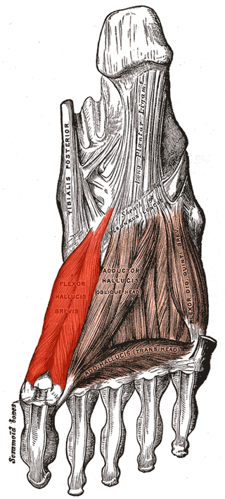
Flexor hallucis brevis muscle is a muscle of the foot that flexes the big toe.

<span class="mw-page-title-main">Flexor digitorum brevis muscle</span> Lies in the middle of the sole of the foot

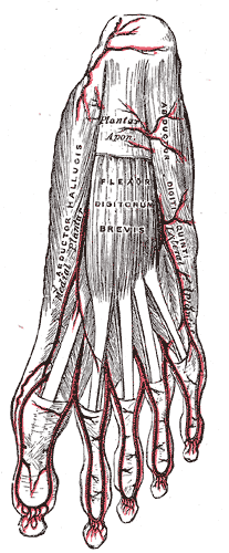
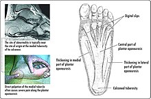
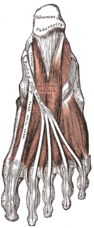
The flexor digitorum brevis is a muscle which lies in the middle of the sole of the foot, immediately above the central part of the plantar aponeurosis, with which it is firmly united.

<span class="mw-page-title-main">Lumbricals of the foot</span> Four small skeletal muscles

The lumbricals are four small skeletal muscles, accessory to the tendons of the flexor digitorum longus muscle. They are numbered from the medial side of the foot.

<span class="mw-page-title-main">Superficial fibular nerve</span> Nerve in the human leg and foot

The superficial fibular nerve is a mixed nerve that provides motor innervation to the fibularis longus and fibularis brevis muscles, and sensory innervation to skin over the antero-lateral aspect of the leg along with the greater part of the dorsum of the foot.

<span class="mw-page-title-main">Plantar nerve</span> Pair of nerves innervating the sole of the foot

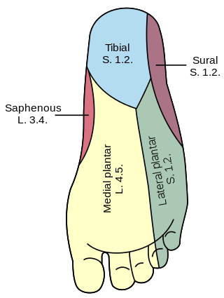
The plantar nerves are a pair of nerves innervating the sole of the foot. They arise from the posterior branch of the tibial nerve.

<span class="mw-page-title-main">Abductor digiti minimi muscle of foot</span> Muscle which lies along the lateral (outer) border of the foot

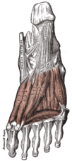
The abductor digiti minimi is a muscle which lies along the lateral (outer) border of the foot, and is in relation by its medial margin with the lateral plantar artery, vein and nerves.

<span class="mw-page-title-main">Medial plantar nerve</span> Larger of the two terminal divisions of the tibial nerve

The medial plantar nerve is the larger of the two terminal divisions of the tibial nerve, which accompanies the medial plantar artery.

<span class="mw-page-title-main">Fibularis muscles</span> Group of muscles in the leg

The fibularis muscles are a group of muscles in the lower leg.

<span class="mw-page-title-main">Outline of human anatomy</span> Overview of and topical guide to human anatomy

The following outline is provided as an overview of and topical guide to human anatomy:

<span class="mw-page-title-main">Cervical spinal nerve 8</span> Spinal nerve of the cervical segment

The cervical spinal nerve 8 (C8) is a spinal nerve of the cervical segment.

References