
The human leg, in the general meaning, is the entire lower limb of the human body, including the foot, thigh and even the hip or gluteal region. However, the definition in human anatomy refers only to the section of the lower limb extending from the knee to the ankle, also known as the crus. Legs are used for standing, and all forms of locomotion including recreational such as dancing, and constitute a significant portion of a person's mass. Female legs generally have greater hip anteversion and tibiofemoral angles, but shorter femur and tibial lengths than those in males.

In the human body, the cuboid bone is one of the seven tarsal bones of the foot.
The posterior tibial artery of the lower limb carries blood to the posterior compartment of the leg and plantar surface of the foot, from the popliteal artery via the tibial-fibular trunk. It is accompanied by a deep vein, the posterior tibial vein, along its course.

The tibial nerve is a branch of the sciatic nerve. The tibial nerve passes through the popliteal fossa to pass below the arch of soleus.
In human anatomy, the dorsal interossei of the foot are four muscles situated between the metatarsal bones.

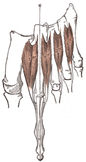
The quadratus plantae is separated from the muscles of the first layer by the lateral plantar vessels and nerve. It acts to aid in flexing the 2nd to 5th toes and is one of the few muscles in the foot with no homolog in the hand.

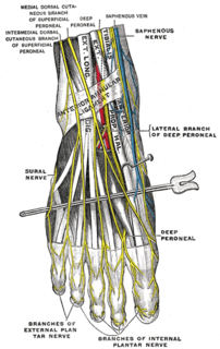
The superficial peroneal nerve or superior fibular nerve, innervates the peroneus longus and peroneus brevis muscles and the skin over the antero-lateral aspect of the leg along with the greater part of the dorsum of the foot.
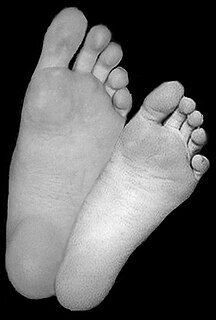
The sole is the bottom of the foot.
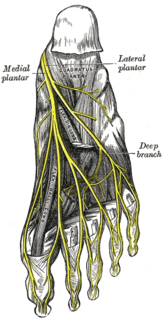
The medial plantar nerve is the larger of the two terminal divisions of the tibial nerve, which accompanies the medial plantar artery.

The lateral plantar nerve is a branch of the tibial nerve, in turn a branch of the sciatic nerve and supplies the skin of the fifth toe and lateral half of the fourth, as well as most of the deep muscles, its distribution being similar to that of the ulnar nerve in the hand.

The plantar metatarsal arteries are four in number, arising from the convexity of the plantar arch. They run forward between the metatarsal bones and in contact with the Interossei. They are located in the fourth layer of the foot.
The medial plantar artery, much smaller than the lateral plantar artery, passes forward along the medial side of the foot.
The deep plantar artery descends into the sole of the foot, between the two heads of the first Interosseous dorsalis, and unites with the termination of the lateral plantar artery, to complete the plantar arch.
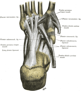
The tarsometatarsal joints are arthrodial joints in the foot. The tarsometatarsal joints involve the first, second and third cuneiform bones, the cuboid bone and the metatarsal bones. The eponym Lisfranc joint is named after 18th-19th century surgeon and gynecologist, Jacques Lisfranc de St. Martin.
The arcuate artery of the foot arises from dorsalis pedis slightly anterior to the lateral tarsal artery, specifically over the naviculocuneiform joint; it passes lateralward, over the bases of the lateral four metatarsal bones, beneath the tendons of the extensor digitorum brevis, its direction being influenced by its point of origin; and it terminates in the lateral tarsal artery. It communicates with the plantar arteries through the perforating arteries of the foot.
The plantar arch is a circulatory anastomosis formed from:
The arcuate artery of the foot gives off the second, third, and fourth dorsal metatarsal arteries, which run forward upon the corresponding Interossei dorsales; in the clefts between the toes, each divides into two dorsal digital branches for the adjoining toes.
The superficial branch of the lateral plantar nerve splits into a proper and a common plantar digital nerve:
The proper plantar digital nerves of medial plantar nerve are nerves of the foot. They primarily arise from the medial plantar nerve's superficial and deep branches. The superficial branch of the medial plantar nerve turns into a proper digital nerve and is responsible for supplies the medial side of the great toe.
The public domain consists of all the creative works to which no exclusive intellectual property rights apply. Those rights may have expired, been forfeited, expressly waived, or may be inapplicable.

Gray's Anatomy is an English language textbook of human anatomy originally written by Henry Gray and illustrated by Henry Vandyke Carter. Earlier editions were called Anatomy: Descriptive and Surgical, Anatomy of the Human Body and Gray's Anatomy: Descriptive and Applied, but the book's name is commonly shortened to, and later editions are titled, Gray's Anatomy. The book is widely regarded as an extremely influential work on the subject, and has continued to be revised and republished from its initial publication in 1858 to the present day. The latest edition of the book, the 41st, was published in September 2015.

