The human leg, in the general word sense, is the entire lower limb of the human body, including the foot, thigh and even the hip or gluteal region. However, the definition in human anatomy refers only to the section of the lower limb extending from the knee to the ankle, also known as the crus or, especially in non-technical use, the shank. Legs are used for standing, and all forms of locomotion including recreational such as dancing, and constitute a significant portion of a person's mass. Female legs generally have greater hip anteversion and tibiofemoral angles, but shorter femur and tibial lengths than those in males.

The radial nerve is a nerve in the human body that supplies the posterior portion of the upper limb. It innervates the medial and lateral heads of the triceps brachii muscle of the arm, as well as all 12 muscles in the posterior osteofascial compartment of the forearm and the associated joints and overlying skin.
The posterior tibial artery of the lower limb is an artery that carries blood to the posterior compartment of the leg and plantar surface of the foot. It branches from the popliteal artery via the tibial-fibular trunk.

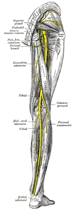
The tibial nerve is a branch of the sciatic nerve. The tibial nerve passes through the popliteal fossa to pass below the arch of soleus.

Tarsal tunnel syndrome (TTS), is a compression neuropathy and painful foot condition in which the tibial nerve is compressed as it travels through the tarsal tunnel. This tunnel is found along the inner leg behind the medial malleolus. The posterior tibial artery, tibial nerve, and tendons of the tibialis posterior, flexor digitorum longus, and flexor hallucis longus muscles travel in a bundle through the tarsal tunnel. Inside the tunnel, the nerve splits into three segments. One nerve (calcaneal) continues to the heel, the other two continue on to the bottom of the foot. The tarsal tunnel is delineated by bone on the inside and the flexor retinaculum on the outside.
The common fibular nerve is a nerve in the lower leg that provides sensation over the posteriolateral part of the leg and the knee joint. It divides at the knee into two terminal branches: the superficial fibular nerve and deep fibular nerve, which innervate the muscles of the lateral and anterior compartments of the leg respectively. When the common fibular nerve is damaged or compressed, foot drop can ensue.

The plantar nerves are a pair of nerves innervating the sole of the foot. They arise from the posterior branch of the tibial nerve.

The deep peroneal nerve begins at the bifurcation of the common peroneal nerve between the fibula and upper part of the peroneus longus, passes infero-medially, deep to extensor digitorum longus, to the anterior surface of the interosseous membrane, and comes into relation with the anterior tibial artery above the middle of the leg; it then descends with the artery to the front of the ankle-joint, where it divides into a lateral and a medial terminal branch.

The popliteal fossa is a shallow depression located at the back of the knee joint. The bones of the popliteal fossa are the femur and the tibia. Like other flexion surfaces of large joints, it is an area where blood vessels and nerves pass relatively superficially, and with an increased number of lymph nodes.

The obturator nerve in human anatomy arises from the ventral divisions of the second, third, and fourth lumbar nerves in the lumbar plexus; the branch from the third is the largest, while that from the second is often very small.
The sural nerve is a sensory nerve in the calf region (sura) of the leg. It is made up of branches of the tibial nerve and common fibular nerve, the medial cutaneous branch from the tibial nerve, and the lateral cutaneous branch from the common fibular nerve. Once formed, the nerves runs down the mid calf to the ankle and along the skin from the mid-posterior popliteal fossa to just behind to the lateral malleolus and then under the malleolus and forward along the lateral aspect of the foot.

The intercostobrachial nerves are cutaneous branches of the intercostal nerves.

The sole is the bottom of the foot.
The saphenous nerve is the largest cutaneous branch of the femoral nerve. It is a strictly sensory nerve, and has no motor function.
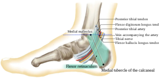
The tarsal tunnel is a passage found along the inner leg underneath the medial malleolus of the ankle.

The medial sural cutaneous nerve is a sensory nerve of the leg. It supplies part of the medial side of the leg.

The anterior compartment of thigh contains muscles which extend the knee and flex the hip.
Occasionally the communicating branch to the anterior cutaneous and saphenous branches of the femoral is continued down, as a cutaneous branch, to the thigh and leg, as the cutaneous branch of the obturator nerve.
Cutaneous innervation refers to the area of the skin which is supplied by a specific nerve.
The subsartorial plexus is a plexus of nerves that is located under the sartorius muscle.