The leg is the entire lower limb of the human body, including the foot, thigh or sometimes even the hip or buttock region. The major bones of the leg are the femur, tibia, and adjacent fibula. The thigh is between the hip and knee, while the calf (rear) and shin (front) are between the knee and foot.

In human anatomy, the fibularis longus is a superficial muscle in the lateral compartment of the leg. It acts to tilt the sole of the foot away from the midline of the body (eversion) and to extend the foot downward away from the body at the ankle.

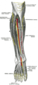
The popliteal artery is a deeply placed continuation of the femoral artery opening in the distal portion of the adductor magnus muscle. It courses through the popliteal fossa and ends at the lower border of the popliteus muscle, where it branches into the anterior and posterior tibial arteries.

The tibial nerve is a branch of the sciatic nerve. The tibial nerve passes through the popliteal fossa to pass below the arch of soleus.

In human anatomy, the fibularis brevis is a muscle that lies underneath the fibularis longus within the lateral compartment of the leg. It acts to tilt the sole of the foot away from the midline of the body (eversion) and to extend the foot downward away from the body at the ankle.
The common fibular nerve is a nerve in the lower leg that provides sensation over the posterolateral part of the leg and the knee joint. It divides at the knee into two terminal branches: the superficial fibular nerve and deep fibular nerve, which innervate the muscles of the lateral and anterior compartments of the leg respectively. When the common fibular nerve is damaged or compressed, foot drop can ensue.

The superficial fibular nerve is a mixed nerve that provides motor innervation to the fibularis longus and fibularis brevis muscles, and sensory innervation to skin over the antero-lateral aspect of the leg along with the greater part of the dorsum of the foot.
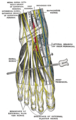
The deep fibular nerve begins at the bifurcation of the common fibular nerve between the fibula and upper part of the fibularis longus, passes infero-medially, deep to the extensor digitorum longus, to the anterior surface of the interosseous membrane, and comes into relation with the anterior tibial artery above the middle of the leg; it then descends with the artery to the front of the ankle-joint, where it divides into a lateral and a medial terminal branch.

The popliteal fossa is a shallow depression located at the back of the knee joint. The bones of the popliteal fossa are the femur and the tibia. Like other flexion surfaces of large joints, it is an area where blood vessels and nerves pass relatively superficially, and with an increased number of lymph nodes.

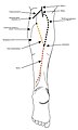
The sural nerve(L4-S1) is generally considered a pure cutaneous nerve of the posterolateral leg to the lateral ankle. The sural nerve originates from a combination of either the sural communicating branch and medial sural cutaneous nerve, or the lateral sural cutaneous nerve. This group of nerves is termed the sural nerve complex. There are eight documented variations of the sural nerve complex. Once formed the sural nerve takes its course midline posterior to posterolateral around the lateral malleolus. The sural nerve terminates as the lateral dorsal cutaneous nerve.
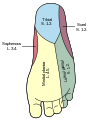
The saphenous nerve is the largest cutaneous branch of the femoral nerve. It is derived from the lumbar plexus (L3-L4). It is a strictly sensory nerve, and has no motor function. It commences in the proximal (upper) thigh and travels along the adductor canal. Upon exiting the adductor canal, the saphenous nerve terminates by splitting into two terminal branches: the sartorial nerve, and the infrapatellar nerve. The saphenous nerve is responsible for providing sensory innervation to the skin of the anteromedial leg.

The intermediate dorsal cutaneous nerve is the smaller and more lateral one of the two terminal branches of the superficial fibular nerve. It passes over the third intermetatarsal space before itself bifurcating into two terminal branches: the lateral dorsal digital nerve of the third toe, and the medial dorsal digital nerve of the fourth toe.
The medial dorsal cutaneous nerve is the more medial one of the two terminal branches of the superficial fibular nerve. Through its branches, it provides innervation to parts of the dorsal aspects of the first, second, and third toes.
The medial sural cutaneous nerve(L4-S3) is a sensory nerve of the leg. It supplies cutaneous innervation the posteromedial leg.

Cutaneous innervation of the lower limbs is the nerve supply to areas of the skin of the lower limbs which are supplied by specific cutaneous nerves.
The lateral dorsal cutaneous nerve is the continuation/terminal sensory branch of the sural nerve, and is ultimately derived from the 1st sacral nerve (S1). It passes distally along the lateral part of the dorsum of foot. It gives rise to the lateral dorsal digital nerve of the 5th toe, and sometimes also the medial dorsal digital nerve of the 5th toe as well as the lateral dorsal digital nerve of the 4th toe.
The sural communicating nerve(SCN) is a separate and independent nerve from both the medial and lateral sural cutaneous nerves, often arising from a common trunk of the common fibular nerve The primary purpose of the sural communicating branch is to provide the structural path for transferring tibial nerve fascicular components to the sural nerve.
Dorsal digital nerves of foot are branches of the intermediate dorsal cutaneous nerve, medial dorsal cutaneous nerve, sural nerve and deep fibular nerve.

In anatomy, the fibular artery, also known as the peroneal artery, supplies blood to the lateral compartment of the leg. It arises from the tibial-fibular trunk.
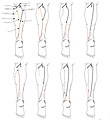
The sural nerve complex are the contributing nerves that form the sural nerve. There are eight documented anatomic variations of the sural nerve complex.