The radial nerve is a nerve in the human body that supplies the posterior portion of the upper limb. It innervates the medial and lateral heads of the triceps brachii muscle of the arm, as well as all 12 muscles in the posterior osteofascial compartment of the forearm and the associated joints and overlying skin.

In human anatomy, the ulnar nerve is a nerve that runs near the ulna bone. The ulnar collateral ligament of elbow joint is in relation with the ulnar nerve. The nerve is the largest in the human body unprotected by muscle or bone, so injury is common. This nerve is directly connected to the little finger, and the adjacent half of the ring finger, innervating the palmar aspect of these fingers, including both front and back of the tips, perhaps as far back as the fingernail beds.
In human anatomy, the dorsalis pedis artery is a blood vessel of the lower limb. It arises from the anterior tibial artery, and ends at the first intermetatarsal space. It carries oxygenated blood to the dorsal side of the foot. It is useful for taking a pulse. It is also at risk during anaesthesia of the deep peroneal nerve.

The tibial nerve is a branch of the sciatic nerve. The tibial nerve passes through the popliteal fossa to pass below the arch of soleus.

In human anatomy, the left gastric artery arises from the celiac artery and runs along the superior portion of the lesser curvature of the stomach before anastomosing with the right gastric artery. It also issues esophageal branches that supply lower esophagus and ascend through the esophageal hiatus to form anastomoses with the esophageal branches of thoracic part of aorta.

In human anatomy, the dorsal interossei of the foot are four muscles situated between the metatarsal bones.
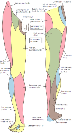
The common fibular nerve is a nerve in the lower leg that provides sensation over the posterolateral part of the leg and the knee joint. It divides at the knee into two terminal branches: the superficial fibular nerve and deep fibular nerve, which innervate the muscles of the lateral and anterior compartments of the leg respectively. When the common fibular nerve is damaged or compressed, foot drop can ensue.

The superficial fibular nerve is a mixed nerve that provides motor innervation to the fibularis longus and fibularis brevis muscles, and sensory innervation to skin over the antero-lateral aspect of the leg along with the greater part of the dorsum of the foot.

The sural nerve(L4-S1) is generally considered a pure cutaneous nerve of the posterolateral leg to the lateral ankle. The sural nerve originates from a combination of either the sural communicating branch and medial sural cutaneous nerve, or the lateral sural cutaneous nerve. This group of nerves is termed the sural nerve complex. There are eight documented variations of the sural nerve complex. Once formed the sural nerve takes its course midline posterior to posterolateral around the lateral malleolus. The sural nerve terminates as the lateral dorsal cutaneous nerve.

The suspensory ligament of the penis is a triangular midline structure anchoring the penis to the pubic symphysis, holding the penis close to the pubic bone and supporting it during erection.

The medial plantar nerve is the larger of the two terminal divisions of the tibial nerve, which accompanies the medial plantar artery.

The dorsal artery of the penis is a bilaterally paired terminal branch of the internal pudendal artery which passes upon the dorsum of the penis to the base of the glans penis, where it unites with its contralateral partner and supply the glans and foreskin.

The cribriform fascia is the portion of the superficial layer of the deep fascia of leg which extends between the sartorius muscle, adductor longus muscle, and inguinal ligament to form the anterior portion of the femoral canal.

The intermediate dorsal cutaneous nerve is the smaller and more lateral one of the two terminal branches of the superficial fibular nerve. It passes over the third intermetatarsal space before itself bifurcating into two terminal branches: the lateral dorsal digital nerve of the third toe, and the medial dorsal digital nerve of the fourth toe.
The medial dorsal cutaneous nerve is the more medial one of the two terminal branches of the superficial fibular nerve. Through its branches, it provides innervation to parts of the dorsal aspects of the first, second, and third toes.
The lateral sural cutaneous nerve of the lumbosacral plexus supplies the skin on the posterior and lateral surfaces of the leg. The lateral sural cutaneous nerve originates from the common fibular nerve(L4-S2) and is the terminal branch of the common fibular nerve.
The medial sural cutaneous nerve(L4-S3) is a sensory nerve of the leg. It supplies cutaneous innervation the posteromedial leg.
The trigeminal tubercle or tuberculum cinereum is a raised area upon the lateral dorsal/posterior aspect of the medulla oblongata produced by the underlying spinal tract of the trigeminal nerve. It is situated just lateral to the tuberculum cuneatus, between the rootlets of the accessory nerve and posterolateral sulcus.
The sural communicating nerve(SCN) is a separate and independent nerve from both the medial and lateral sural cutaneous nerves, often arising from a common trunk of the common fibular nerve The primary purpose of the sural communicating branch is to provide the structural path for transferring tibial nerve fascicular components to the sural nerve.
Dorsal digital nerves of foot are branches of the intermediate dorsal cutaneous nerve, medial dorsal cutaneous nerve, sural nerve and deep fibular nerve.