Superficial fibular nerve

Last updated
Superficial fibular (peroneal) nerve
Gray835.png
Deep nerves of the front of the leg.
Details
From Common fibular nerve
To Medial dorsal cutaneous nerve, intermediate dorsal cutaneous nerve
Identifiers
Latin nervus fibularis superficialis,
nervus peroneus superficialis
TA98 A14.2.07.050
TA2 6574
FMA 44699
Anatomical terms of neuroanatomy

The superficial fibular nerve (also known as superficial peroneal nerve) is a mixed (motor and sensory) nerve that provides motor innervation to the fibularis longus and fibularis brevis muscles, and sensory innervation to skin over the antero-lateral aspect of the leg along with the greater part of the dorsum of the foot (with the exception of the first web space, which is innervated by the deep fibular nerve).

Contents

Structure

Lateral side of the leg

The superficial fibular nerve is the main nerve of the lateral compartment of the leg. It begins at the lateral side of the neck of fibula, and runs through the fibularis longus and fibularis brevis muscles. In the middle third of the leg, it descends between the fibularis longus and fibularis brevis, and then reaches the anterior border of the fibularis brevis to enter the groove between the fibularis brevis and the extensor digitorum longus under the deep fascia of leg. It becomes superficial at the junction of upper two-thirds and lower one-thirds of the leg by piercing the deep fascia. The superficial fibular nerve gives off several branches in the leg. [1]

At the junction between the upper two-thirds and lower one-thirds of the leg, the superficial fibular nerve is divided into medial dorsal cutaneous nerve (medial branch) and intermediate dorsal cutaneous nerve (lateral branch). [1]

Distribution

The branches of the superficial fibular nerve supply the skin of the dorsal surfaces of all the toes excepting the lateral side of the little toe, and the adjoining sides of the great and second toes, the former being supplied by the lateral dorsal cutaneous nerve from the sural nerve, and the latter by the medial branch of the deep peroneal nerve.

Frequently some of the lateral branches of the superficial fibular nerve are absent, and their places are then taken by branches of the sural nerve.

Clinical significance

Injury to the nerve can result in an inability to evert the foot and loss of sensation over the dorsum of the foot (with the exception of the first web space between the great toe and the second toe, where the deep fibular nerve innervates).[ citation needed ]

Additional images

Related Research Articles

<span class="mw-page-title-main">Human leg</span> Lower extremity or limb of the human body (foot, lower leg, thigh and hip)

The leg is the entire lower limb of the human body, including the foot, thigh or sometimes even the hip or buttock region. The major bones of the leg are the femur, tibia, and adjacent fibula. The thigh is between the hip and knee, while the calf (rear) and shin (front) are between the knee and foot.

<span class="mw-page-title-main">Radial nerve</span> Nerve in the human body that supplies the posterior portion of the upper limb

The radial nerve is a nerve in the human body that supplies the posterior portion of the upper limb. It innervates the medial and lateral heads of the triceps brachii muscle of the arm, as well as all 12 muscles in the posterior osteofascial compartment of the forearm and the associated joints and overlying skin.

<span class="mw-page-title-main">Fibularis longus</span> Superficial muscle in the lateral compartment of the leg

In human anatomy, the fibularis longus is a superficial muscle in the lateral compartment of the leg. It acts to tilt the sole of the foot away from the midline of the body (eversion) and to extend the foot downward away from the body at the ankle.

<span class="mw-page-title-main">Tibial nerve</span> Branch of the sciatic nerve

The tibial nerve is a branch of the sciatic nerve. The tibial nerve passes through the popliteal fossa to pass below the arch of soleus.

<span class="mw-page-title-main">Dorsal interossei of the foot</span> Four muscles situated between the metatarsal bones

In human anatomy, the dorsal interossei of the foot are four muscles situated between the metatarsal bones.

<span class="mw-page-title-main">Extensor digitorum longus muscle</span> Pennate muscle, situated at the lateral part of the front of the leg

The extensor digitorum longus is a pennate muscle, situated at the lateral part of the front of the leg.

<span class="mw-page-title-main">Extensor digitorum brevis muscle</span> Muscle on the upper surface of the foot that helps extend digits 2 through 4

The extensor digitorum brevis muscle is a muscle on the upper surface of the foot that helps extend digits 2 through 4.

<span class="mw-page-title-main">Fibularis brevis</span> Shorter and smaller of the fibularis (peroneus) muscles

In human anatomy, the fibularis brevis is a muscle that lies underneath the fibularis longus within the lateral compartment of the leg. It acts to tilt the sole of the foot away from the midline of the body (eversion) and to extend the foot downward away from the body at the ankle.

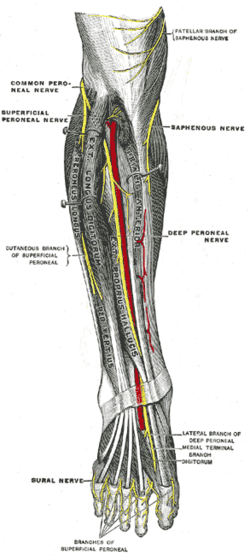
The common fibular nerve is a nerve in the lower leg that provides sensation over the posterolateral part of the leg and the knee joint. It divides at the knee into two terminal branches: the superficial fibular nerve and deep fibular nerve, which innervate the muscles of the lateral and anterior compartments of the leg respectively. When the common fibular nerve is damaged or compressed, foot drop can ensue.

<span class="mw-page-title-main">Extensor hallucis brevis muscle</span> Muscle on the top of the foot that helps to extend the big toe

The extensor hallucis brevis is a muscle on the top of the foot that helps to extend the big toe.

<span class="mw-page-title-main">Plantar nerve</span> Pair of nerves innervating the sole of the foot

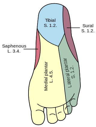
The plantar nerves are a pair of nerves innervating the sole of the foot. They arise from the posterior branch of the tibial nerve.

<span class="mw-page-title-main">Deep fibular nerve</span> Type of nerve

The deep fibular nerve begins at the bifurcation of the common fibular nerve between the fibula and upper part of the fibularis longus, passes infero-medially, deep to the extensor digitorum longus, to the anterior surface of the interosseous membrane, and comes into relation with the anterior tibial artery above the middle of the leg; it then descends with the artery to the front of the ankle-joint, where it divides into a lateral and a medial terminal branch.

<span class="mw-page-title-main">Sole (foot)</span> Bottom part of foot

In humans, the sole of the foot is anatomically referred to as the plantar aspect.

<span class="mw-page-title-main">Fibularis muscles</span> Group of muscles in the leg

The fibularis muscles are a group of muscles in the lower leg.

The saphenous nerve is the largest cutaneous branch of the femoral nerve. It is derived from the lumbar plexus (L3-L4). It is a strictly sensory nerve, and has no motor function. It commences in the proximal (upper) thigh and travels along the adductor canal. Upon exiting the adductor canal, the saphenous nerve terminates by splitting into two terminal branches: the sartorial nerve, and the infrapatellar nerve. The saphenous nerve is responsible for providing sensory innervation to the skin of the anteromedial leg.

<span class="mw-page-title-main">Intermediate dorsal cutaneous nerve</span>

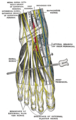
The intermediate dorsal cutaneous nerve is the smaller and more lateral one of the two terminal branches of the superficial fibular nerve. It passes over the third intermetatarsal space before itself bifurcating into two terminal branches: the lateral dorsal digital nerve of the third toe, and the medial dorsal digital nerve of the fourth toe.

<span class="mw-page-title-main">Medial dorsal cutaneous nerve</span>

The medial dorsal cutaneous nerve is the more medial one of the two terminal branches of the superficial fibular nerve. Through its branches, it provides innervation to parts of the dorsal aspects of the first, second, and third toes.

<span class="mw-page-title-main">Lateral sural cutaneous nerve</span> Sensory nerve of the leg that supplies part of the posterolateral side of the leg

The lateral sural cutaneous nerve of the lumbosacral plexus supplies the skin on the posterior and lateral surfaces of the leg. The lateral sural cutaneous nerve originates from the common fibular nerve(L4-S2) and is the terminal branch of the common fibular nerve.

<span class="mw-page-title-main">Cutaneous innervation of the lower limbs</span> Area of the skin

Cutaneous innervation of the lower limbs is the nerve supply to areas of the skin of the lower limbs which are supplied by specific cutaneous nerves.

<span class="mw-page-title-main">Dorsal digital nerves of foot</span> Various nerve branches that supply the toes

Dorsal digital nerves of foot are branches of the intermediate dorsal cutaneous nerve, medial dorsal cutaneous nerve, sural nerve and deep fibular nerve.

References

PD-icon.svgThis article incorporates text in the public domain from page 966 of the 20th edition of Gray's Anatomy (1918)

  1. 1 2 3 4 5 6 7 Krishna, Garg (2010). "Front, lateral, and medial sides of leg and dorsum of foot (Chapter 8)". BD Chaurasia's Human Anatomy (Regional and Applied Dissection and Clinical) Volume 2 - Lower limb, abdomen, and pelvis (Fifth ed.). India: CBS Publishers and Distributors Pvt Ltd. p. 109,110. ISBN   978-81-239-1864-8.