Sural communicating branch of common peroneal nerve

Last updated
Sural communicating nerve
RobertSteele SuralNerve GraysMODified.png
Dissection of popliteal space to show the formation of a type 1 sural nerve
Sural nerve formation.jpg
Cartoon version adapted from Steele et al. depicting type 1 sural nerve
Details
From Lateral sural cutaneous nerve
To Medial sural cutaneous nerve
Identifiers
Latin R. communicans fibularis
TA98 A14.2.07.049
TA2 6573
FMA 77602
Anatomical terms of neuroanatomy

The sural communicating nerve(SCN) (peroneal communicating branch of the common fibular nerve) is a separate and independent nerve from both the medial and lateral sural cutaneous nerves, often arising from a common trunk of the common fibular nerve [1] [2] The primary purpose of the sural communicating branch is to provide the structural path for transferring tibial nerve fascicular components to the sural nerve. [3]

Contents

Anatomy

The sural communicating nerve (colloquially the peroneal communicating nerve) is one of the components of the sural nerve complex (MSCN, LSCN,SCN). It travels in an inferomedial direction from its origins either as a terminal component of the LSCN or is considered a nerve that originates along a common trunk of the lateral sural cutaneous nerve.

This particular nerve is best described by Dr. Huelke in 1958. Dr. Huelke took the original modern cadaveric studies (from the turn of the 20th century) and wrote a treatise titled "The origin of the peroneal communicating nerve" that describes the origins and pathways of the peroneal communicating nerve. He did this research in such a way that would today be called a systematic review with the addition of his own cadaveric samples. He reported that the peroneal communicating nerve was only considered named if it 'originated from the lateral sural cutaneous nerve to anastomose with the medial sural cutaneous nerve' to form the sural nerve proper.

Huelke found of the 159 samples that the peroneal communicating nerve arose from the common peroneal nerve with the lateral sural cutaneous nerve around 58.5% of the time and originated separately from the common peroneal nerve the remaining 41.5% of the time. In fact, Huelkes work is directly observed and fortified in a recent cadaveric study by Steele et al. in which further origins of the sural communicating nerve arise lending the name nerve over branch, however even now there is no firm definition of the communication from the common peroneal nerve or from the lateral sural cutaneous nerve [ citation needed ]. Steele et al. found that the sural communicating branch originated from the common peroneal nerve 50.79%, common trunk 16.67%, the lateral sural cutaneous nerve 30.16%, and sciatic nerve 2.38% of the 126 total samples that contained a sural communicating branch. [4] [5]

Essential component to the Sural Nerve Complex

The sural nerve complex was first used by Mayo Clinic orthopedic surgeon Dr. Ortiguela in a 1987 cadaveric study of the contributing nerves that is the genesis before the sural nerve moves from deep fascia of the gastrocnemius into the subcutaneous of the posterior sura. [6] Later, meta-analysis [7] and follow on large sample cadaveric confirmation study [1] make reference to the sural communicating branch and both reference this nerve complex as the components contributing to the formation of the sural nerve.

In Steele et al. [1] study identifying eight variations of the sural nerve formation. Types 1 and 7 sural nerve complex formations possess a sural communicating branch. This recent cadaveric study a 50.79% prevalence of nerves with a sural communicating nerve. Ramakrishnan et al. found that type 1 nerves (only sural nerve complex types that contain SCN as defined by his figures) were globally present in 51.50% of all cadaveric studies captured in their systematic review.

Additional images

Related Research Articles

<span class="mw-page-title-main">Sciatic nerve</span> Large nerve in humans and other animals

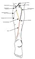
The sciatic nerve, also called the ischiadic nerve, is a large nerve in humans and other vertebrate animals which is the largest branch of the sacral plexus and runs alongside the hip joint and down the lower limb. It is the longest and widest single nerve in the human body, going from the top of the leg to the foot on the posterior aspect. The sciatic nerve has no cutaneous branches for the thigh. This nerve provides the connection to the nervous system for the skin of the lateral leg and the whole foot, the muscles of the back of the thigh, and those of the leg and foot. It is derived from spinal nerves L4 to S3. It contains fibers from both the anterior and posterior divisions of the lumbosacral plexus.

<span class="mw-page-title-main">Popliteal artery</span> Continuation of the femoral artery that supplies the lower leg

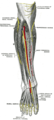
The popliteal artery is a deeply placed continuation of the femoral artery opening in the distal portion of the adductor magnus muscle. It courses through the popliteal fossa and ends at the lower border of the popliteus muscle, where it branches into the anterior and posterior tibial arteries.

<span class="mw-page-title-main">Tibial nerve</span> Branch of the sciatic nerve

The tibial nerve is a branch of the sciatic nerve. The tibial nerve passes through the popliteal fossa to pass below the arch of soleus.

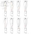
The common fibular nerve is a nerve in the lower leg that provides sensation over the posterolateral part of the leg and the knee joint. It divides at the knee into two terminal branches: the superficial fibular nerve and deep fibular nerve, which innervate the muscles of the lateral and anterior compartments of the leg respectively. When the common fibular nerve is damaged or compressed, foot drop can ensue.

<span class="mw-page-title-main">Superficial fibular nerve</span> Nerve in the human leg and foot

The superficial fibular nerve is a mixed nerve that provides motor innervation to the fibularis longus and fibularis brevis muscles, and sensory innervation to skin over the antero-lateral aspect of the leg along with the greater part of the dorsum of the foot.

<span class="mw-page-title-main">Deep fibular nerve</span> Type of nerve

The deep fibular nerve begins at the bifurcation of the common fibular nerve between the fibula and upper part of the fibularis longus, passes infero-medially, deep to the extensor digitorum longus, to the anterior surface of the interosseous membrane, and comes into relation with the anterior tibial artery above the middle of the leg; it then descends with the artery to the front of the ankle-joint, where it divides into a lateral and a medial terminal branch.

<span class="mw-page-title-main">Popliteal fossa</span> Depression at back of knee joint

The popliteal fossa is a shallow depression located at the back of the knee joint. The bones of the popliteal fossa are the femur and the tibia. Like other flexion surfaces of large joints, it is an area where blood vessels and nerves pass relatively superficially, and with an increased number of lymph nodes.

<span class="mw-page-title-main">Medial cutaneous nerve of arm</span>

The medial brachial cutaneous nerve is a sensory branch of the medial cord of the brachial plexus derived from spinal nerves C8-T1. It provides sensory innervation to the medial arm. It descends accompanied by the basilic vein.

<span class="mw-page-title-main">Sural nerve</span> Pure cutaneous nerve of the posterolateral leg to the lateral ankle

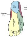
The sural nerve(L4-S1) is generally considered a pure cutaneous nerve of the posterolateral leg to the lateral ankle. The sural nerve originates from a combination of either the sural communicating branch and medial sural cutaneous nerve, or the lateral sural cutaneous nerve. This group of nerves is termed the sural nerve complex. There are eight documented variations of the sural nerve complex. Once formed the sural nerve takes its course midline posterior to posterolateral around the lateral malleolus. The sural nerve terminates as the lateral dorsal cutaneous nerve.

<span class="mw-page-title-main">Sole (foot)</span> Bottom part of foot

The sole is the bottom of the foot.

The saphenous nerve is the largest cutaneous branch of the femoral nerve. It is derived from the lumbar plexus (L3-L4). It is a strictly sensory nerve, and has no motor function. It commences in the proximal (upper) thigh and travels along the adductor canal. Upon exiting the adductor canal, the saphenous nerve terminates by splitting into two terminal branches: the sartorial nerve, and the infrapatellar nerve. The saphenous nerve is responsible for providing sensory innervation to the skin of the anteromedial leg.

<span class="mw-page-title-main">Intermediate dorsal cutaneous nerve</span>

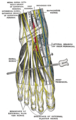
The intermediate dorsal cutaneous nerve, the smaller, passes along the lateral part of the dorsum of the foot, and divides into dorsal digital branches, which supply the contiguous sides of the third and fourth, and of the fourth and fifth toes.

<span class="mw-page-title-main">Lateral sural cutaneous nerve</span> Sensory nerve of the leg that supplies part of the posterolateral side of the leg

The lateral sural cutaneous nerve of the lumbosacral plexus supplies the skin on the posterior and lateral surfaces of the leg. The lateral sural cutaneous nerve originates from the common fibular nerve(L4-S2) and is the terminal branch of the common fibular nerve.

<span class="mw-page-title-main">Medial sural cutaneous nerve</span> Sensory nerve of the leg that supplies part of the medial side of the leg

The medial sural cutaneous nerve(L4-S3) is a sensory nerve of the leg. It supplies cutaneous innervation the posteromedial leg.

<span class="mw-page-title-main">Cutaneous innervation of the lower limbs</span> Area of the skin

Cutaneous innervation of the lower limbs is the nerve supply to areas of the skin of the lower limbs which are supplied by specific cutaneous nerves.

A neurovascular bundle is a structure that binds nerves and veins with connective tissue so that they travel in tandem through the body.

<span class="mw-page-title-main">Outline of human anatomy</span> Overview of and topical guide to human anatomy

The following outline is provided as an overview of and topical guide to human anatomy:

<span class="mw-page-title-main">Lateral dorsal cutaneous nerve</span>

The lateral dorsal cutaneous nerve is a cutaneous branch of the foot.

<span class="mw-page-title-main">Fibular artery</span>

In anatomy, the fibular artery, also known as the peroneal artery, supplies blood to the lateral compartment of the leg. It arises from the tibial-fibular trunk.

The sural nerve complex are the contributing nerves that form the sural nerve. There are eight documented anatomic variations of the sural nerve complex.

References

PD-icon.svgThis article incorporates text in the public domain from the 20th edition of Gray's Anatomy (1918)

  1. 1 2 3 Robert Steele DO MS, et al. (2021). "Anatomy of the sural nerve complex: Unaccounted anatomic variations and morphometric data". Annals of Anatomy . 238 (151742): 151742. doi:10.1016/j.aanat.2021.151742. PMID   33932499.
  2. Sekiya, S; Tokita, K; Banneheka, SK; Suzuki, R; Miyawaki, M; Chiba, S; Kumaki, K (September 2006). "[Reexamination of the communicating branch between the sural and tibial nerves]". Kaibogaku Zasshi. Journal of Anatomy. 81 (3): 83–8. PMID   16999334.
  3. Sekiya, S; Tokita, K; Banneheka, SK; Suzuki, R; Miyawaki, M; Chiba, S; Kumaki, K (September 2006). "[Reexamination of the communicating branch between the sural and tibial nerves]". Kaibogaku Zasshi. Journal of Anatomy. 81 (3): 83–8. PMID   16999334.
  4. Huelke, Donald (1958). "The origin of the peroneal communicating nerve in adult man". The Anatomical Record. 132 (1): 81–92. doi:10.1002/ar.1091320106. hdl: 2027.42/49782 . PMID   13627490. S2CID   12840788.
  5. Steele, Robert; Coker, Charles; Freed, Blair; Wright, Barth; Brauer, Philip (November 2021). "Anatomy of the sural nerve complex: Unaccounted anatomic variations and morphometric data". Annals of Anatomy. 238: 151742. doi:10.1016/j.aanat.2021.151742. PMID   33932499.
  6. Ortigüela ME, Wood MB, Cahill DR (1987). "Anatomy of the sural nerve complex". J Hand Surg . 12 (6): 1119–23. doi:10.1016/s0363-5023(87)80129-6. PMID   3693848.
  7. Ramakrishnan, Piravin Kumar; Henry, Brandon Michael; Vikse, Jens; Roy, Joyeeta; Saganiak, Karolina; Mizia, Ewa; Tomaszewski, Krzysztof A. (November 2015). "Anatomical variations of the formation and course of the sural nerve: A systematic review and meta-analysis". Annals of Anatomy - Anatomischer Anzeiger. 202: 36–44. doi:10.1016/j.aanat.2015.08.002. PMID   26342158.

Referenced papers:

Anatomy web references