The human leg, in the general word sense, is the entire lower limb of the human body, including the foot, thigh or sometimes even the hip or gluteal region. However, the definition in human anatomy refers only to the section of the lower limb extending from the knee to the ankle, also known as the crus or, especially in non-technical use, the shank. Legs are used for standing, and all forms of locomotion including recreational such as dancing, and constitute a significant portion of a person's mass. Female legs generally have greater hip anteversion and tibiofemoral angles, but shorter femur and tibial lengths than those in males.

The femoral triangle is an anatomical region of the upper third of the thigh. It is a subfascial space which appears as a triangular depression below the inguinal ligament when the thigh is flexed, abducted and laterally rotated.

The great saphenous vein(GSV), or long saphenous vein; ) is a large, subcutaneous, superficial vein of the leg. It is the longest vein in the body, running along the length of the lower limb, returning blood from the foot, leg and thigh to the deep femoral vein at the femoral triangle.

The genitofemoral nerve is a mixed branch of the lumbar plexus derived from anterior rami of L1-L2. It splits a genital branch and a femoral branch. It provides sensory innervation to the upper anterior thigh, as well as the skin of the anterior scrotum in males and mons pubis in females. It also provides motor innervation to the cremaster muscle.

The pectineus muscle is a flat, quadrangular muscle, situated at the anterior (front) part of the upper and medial (inner) aspect of the thigh. The pectineus muscle is the most anterior adductor of the hip. The muscle does adduct and internally rotate the thigh but its primary function is hip flexion.

The femoral nerve is a nerve in the thigh that supplies skin on the upper thigh and inner leg, and the muscles that extend the knee. It is the largest branch of the lumbar plexus.

The popliteal fossa is a shallow depression located at the back of the knee joint. The bones of the popliteal fossa are the femur and the tibia. Like other flexion surfaces of large joints, it is an area where blood vessels and nerves pass relatively superficially, and with an increased number of lymph nodes.

The posterior cutaneous nerve of the thigh is a sensory nerve of the thigh. It is a branch of the sacral plexus. It supplies the skin of the posterior surface of the thigh, leg, buttock, and also the perineum.
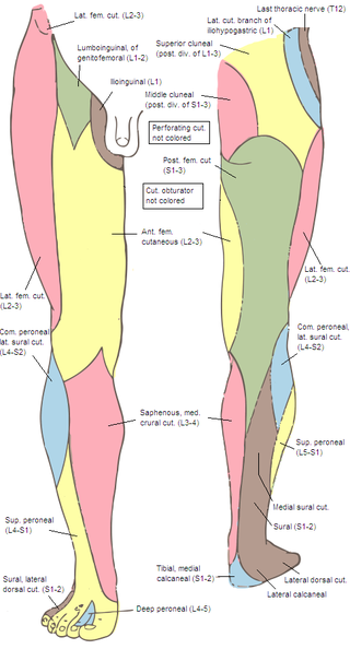
The lumbar plexus is a web of nerves in the lumbar region of the body which forms part of the larger lumbosacral plexus. It is formed by the divisions of the first four lumbar nerves (L1-L4) and from contributions of the subcostal nerve (T12), which is the last thoracic nerve. Additionally, the ventral rami of the fourth lumbar nerve pass communicating branches, the lumbosacral trunk, to the sacral plexus. The nerves of the lumbar plexus pass in front of the hip joint and mainly support the anterior part of the thigh.

The obturator nerve in human anatomy arises from the ventral divisions of the second, third, and fourth lumbar nerves in the lumbar plexus; the branch from the third is the largest, while that from the second is often very small.
The lateral cutaneous nerve of the thigh is a cutaneous nerve of the thigh. It originates from the dorsal divisions of the second and third lumbar nerves from the lumbar plexus. It passes under the inguinal ligament to reach the thigh. It supplies sensation to the skin on the lateral part of the thigh by an anterior branch and a posterior branch.

The descending genicular artery arises from the femoral artery just before its passage through the adductor hiatus.

The patellar plexus is a plexus of fine nerves situated in front of the patella, the ligamentum patellae and the upper end of the tibia. It is formed by contribution from the following:
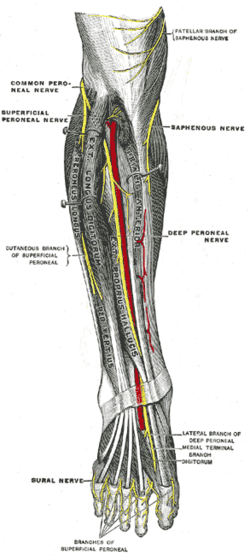
The saphenous nerve is the largest cutaneous branch of the femoral nerve. It is derived from the lumbar plexus (L3-L4). It is a strictly sensory nerve, and has no motor function. It commences in the proximal (upper) thigh and travels along the adductor canal. Upon exiting the adductor canal, the saphenous nerve terminates by splitting into two terminal branches: the sartorial nerve, and the infrapatellar nerve. The saphenous nerve is responsible for providing sensory innervation to the skin of the anteromedial leg.
The anterior branch of the obturator nerve is a branch of the obturator nerve found in the pelvis and leg.
The anterior cutaneous branches of the femoral nerve consist of the following nerves: intermediate cutaneous nerve and medial cutaneous nerve.
Occasionally the communicating branch to the anterior cutaneous and saphenous branches of the femoral is continued down, as a cutaneous branch, to the thigh and leg, as the cutaneous branch of the obturator nerve.
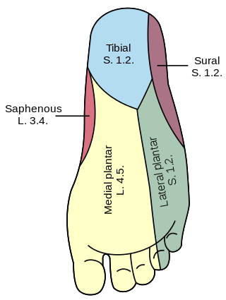
Cutaneous innervation of the lower limbs is the nerve supply to areas of the skin of the lower limbs which are supplied by specific cutaneous nerves.
The subsartorial plexus is a plexus of nerves that is located under the sartorius muscle.