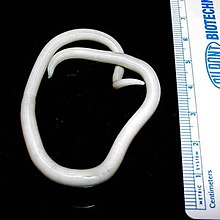
Trichuris trichiura, Trichocephalus trichiuris or whipworm, is a parasitic roundworm that causes trichuriasis when it infects a human large intestine. It is commonly known as the whipworm which refers to the shape of the worm; it looks like a whip with wider "handles" at the posterior end.

Trichuriasis, also known as whipworm infection, is an infection by the parasitic worm Trichuris trichiura (whipworm). If infection is only with a few worms, there are often no symptoms. In those who are infected with many worms, there may be abdominal pain, fatigue and diarrhea. The diarrhea sometimes contains blood. Infections in children may cause poor intellectual and physical development. Low red blood cell levels may occur due to loss of blood.

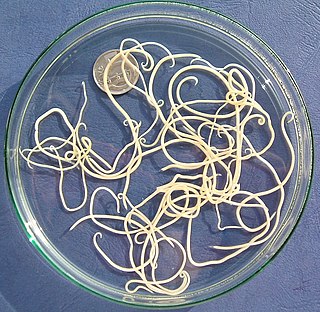
Ascariasis is a disease caused by the parasitic roundworm Ascaris lumbricoides. Infections have no symptoms in more than 85% of cases, especially if the number of worms is small. Symptoms increase with the number of worms present and may include shortness of breath and fever in the beginning of the disease. These may be followed by symptoms of abdominal swelling, abdominal pain, and diarrhea. Children are most commonly affected, and in this age group the infection may also cause poor weight gain, malnutrition, and learning problems.

Helminthiasis, also known as worm infection, is any macroparasitic disease of humans and other animals in which a part of the body is infected with parasitic worms, known as helminths. There are numerous species of these parasites, which are broadly classified into tapeworms, flukes, and roundworms. They often live in the gastrointestinal tract of their hosts, but they may also burrow into other organs, where they induce physiological damage.

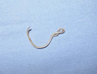
Necator americanus is a species of hookworm commonly known as the New World hookworm. Like other hookworms, it is a member of the phylum Nematoda. It is an obligatory parasitic nematode that lives in the small intestine of human hosts. Necatoriasis—a type of helminthiasis—is the term for the condition of being host to an infestation of a species of Necator. Since N. americanus and Ancylostoma duodenale are the two species of hookworms that most commonly infest humans, they are usually dealt with under the collective heading of "hookworm infection". They differ most obviously in geographical distribution, structure of mouthparts, and relative size.

Ascaris suum, also known as the large roundworm of pig, is a parasitic nematode that causes ascariasis in pigs. While roundworms in pigs and humans are today considered as two species with different hosts, cross-infection between humans and pigs is possible; some researchers have thus argued they are the same species. Ascariasis is associated with contact to pigs and pig manure in Denmark.

Ascaris is a nematode genus of parasitic worms known as the "small intestinal roundworms", which is a type of parasitic worm. One species, Ascaris lumbricoides, affects humans and causes the disease ascariasis. Another species, Ascaris suum, typically infects pigs. Other ascarid genera infect other animals, such as Parascaris equorum, the equine roundworm, and Toxocara and Toxascaris, which infect dogs and cats.

Parasitic worms, also known as helminths, are large macroparasites; adults can generally be seen with the naked eye. Many are intestinal worms that are soil-transmitted and infect the gastrointestinal tract. Other parasitic worms such as schistosomes reside in blood vessels.
The soil-transmitted helminths are a group of intestinal parasites belonging to the phylum Nematoda that are transmitted primarily through contaminated soil. They are so called because they have a direct life cycle which requires no intermediate hosts or vectors, and the parasitic infection occurs through faecal contamination of soil, foodstuffs and water supplies. The adult forms are essentially parasites of humans, causing soil-transmitted helminthiasis (STH), but also infect domesticated mammals. The juveniles are the infective forms and they undergo tissue-migratory stages during which they invade vital organs such as lungs and liver. Thus the disease manifestations can be both local and systemic. The geohelminths together present an enormous infection burden on humanity, amounting to 135,000 deaths every year, and persistent infection of more than two billion people.
Mammomonogamus is a genus of parasitic nematodes of the family Syngamidae that parasitise the respiratory tracts of cattle, sheep, goats, deer, cats, orangutans, and elephants. The nematodes can also infect humans and cause the disease called mammomonogamiasis. Several known species fall under the genus Mammomonogamus, but the most common species found to infest humans is M. laryngeus. Infection in humans is very rare, with only about 100 reported cases worldwide, and is assumed to be largely accidental. Cases have been reported from the Caribbean, China, Korea, Thailand, and Philippines.
Toxocara canis is a worldwide-distributed helminth parasite that primarily infects dogs and other canids, but can also infect other animals including humans. The name is derived from the Greek word "toxon," meaning bow or quiver, and the Latin word "caro," meaning flesh. T. canis live in the small intestine of the definitive host. This parasite is very common in puppies and somewhat less common in adult dogs. In adult dogs, infection is usually asymptomatic but may be characterized by diarrhea. By contrast, untreated infection with Toxocara canis can be fatal in puppies, causing diarrhea, vomiting, pneumonia, enlarged abdomen, flatulence, poor growth rate, and other complications.

Toxocara cati, also known as the feline roundworm, is a parasite of cats and other felids. It is one of the most common nematodes of cats, infecting both wild and domestic felids worldwide. Adult worms are localised in the gut of the host. In adult cats, the infection – which is called toxocariasis – is usually asymptomatic. However, massive infection in juvenile cats can be fatal.

Toxascaris leonina is a common parasitic roundworm found in dogs, cats, foxes, and related host species. T. leonina is an ascarid nematode, a worldwide distributed helminth parasite which is in a division of eukaryotic parasites that, unlike external parasites such as lice and fleas, live inside their host. The definitive hosts of T. leonina include canids and felines (cats), while the intermediate hosts are usually rodents, such as mice or rats. Infection occurs in the definitive host when the animal eats an infected rodent. While T. leonina can occur in either dogs or cats, it is far more frequent in cats.
Ascaricides are drugs to treat ascariasis that is caused by infections with parasitic nematodes (roundworms) of the genus Ascaris. The large roundworm of pigs typically infects pigs while Ascaris lumbricoides affects human populations, typically in sub-tropical and tropical areas with poor sanitation. Ascaricides belong to the group of drugs collectively called anthelmintics which expel parasitic worms (helminths) and other internal parasites from the body by either stunning or killing them and without causing significant damage to the host.
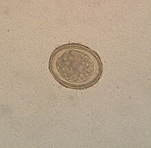
Trichuris vulpis is a whipworm that lives in the large intestine of canines in its adult stages. Out of different types of worms, Trichuris vulpis is one of the smaller worms with a size ranging from 30–50 mm in length. As the name suggests, the worm has a whip-like shape with distinct features including a small, narrow anterior head, which is the digestive part of the worm, and a larger posterior tail, which is the reproductive part of the worm. Eggs from T. vulpis are oval shaped with bipolar plugs and contain a thick outer shell. Their sizes range from 72–90 μm in length and 32–40 μm in width. Because of their thick outer shell, T. vulpis eggs are very resistant to environmental extremes such as freezing or hot temperatures, thus allowing for their long viability in the outside world.

Baylisascaris procyonis, also known by the common name raccoon roundworm, is a roundworm nematode, found ubiquitously in raccoons, the definitive hosts. It is named after H. A. Baylis, who studied them in the 1920s–30s, and Greek askaris. Baylisascaris larvae in paratenic hosts can migrate, causing larva migrans. Baylisascariasis as the zoonotic infection of humans is rare, though extremely dangerous due to the ability of the parasite's larvae to migrate into brain tissue and cause damage. Concern for human infection has been increasing over the years due to the urbanization of rural areas, resulting in the increase in proximity and potential human interaction with raccoons.

Soil-transmitted helminthiasis is a type of worm infection (helminthiasis) caused by different species of roundworms. It is caused specifically by those worms which are transmitted through soil contaminated with faecal matter and are therefore called soil-transmitted helminths. Three types of soil-transmitted helminthiasis can be distinguished: ascariasis, hookworm infection and whipworm infection. These three types of infection are therefore caused by the large roundworm A. lumbricoides, the hookworms Necator americanus or Ancylostoma duodenale and by the whipworm Trichuris trichiura.

Hookworms are intestinal, blood-feeding, parasitic roundworms that cause types of infection known as helminthiases. Hookworm infection is found in many parts of the world, and is common in areas with poor access to adequate water, sanitation, and hygiene. In humans, infections are caused by two main species of roundworm, belonging to the genera Ancylostoma and Necator. In other animals the main parasites are species of Ancylostoma. Hookworm is closely associated with poverty because it is most often found in impoverished areas, and its symptoms promote poverty through the educational and health effects it has on children. It is the leading cause of anemia and undernutrition in developing countries, while being one of the most commonly occurring diseases among poor people. Hookworm thrives in areas where rainfall is sufficient and keeps the soil from drying out, and where temperatures are higher, making rural, coastal areas prime conditions for the parasite to breed.
Cat worm infections, the infection of cats (Felidae) with parasitic worms, occur frequently. Most worm species occur worldwide in both domestic and other cats, but there are regional, species and lifestyle differences in the frequency of infestation. According to the classification of the corresponding parasites in the zoological system, infections can be divided into those caused by nematode and flatworms - in the case of the latter, mainly cestoda and trematoda - while other strains are of no veterinary significance. While threadworms usually do not require an intermediate host for their reproduction, the development cycle of flatworms always proceeds via alternate hosts.
Nematode infection in dogs - the infection of dogs with parasitic nemamotodes - are, along with tapeworm infections and infections with protozoa, frequent parasitoses in veterinary practice. Nematodes, as so-called endoparasites, colonize various internal organs - most of them the digestive tract - and the skin. To date, about 30 different species of nematode have been identified in domestic dogs; they are essentially also found in wild dog species. However, the majority of them often cause no or only minor symptoms of disease in adult animals. The infection therefore does not necessarily have to manifest itself in a worm disease (helminthosis). For most nematodes, an infection can be detected by examining the feces for eggs or larvae. Roundworm infection in dogs and the hookworm in dogs is of particular health significance in Central Europe, as they can also be transmitted to humans (zoonosis). Regular deworming can significantly reduce the frequency of infection and thus the risk of infection for humans and dogs.