Schistosoma haematobium

Last updated

Schistosoma haematobium
Schistosomiasis haematobia.jpg
Eggs of S. haematobium surrounded by intense infiltrates of eosinophils in bladder tissue.
Scientific classification OOjs UI icon edit-ltr.svg
Domain: Eukaryota
Kingdom: Animalia
Phylum: Platyhelminthes
Class: Trematoda
Order: Diplostomida
Family: Schistosomatidae
Genus: Schistosoma
Species:
S. haematobium
Binomial name
Schistosoma haematobium
(Bilharz, 1852)

Schistosoma haematobium (urinary blood fluke) is a species of digenetic trematode, belonging to a group (genus) of blood flukes ( Schistosoma ). It is found in Africa and the Middle East. It is the major agent of schistosomiasis, the most prevalent parasitic infection in humans. [1] It is the only blood fluke that infects the urinary tract, causing urinary schistosomiasis, and is the leading cause of bladder cancer (only next to tobacco smoking). [2] [3] The diseases are caused by the eggs.

Contents

Adults are found in the venous plexuses around the urinary bladder and the released eggs travels to the wall of the urine bladder causing haematuria and fibrosis of the bladder. The bladder becomes calcified, and there is increased pressure on ureters and kidneys otherwise known as hydronephrosis. Inflammation of the genitals due to S. haematobium may contribute to the propagation of HIV. [4]

S. haematobium was the first blood fluke discovered. Theodor Bilharz, a German surgeon working in Cairo, identified the parasite as a causative agent of urinary infection in 1851. After the discoverer, the infection (generally including all schistosome infections) was called bilharzia or bilharziasis. [5] Along with other helminth parasites Clonorchis sinensis and Opisthorchis viverrini , S. haematobium was declared as Group 1 (extensively proven) carcinogens by the WHO International Agency for Research on Cancer (IARC) Working Group on the Evaluation of Carcinogenic Risks to Humans in 2009. [6]

History

Bloody urine (haematuria) was recorded by Ancient Egyptians in papyri 5,000 years ago. [7] The first scientific report was by Marc Armand Ruffer, a British physician in Egypt, in 1910. He discovered parasite eggs from two mummies, which were dated to around 1250–1000 BC. [8] The oldest infection known to date was revealed using ELISA, and is more than 5,000 years old. [9] Since the cause of the disease was unknown, Napoleon's army in 1798 called Egypt as "the land of menstruating men." [10]

In 1851, Theodor Maximillian Bilharz, a German physician at the Kasr el-Aini Hospital in Cairo recovered the adult fluke from a dead soldier. He named it Distomum haematobium, for its apparent two mouths (now called ventral and oral suckers) and habitat of the blood vessel. [11] He published the formal description in 1852. [12] The genus Distomum (literally "two-mouthed") was created by Carl Linnaeus in 1758 for all flukes; hence, it was not specific. Another German physician Heinrich Meckel von Hemsbach introduced a new name Bilharzia haematobium in 1856 to honour the discoverer. He also introduced the medical term bilharzia or bilharziasis to describe the infection. [13] Unbeknown to von Hemsbach, a German zoologist David Friedrich Weinland established a new genus Schistosoma in 1858. After almost a century of taxonomic dispute, Schistosoma was validated by ICZN in 1954; [14] thereby validating the name Schistosoma haematobium.

The infectious nature was discovered by British physician Robert Thomson Leiper in 1915. [15] He successfully infected mice, rats, guinea pigs, and monkey using cercariae from four species of snails, belonging to Bullinus (now Bulinus ) and Planorbis , which were collected from El Marg canal near Cairo; proving that snails are the intermediate hosts. [16]

Its role in cancer was first noted by a British Surgeon Reginald Harrison, at the Liverpool Royal Infirmary, in 1889. He recorded that four people out of five cancer patients had bilharzia. A German physician Carl Goebel confirmed in 1903 that bladder tumour occurred in most bilharzia patients. By 1905, he was convinced that carcinoma of bladder was due to bilharzia. [17] After decades of assessing the medical reports, it was finally declared by the WHO International Agency for Research on Cancer (IARC) Working Group on the Evaluation of Carcinogenic Risks to Humans in 2009 that S. haematobium is Group 1 carcinogen. [6]

Structure

Adult Schistosoma haematobium has male and female, which are permanently paired (a condition called in copula) as what looks like an individual. The male forms the flatworm part, measuring 10–18 mm in length and 1 mm in width. It bears oral and ventral suckers towards its anterior end. Its leaf-like flat body is curled up from both sides to form a channel or groove called gynaecophoric canal in which the female is wrapped up. [18] Thus, it gives the general appearance of a cylindrical roundworm body. Only the extreme anterior and posterior ends of the female are exposed. In contrast to the male, a female exhibits every feature of a roundworm. It is cylindrical and elongated, measuring about 20 mm in length and 0.25 mm in width. Its pathogenic armament, the eggs are oval-shaped, measuring 144 × 58 µm in diameter, with characteristic terminal spine. This is an important diagnostic tool because co-infection with S. mansoni (having a lateral-spined eggs) is common. [19] [20]

The miracidium measures about 136 μm long and 55 μm wide. The body is covered by anucleate epidermal plates separated by epidermal ridges. The epidermal cells give off numerous hair-like cilia on the body surface. Epidermal plate is absent only at the extreme anterior called apical papilla, or terebratorium, which contains numerous sensory organelles. Its internal body is almost fully filled with glycogen particles and vesicles. [21]

The cercaria has a characteristic bifurcated tail, classically called furcae (Latin for fork); hence, the name (derived from a Greek word κέρκος, kerkos, meaning tail). The body is pear-shaped and measures 0.24 mm in length and 0.1 mm in width. [22] Its tegument is fully covered with spine. A conspicuous oral sucker is at the tip of the body. [23] [24]

Life cycle

Life cycle of S. haematobium. S. haematobium life cycle.png
Life cycle of S. haematobium.

S. haematobium completes it life cycle in humans, as definitive hosts, and freshwater snails, as intermediate hosts, just like other schistosomes. But unlike other schistosomes that release eggs in the intestine, it releases its eggs in the urinary tract and excrete along with the urine. [15] In stagnant freshwater, the eggs hatch within 15 minutes into the larvae called miracidia. Each miracidium is either male or female. [25] Miracidia are covered with hair-like cilia with which actively swims searching for snails. Unless they infect a snail within 24–28 hours, they run out of energy (glycogen) reserves and die. Species of snail belonging to the genus Bulinus, including B. globosus , B. forskalii , B. nasutus , B. nyassanus , and B. truncatus , can harbour the miracidia. [26] The miracidia simply pierce through the soft skin of the snail and move to the liver. Inside the snail, their cilia is cast off and extra-epithelial covering forms within 24 hours. Then they transform into sporocysts and undergo active cell division after two weeks. The mother sporocyst produces many daughter sporocysts. Each daughter sporocyst forms new larvae called cercariae. One mother sporocyst produces half a million cercariae. After a month, the sporocysts rupture and cercariae are liberated. Free cercariae penetrate the liver and move out of the snail into water. Each cercaria has a biforked tail with which it swims to find a human host. Again the cercariae are short lived and can survive in water for 4–6 days unless they find a human host. [27]

The involvement of the Planorbarius metidjensis snail, which is native to Northwestern Africa and the Iberian peninsula is possible, though clear evidence only stems from experimental infections.

When human comes in contact with an infested water, the cercariae attach themselves on the skin using their suckers. After proper orientation, they start piercing the skin by secreting proteolytic enzymes that widen the skin pores (hair follicles). This process takes about 3–5 minutes and produces itching, but by then, they have penetrated the skin. Their tails are removed during the penetration such that only the head parts enter. When they enter the blood vessels, they are known as schisotomulae. [25] They enter the systemic system to reach the heart and then the liver, and along the way many are killed by the immune cells. Survivors enter the liver within 24 hours. From the liver they enter the portal vein to reach different parts of the body. Unlike other species again, the schistosomulae of S. haematobium reach the vesical vessels through anastomotic channels between radicles of the inferior mesenteric vein and pelvic veins. After living inside small venules in the submucosa and wall of the bladder, they migrate to the perivesical venous plexus (a group of veins at the lower portion of the bladder) to attain full maturation. [28] To evade detection by the host's immune system, the adults have the ability to coat themselves with host antigen. [29]

Individuals sort out opposite sexes. The female body becomes enveloped within the rolled-up gynaecophoric canal of the male; thus, becoming partners for life. Sexual maturation is attained after 4–6 weeks of initial infection. A female generally lays 500–1,000 eggs in a day. [27] The female only leaves the male briefly for laying eggs. It has to because only it can enter the small and narrow peripheral venule in the submucosa so that the eggs can be released into the bladder. The embryonated eggs penetrate the bladder mucosa using proteolytic enzymes, aided by their terminal spines and by the contraction of the bladder. The enzyme is a toxin specifically for damaging (necrosis) the tissue. Under normal situation, the eggs released into the bladder do not cause pathological symptoms. But eggs often fail to penetrate the bladder mucosa and remain trapped in the bladder wall; it is these which produce the lesions by releasing their antigens and provoking granuloma formation. Granulomata in turn coalesce to form tubercles, nodules or masses that often ulcerate. This is the condition behind the pathological lesions found in the bladder wall, ureter and renal; and also tumour, both benign and malignant. [30] [31] The fluke continuously lays eggs throughout their life. An average lifespan is 3–4 years. [32]

Diagnosis

Traditionally, diagnoses has been made by examination of the urine for eggs. In chronic infections, or if eggs are difficult to find, an intradermal injection of schistosome antigen to form a wheal is effective in determining infection. Alternatively diagnosis can be made by complement fixation tests. [29] As of 2012, commercial blood tests included ELISA and an Indirect immunofluorescence test, but these have low sensitivity ranging from 21% to 71%. [33]

Prevention

The main cause of schistomiasis is the dumping of human waste into water supplies. Hygienic disposal of waste would be sufficient to eliminate the disease. [29] Water for drinking and bathing should be boiled in endemic regions. Infested water should be avoided. However, agricultural activities such as fishing and rice cultivation involve long contact with water, making avoidance impractical. [34] Systematic eradication of snails is an effective method. [35]

Pathology

Egg of S. haematobium. Note the pointed spine on the left tip. Schistosoma haematobium egg 4843 lores.jpg
Egg of S. haematobium. Note the pointed spine on the left tip.

Normal infection of adults does not produce symptoms. When eggs are released, they sometimes become permanently stuck in the bladder and cause pathological symptoms. The eggs are initially deposited in the muscularis propria which leads to ulceration of the overlaying tissue. Infections are characterized by pronounced acute inflammation, squamous metaplasia, blood and reactive epithelial changes. Granulomas and multinucleated giant cells may be seen. The eggs induce a granulomatous host immune response which is indicated by lymphocytes (which mainly produce T-helper-2 cytokines such as interleukins 4, 5, and 13), eosinophils, and activated macrophages. This granuloma formation induces chronic inflammation. [36]

In response to infection, the hosts' antibodies bind to the tegument of the schistosome. But they are quickly removed as the tegument itself is shed every few hours. The schistosome can also take on host proteins. Schistomiasis can be divided into three phases: (1) the migratory phase lasting from penetration to maturity,(2) the acute phase which occurs when the schistosomes begin producing eggs, and (3) the chronic phase which occurs mainly in endemic areas. [29] In late stage, the infection may lead to extra-urinary complication named Bilharzial cor pulmonale. The distinct symptom for urogenital schistosomiasis is blood in the urine (haematuria), which is often associated with frequent urination, painful micturition, and discomfort in the groin. In endemic regions, haematuria is so widespread that it is thought a natural sign of puberty for boys, and is confused with menses in girls. [37] Under serious infection, urinary tract can be blocked leading to obstructive uropathy (hydroureter and hydronephrosis), which can be further complicated by bacterial infection and kidney failure. In the most severe condition, chronic bladder ulcers and bladder carcinoma develop. [38]

Treatment

The drug of choice is praziquantel, a quinolone derivative. But it has low cure rate (only 82-88%). [39]

Epidemiology

S. hematobium is found in Africa and the Middle East, where infants and young children are most infected. [20] Infection is most prevalent in both the Nile Delta and the Nile Valley South of Cairo. The first epidemiological survey in 1937 indicated that infection rate was as high as 85% among people in the Northern and Eastern parts of the Delta. Following construction of the Aswan Dam, basin irrigation is converted to perennial irrigation system, and this has significantly reduced the infection. [15]

Related Research Articles

<span class="mw-page-title-main">Schistosomiasis</span> Human disease caused by parasitic worms called schistosomes

Schistosomiasis, also known as snail fever, bilharzia, and Katayama fever, is a disease caused by parasitic flatworms called schistosomes. The urinary tract or the intestines may be infected. Symptoms include abdominal pain, diarrhea, bloody stool, or blood in the urine. Those who have been infected for a long time may experience liver damage, kidney failure, infertility, or bladder cancer. In children, it may cause poor growth and learning difficulty.

<span class="mw-page-title-main">Trematoda</span> Class of parasitic flatworms

Trematoda is a class of flatworms known as flukes or trematodes. They are obligate internal parasites with a complex life cycle requiring at least two hosts. The intermediate host, in which asexual reproduction occurs, is usually a snail. The definitive host, where the flukes sexually reproduce, is a vertebrate. Infection by trematodes can cause disease in all five traditional vertebrate classes: mammals, birds, amphibians, reptiles, and fish.

<i>Schistosoma</i> Genus of flukes

Schistosoma is a genus of trematodes, commonly known as blood flukes. They are parasitic flatworms responsible for a highly significant group of infections in humans termed schistosomiasis, which is considered by the World Health Organization as the second-most socioeconomically devastating parasitic disease, with hundreds of millions infected worldwide.

<i>Clonorchis sinensis</i> Species of fluke

Clonorchis sinensis, the Chinese liver fluke, is a liver fluke belonging to the class Trematoda, phylum Platyhelminthes. It infects fish-eating mammals, including humans. In humans, it infects the common bile duct and gall bladder, feeding on bile. It was discovered by British physician James McConnell at the Medical College Hospital in Calcutta (Kolkata) in 1874. The first description was given by Thomas Spencer Cobbold, who named it Distoma sinense. The fluke passes its lifecycle in three different hosts, namely freshwater snail as first intermediate hosts, freshwater fish as second intermediate host, and mammals as definitive hosts.

<span class="mw-page-title-main">Trematode life cycle stages</span>

Trematodes are parasitic flatworms of the class Trematoda, specifically parasitic flukes with two suckers: one ventral and the other oral. Trematodes are covered by a tegument, that protects the organism from the environment by providing secretory and absorptive functions.

Schistosoma japonicum is an important parasite and one of the major infectious agents of schistosomiasis. This parasite has a very wide host range, infecting at least 31 species of wild mammals, including nine carnivores, 16 rodents, one primate (human), two insectivores and three artiodactyls and therefore it can be considered a true zoonosis. Travelers should be well-aware of where this parasite might be a problem and how to prevent the infection. S. japonicum occurs in the Far East, such as China, the Philippines, Indonesia and Southeast Asia.

<i>Trichobilharzia regenti</i> Species of fluke

Trichobilharzia regenti is a neuropathogenic parasitic flatworm of birds which also causes cercarial dermatitis in humans. The species was originally described in 1998 in the Czech Republic and afterwards it was detected also in other European countries, e.g. Denmark, Germany, France, Iceland, Poland, Switzerland, or Russia, and even in Iran. For its unique neurotropic behaviour in vertebrate hosts, the host-parasite interactions are extensively studied in terms of molecular biology, biochemistry and immunology.

<i>Schistosoma mansoni</i> Species of fluke

Schistosoma mansoni is a water-borne parasite of humans, and belongs to the group of blood flukes (Schistosoma). The adult lives in the blood vessels near the human intestine. It causes intestinal schistosomiasis. Clinical symptoms are caused by the eggs. As the leading cause of schistosomiasis in the world, it is the most prevalent parasite in humans. It is classified as a neglected tropical disease. As of 2021, the World Health Organization reports that 251.4 million people have schistosomiasis and most of it is due to S. mansoni. It is found in Africa, the Middle East, the Caribbean, Brazil, Venezuela and Suriname.

<span class="mw-page-title-main">Swimmer's itch</span> Medical condition

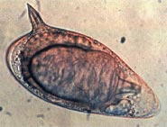
Swimmer's itch, cercarial dermatitis or schistosome dermatitis is a short-term allergic contact dermatitis occurring in the skin of humans that have been infected by water-borne schistosomes, a type of flatworm. It is common in freshwater, brackish and marine habitats worldwide. The incidence of this condition may be increasing, although this may be attributed to better monitoring and reporting. Nevertheless, the condition is considered to be an emerging infectious disease.

<i>Schistosoma intercalatum</i> Species of fluke

Schistosoma intercalatum is a parasitic worm found in parts of western and central Africa. There are two strains: the Lower Guinea strain and the Zaire strain. S. intercalatum is one of the major agents of the rectal form of schistosomiasis, also called bilharzia. It is a trematode, and being part of the genus Schistosoma, it is commonly referred to as a blood-fluke since the adult resides in blood vessels.

<span class="mw-page-title-main">Theodor Bilharz</span> German physician

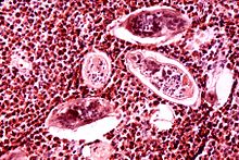
Theodor Maximilian Bilharz was a German physician who made pioneering discoveries in the field of parasitology. His contributions led to the foundation of tropical medicine. He is best remembered as the discoverer of the blood fluke Schistosoma haematobium, the causative parasite of bloody urine (haematuria) known since ancient times in Egypt. The parasite, as the cause of bladder cancer, is declared by the International Agency for Research on Cancer as Group 1 carcinogen. The infection is known by an eponymous term bilharzia or bilharziasis, as well as by schistosomiasis.

Schistosoma indicum is a species of digenetic trematode in the family Schistosomatidae. The parasite is widespread in domestic animals in India and other Asian countries.

<span class="mw-page-title-main">Schistosomiasis vaccine</span>

A Schistosomiasis vaccine is a vaccine against Schistosomiasis, a parasitic disease caused by several species of fluke of the genus Schistosoma. No effective vaccine for the disease exists yet. Schistosomiasis affects over 200 million people worldwide, mainly in rural agricultural and peri-urban areas of the third world, and approximately 10% suffer severe health complications from the infection. While chemotherapeutic drugs, such as praziquantel, oxamniquine and metrifonate both no longer on the market, are currently considered safe and effective for the treatment of schistosomiasis, reinfection occurs frequently following drug treatment, thus a vaccine is sought to provide long-term treatment. Additionally, experimental vaccination efforts have been successful in animal models of schistosomiasis.

Schistosoma mekongi is a species of trematodes, also known as flukes. It is one of the five major schistosomes that account for all human infections, the other four being S. haematobium, S. mansoni, S. japonicum, and S. intercalatum. This trematode causes schistosomiasis in humans.

<i>Schistosoma spindale</i> Species of fluke

Schistosoma spindale is a species of digenetic trematode in the family Schistosomatidae. It causes intestinal schistosomiasis in the ruminants.

<span class="mw-page-title-main">Bivitellobilharzia nairi</span> Species of fluke

Bivitellobilharzia nairi is a species of trematodes, part of the family Schistosomatidae. This is a fairly new identified endoparasite that was found in 1945 by Mudaliar and Ramanujachari, who first recorded the parasite in India. Researchers collected fecal samples of the Indian rhinoceros and were startled to find B. nairi eggs.

<i>Metagonimus yokogawai</i> Species of fluke

Metagonimus yokogawai, or the Yokogawa fluke, is a species of a trematode, or fluke worm, in the family Heterophyidae.

Schistosoma bovis is a two-host blood fluke, that causes intestinal schistosomiasis in ruminants in North Africa, Mediterranean Europe and the Middle East. S. bovis is mostly transmitted by Bulinus freshwater snail species. It is one of nine haematobium group species and exists in the same geographical areas as Schistosoma haematobium, with which it can hybridise. S. bovis-haematobium hybrids can infect humans, and have been reported in Senegal since 2009, and a 2013 outbreak in Corsica.

Carcinogenic parasites are parasitic organisms that depend on other organisms for their survival, and cause cancer in such hosts. Three species of flukes (trematodes) are medically-proven carcinogenic parasites, namely the urinary blood fluke, the Southeast Asian liver fluke and the Chinese liver fluke. S. haematobium is prevalent in Africa and the Middle East, and is the leading cause of bladder cancer. O. viverrini and C. sinensis are both found in eastern and southeastern Asia, and are responsible for cholangiocarcinoma. The International Agency for Research on Cancer declared them in 2009 as a Group 1 biological carcinogens in humans.

Schistosoma hippopotami is a species of digenetic trematode that belongs to the genus of blood flukes (Schistosoma) that is found in sub-Saharan Africa. It primarily infects African hippopotamuses and has a more limited host range compared to other Schistosoma species.

References

  1. Anon (2017). "Schistosomiasis". WHO Fact Sheet. WHO Media Centre. Retrieved 12 December 2017.
  2. Antoni, S.; Ferlay, J.; Soerjomataram, I.; Znaor, A.; Jemal, A.; Bray, F. (2017). "Bladder Cancer incidence and mortality: A global overview and recent trends". European Urology. 71 (1): 96–108. doi:10.1016/j.eururo.2016.06.010. PMID   27370177.
  3. Khurana S, Dubey ML, Malla N (April 2005). "Association of parasitic infections and cancers". Indian J Med Microbiol. 23 (2): 74–79. doi:10.1016/S0300-483X(01)00357-2. PMID   15928434.
  4. Leutscher PD, Pedersen M, Raharisolo C, et al. (2005). "Increased prevalence of leukocytes and elevated cytokine levels in semen from Schistosoma haematobium-infected individuals". J Infect Dis. 191 (10): 1639–47. doi: 10.1086/429334 . PMID   15838790.
  5. Tan, SY; Ahana, A (2007). "Theodor Bilharz (1825–1862): discoverer of schistosomiasis" (PDF). Singapore Medical Journal. 48 (3): 184–185. PMID   17342284.
  6. 1 2 van Tong, Hoang; Brindley, Paul J.; Meyer, Christian G.; Velavan, Thirumalaisamy P. (2017). "Parasite Infection, Carcinogenesis and Human Malignancy". eBioMedicine. 15: 12–23. doi:10.1016/j.ebiom.2016.11.034. PMC   5233816 . PMID   27956028.
  7. Contis, G.; David, A.R. (1996). "The epidemiology of Bilharzia in Ancient Egypt: 5000 years of schistosomiasis". Parasitology Today. 12 (7): 253–255. doi:10.1016/0169-4758(96)30007-0.
  8. Ruffer, M.A. (1910). "Note on the presence of Bilharzia haematobia in Egyptian mummies of the twentieth dynasty [1250–1000 BC]". British Medical Journal. 1 (2557): 16. doi:10.1136/bmj.1.2557.16-a. PMC   2330583 . PMID   20764829.
  9. Deelder, A.M.; Miller, R.L.; de Jonge, N.; Krijger, F.W. (1990). "Detection of schistosome antigen in mummies". The Lancet. 335 (8691): 724–5. doi:10.1016/0140-6736(90)90838-v. PMID   1969079. S2CID   12113260.
  10. Khanna, Kanika (2021-10-28). "How a Schistosoma Parasite Prevented a War". The American Society for Microbiology. Retrieved 2022-01-24.
  11. Grove, D.I. (1990). A History of Human Helminthology. Wallingford, Oxon (UK): C.A.B. International. p. 188. ISBN   978-0-85198-689-0.
  12. Bilharz, T. (1852). "Fernere Mittheilungen uber Distomum haematobium". Zeitschrift für Wissenschaftliche Zoologie. 4 (454–456).
  13. Mutapi, F. (2016). "Getting a GRiPP on everyday schistosomiasis: experience from Zimbabwe" (PDF). Parasitology. 144 (12): 1624–1632. doi:10.1017/S0031182016001724. PMID   27729092. S2CID   22153134.
  14. Hemming, F., ed. (1954). Opinions and Declarations Rendered by the International Commission on Zoological Nomenclature Volume 4 Part 16. London (UK): International Trust for Zoological Nomenclature. pp. 177–200.
  15. 1 2 3 Barakat, Rashida M.R. (2013). "Epidemiology of Schistosomiasis in Egypt: Travel through Time: Review". Journal of Advanced Research. 4 (5): 425–432. doi:10.1016/j.jare.2012.07.003. PMC   4293883 . PMID   25685449.
  16. Leiper, R.T. (1915). "Report on the results of the Bilharzia Mission in Egypt, 1915". Journal of the Royal Army Medical Corps. 25 (2): 1–55, 147–192.
  17. Berry, A.; Iriart, X.; Fillaux, J.; Magnaval, J.-F. (2017). "Schistosomose urogénitale et cancer [Urinary schistosomiasis and cancer]". Bulletin de la Société de Pathologie Exotique. 110 (1): 68–75. doi:10.1007/s13149-017-0547-4. PMID   28185084. S2CID   195078476.
  18. Hicks, R; Newman, J (1977). "The surface structure of the tegument of Schistosoma haematobium". Cell Biology International Reports. 1 (2): 157–167. doi:10.1016/0309-1651(77)90036-4 (inactive 2024-04-12). PMID   608178.{{cite journal}}: CS1 maint: DOI inactive as of April 2024 (link)
  19. Hutchison, H.S. (1928). "The pathology of bilharziasis". The American Journal of Pathology. 4 (1): 1–16. PMC   2006716 . PMID   19969774.
  20. 1 2 Colley, Daniel G; Bustinduy, Amaya L; Secor, W Evan; King, Charles H (2014). "Human schistosomiasis". The Lancet. 383 (9936): 2253–2264. doi:10.1016/S0140-6736(13)61949-2. PMC   4672382 . PMID   24698483.
  21. Cort, W.W. (1919). "Notes on the eggs and miracidia of the human schistosomes". Univiversity of California Publications in Zoology. 18 (18): 509–519.
  22. Faust, E.C. (1919). "Notes on South African cercariae". The Journal of Parasitology. 5 (4): 164–175. doi:10.2307/3271082. JSTOR   3271082. S2CID   87277224.
  23. Faust, C.E. (1920). "Criteria for the differentiation of Schistosome larvae". The Journal of Parasitology. 6 (4): 192–194. doi:10.2307/3270844. JSTOR   3270844. S2CID   85212596.
  24. Mohammed, A.S. (1931). "The secretory glands of the cercariae of S. Haematobium and S. Mansoni from Egypt". Annals of Tropical Medicine & Parasitology. 26 (1): 7–22. doi:10.1080/00034983.1932.11684702.
  25. 1 2 Despommier, Dickson D.; Karapelou, John W. (1987). Parasite Life Cycles. New York, NY: Springer Verlag. pp. 76–77. ISBN   978-1-4612-3722-8.
  26. Rollinson, D; Stothard, JR; Southgate, VR (2001). "Interactions between intermediate snail hosts of the genus Bulinus and schistosomes of the Schistosoma haematobium group". Parasitology. 123 (Suppl): S245–60. doi:10.1017/S0031182001008046. PMID   11769287. S2CID   8361773.
  27. 1 2 Akl, M.M. (2009). "Bilharziasis: A Granulomatous Parasitic Disorder with Grave Implications". In Mansourian, B.P.; Wojtczak, A.; Sayers, B.M (eds.). Medical Sciences – Volume I. Oxford (UK): Eolss Publishers Co. Ltd. pp. 374–400. ISBN   978-1-84826-733-6.
  28. Dew, H.R. (1923). "Observations on the pathology of schistosomiasis (S. haematobium and S. mansoni) in the human subject". The Journal of Pathology and Bacteriology. 26 (1): 27–39. doi:10.1002/path.1700260104.
  29. 1 2 3 4 Black, J. (2005). Microbiology: Principles and Explorations (6 ed.). Wiley. ISBN   978-0471743255.
  30. Mills, E.A.; Machattie, C.; Chadwick, C.R. (1936). "Schistosoma haematobium and its life cycle in Iraq". Transactions of the Royal Society of Tropical Medicine and Hygiene. 30 (3): 317–334. doi:10.1016/S0035-9203(36)90068-8.
  31. Ibrahim, H. (1948). "Bilharziasis and bilharzial cancer of the bladder". Annals of the Royal College of Surgeons of England. 2 (3): 129–141. PMC   1940191 . PMID   18908968.
  32. Wilkins, H.A.; Goll, P.H.; de C. Marshall, T.F.; Moore, P.J. (1984). "Dynamics of Schistosoma haematobium infection in a Gambian community. III. Acquisition and loss of infection". Transactions of the Royal Society of Tropical Medicine and Hygiene. 78 (2): 227–232. doi:10.1016/0035-9203(84)90283-9. PMID   6464111.
  33. Kinkel HF, Dittrich S, Bäumer B, Weitzel T (2012). "Evaluation of eight serological tests for diagnosis of imported schistosomiasis". Clin Vaccine Immunol. 19 (6): 948–53. doi:10.1128/CVI.05680-11. PMC   3370443 . PMID   22441394.
  34. Ross, Allen; Inobaya, Marianette; Olveda, Remigio; Chau, Thao; Olveda, David (2014). "Prevention and control of schistosomiasis: a current perspective". Research and Reports in Tropical Medicine. 2014 (5): 65–75. doi: 10.2147/RRTM.S44274 . PMC   4231879 . PMID   25400499.
  35. King, Charles H.; Sutherland, Laura J.; Bertsch, David; Loker, Eric S (2015). "Systematic Review and Meta-analysis of the Impact of Chemical-Based Mollusciciding for Control of Schistosoma mansoni and S. haematobium Transmission". PLOS Neglected Tropical Diseases. 9 (12): e0004290. doi: 10.1371/journal.pntd.0004290 . PMC   4692485 . PMID   26709922.
  36. Pearce, Edward J.; MacDonald, Andrew S. (2002). "The immunobiology of schistosomiasis". Nature Reviews Immunology. 2 (7): 499–511. doi:10.1038/nri843. PMID   12094224. S2CID   4695994.
  37. Ouma, John H.; King, Charles H.; Mahmoud, Adel A. F.; Keating, Catherine E.; Houser, Harold; Muruka, Jagon F.; Siongok, Timothy K. Arap (1988). "Urinary Tract Morbidity in Schistosomiasis Haematobia: Associations with Age and Intensity of Infection in an Endemic Area of Coast Province, Kenya". The American Journal of Tropical Medicine and Hygiene. 39 (4): 361–368. doi:10.4269/ajtmh.1988.39.361. PMID   3142286.
  38. Khalaf, Ismail; Shokeir, Ahmed; Shalaby, Mohamed (2011). "Urologic complications of genitourinary schistosomiasis". World Journal of Urology. 30 (1): 31–38. doi:10.1007/s00345-011-0751-7. PMID   21909645. S2CID   21413277.
  39. Kabuyaya, Muhubiri; Chimbari, Moses John; Manyangadze, Tawanda; Mukaratirwa, Samson (2017). "Efficacy of praziquantel on Schistosoma haematobium and re-infection rates among school-going children in the Ndumo area of uMkhanyakude district, KwaZulu-Natal, South Africa". Infectious Diseases of Poverty. 6 (1): 83. doi: 10.1186/s40249-017-0293-3 . PMC   5383960 . PMID   28385154.

Further reading