Asymmetric dimethylarginine

Last updated
Asymmetric dimethylarginine
Asymmetric dimethylarginine.svg
ADMA-3D-balls-by-AHRLS-2012.png
Names
IUPAC name
2-Amino-5-[[amino(dimethylamino)methylidene]amino]pentanoic acid
Other names
N(G),N(G)-Dimethylarginine
Identifiers
3D model (JSmol)
3DMet
2261521 S
ChEBI
ChEMBL
ChemSpider
DrugBank
KEGG
MeSH N,N-dimethylarginine
PubChem CID
UNII
  • InChI=1S/C8H18N4O2/c1-12(2)8(10)11-5-3-4-6(9)7(13)14/h6H,3-5,9H2,1-2H3,(H2,10,11)(H,13,14) X mark.svgN
    Key: YDGMGEXADBMOMJ-UHFFFAOYSA-N X mark.svgN
  • CN(C)C(=N)NCCC[C@H](N)C(=O)O
Properties
C8H18N4O2
Molar mass 202.258 g·mol−1
log P −0.716
Acidity (pKa)2.497
Basicity (pKb)11.500
Related compounds
Related alkanoic acids
Related compounds
Except where otherwise noted, data are given for materials in their standard state (at 25 °C [77 °F], 100 kPa).
X mark.svgN  verify  (what is  Yes check.svgYX mark.svgN ?)

Asymmetric dimethylarginine (ADMA) is a naturally occurring chemical found in blood plasma. It is a metabolic by-product of continual protein modification processes in the cytoplasm of all human cells. It is closely related to L-arginine, a conditionally essential amino acid. ADMA interferes with L-arginine in the production of nitric oxide (NO), a key chemical involved in normal endothelial function and, by extension, cardiovascular health.

Contents

Discovery

Patrick Vallance and his London co-workers first noted the interference role for asymmetric dimethylarginine in the early 1990s. [1] Today biochemical and clinical research continues into the role of ADMA in cardiovascular disease, diabetes mellitus, erectile dysfunction and certain forms of kidney disease.

Synthesis and regulation in the body

Cardiovascular effects have been linked to ADMA Heart-and-lungs.jpg
Cardiovascular effects have been linked to ADMA

Asymmetric dimethylarginine is created in protein methylation, a common mechanism of post-translational protein modification. This reaction is catalyzed by an enzyme set called protein arginine N-methyltransferases 1 and 2 (also known as S-adenosylmethionine protein N-methyltransferases I and II). [2] The methyl groups transferred to create ADMA are derived from the methyl group donor S-adenosylmethionine, an intermediate in the metabolism of homocysteine. (Homocysteine is an important blood chemical because it is also a marker of cardiovascular disease). After synthesis, ADMA migrates into the extracellular space and thence into blood plasma. Asymmetric dimethylarginine is measured using high-performance liquid chromatography.

ADMA concentrations are substantially elevated by native or oxidized LDL cholesterol. [3] Thus a spiralling effect occurs with high endothelial LDL levels causing greater ADMA values, which in turn inhibit NO production needed to promote vasodilation. The elimination of ADMA occurs through urine excretion and metabolism by the enzyme dimethylarginine dimethylaminohydrolase (DDAH). The role of homocysteine as a risk factor for cardiovascular disease is suggested to be mediated by homocysteine down-regulating production of DDAH in the body. Polyphenol antioxidants also play a role in down-regulating homocysteine.

ADMA and suggested lines of therapeutic research

ADMA may play a role in certain forms of kidney disease Kidney section.jpg
ADMA may play a role in certain forms of kidney disease

With raised levels of ADMA seemingly to be associated with adverse human health consequences for cardiovascular disease, metabolic diseases, and also a wide range of diseases of the elderly, the possible lowering of ADMA levels may have important therapeutic effects. However, it has yet to be established whether ADMA levels can be manipulated and, more important, if this results in useful clinical benefits.

The association of ADMA with abnormalities of lipid regulation suggested that supplements of free fatty acids might manipulate ADMA levels. However, research has failed to show that these have an effect. [4] [5]

ADMA's role has been linked with elevated levels of homocysteine. [6] [7] [8] Whilst approaches at modifying the latter with oral supplements of folic acid were strongly suggested, studies have shown this fails to give any clinical benefit and suggested that B vitamins might instead increase some cardiovascular risks. [9] [10] [11]

Direct alteration of ADMA levels with supplements of L-arginine have been suggested. [12] [13] The hope is that such intervention might not only improve endothelial function but also reduce clinical symptoms of overt cardiovascular disease. [14] [15] However studies show inconsistency in results in a clinical context, [16] and the recent results with manipulating homocysteine levels warrant extreme care with what clinical outcomes might arise from this approach.

Statins, as well as affecting circulating cholesterol levels, also increase nitric oxide levels and so have a direct effect on blood supply to the heart. Elevated levels of ADMA seems to modify this effect and so may have consequences for patients' responsiveness to taking statins. [17]

Repeated administration of d-amphetamine may decrease ADMA. [18]

See also

Related Research Articles

High-density lipoprotein (HDL) is one of the five major groups of lipoproteins. Lipoproteins are complex particles composed of multiple proteins which transport all fat molecules (lipids) around the body within the water outside cells. They are typically composed of 80–100 proteins per particle. HDL particles enlarge while circulating in the blood, aggregating more fat molecules and transporting up to hundreds of fat molecules per particle.

<span class="mw-page-title-main">Folate</span> Vitamin B9; nutrient essential for DNA synthesis

Folate, also known as vitamin B9 and folacin, is one of the B vitamins. Manufactured folic acid, which is converted into folate by the body, is used as a dietary supplement and in food fortification as it is more stable during processing and storage. Folate is required for the body to make DNA and RNA and metabolise amino acids necessary for cell division. As the human body cannot make folate, it is required in the diet, making it an essential nutrient. It occurs naturally in many foods. The recommended adult daily intake of folate in the U.S. is 400 micrograms from foods or dietary supplements.

<span class="mw-page-title-main">Methionine</span> Sulfur-containing amino acid

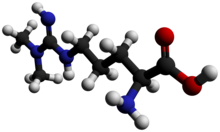
Methionine is an essential amino acid in humans.

<span class="mw-page-title-main">Arginine</span> Amino acid

Arginine is the amino acid with the formula (H2N)(HN)CN(H)(CH2)3CH(NH2)CO2H. The molecule features a guanidino group appended to a standard amino acid framework. At physiological pH, the carboxylic acid is deprotonated (−CO2) and both the amino and guanidino groups are protonated, resulting in a cation. Only the l-arginine (symbol Arg or R) enantiomer is found naturally. Arg residues are common components of proteins. It is encoded by the codons CGU, CGC, CGA, CGG, AGA, and AGG. The guanidine group in arginine is the precursor for the biosynthesis of nitric oxide. Like all amino acids, it is a white, water-soluble solid.

<span class="mw-page-title-main">Homocysteine</span> Chemical compound

Homocysteine or Hcy: is a non-proteinogenic α-amino acid. It is a homologue of the amino acid cysteine, differing by an additional methylene bridge (-CH2-). It is biosynthesized from methionine by the removal of its terminal Cε methyl group. In the body, homocysteine can be recycled into methionine or converted into cysteine with the aid of vitamin B6, B9, and B12.

<span class="mw-page-title-main">Histone methyltransferase</span> Histone-modifying enzymes

Histone methyltransferases (HMT) are histone-modifying enzymes, that catalyze the transfer of one, two, or three methyl groups to lysine and arginine residues of histone proteins. The attachment of methyl groups occurs predominantly at specific lysine or arginine residues on histones H3 and H4. Two major types of histone methyltranferases exist, lysine-specific and arginine-specific. In both types of histone methyltransferases, S-Adenosyl methionine (SAM) serves as a cofactor and methyl donor group.
The genomic DNA of eukaryotes associates with histones to form chromatin. The level of chromatin compaction depends heavily on histone methylation and other post-translational modifications of histones. Histone methylation is a principal epigenetic modification of chromatin that determines gene expression, genomic stability, stem cell maturation, cell lineage development, genetic imprinting, DNA methylation, and cell mitosis.

<i>S</i>-Adenosyl methionine Chemical compound found in all domains of life with largely unexplored effects

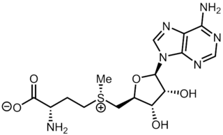
S-Adenosyl methionine (SAM), also known under the commercial names of SAMe, SAM-e, or AdoMet, is a common cosubstrate involved in methyl group transfers, transsulfuration, and aminopropylation. Although these anabolic reactions occur throughout the body, most SAM is produced and consumed in the liver. More than 40 methyl transfers from SAM are known, to various substrates such as nucleic acids, proteins, lipids and secondary metabolites. It is made from adenosine triphosphate (ATP) and methionine by methionine adenosyltransferase. SAM was first discovered by Giulio Cantoni in 1952.

<span class="mw-page-title-main">Methyltransferase</span> Group of methylating enzymes

Methyltransferases are a large group of enzymes that all methylate their substrates but can be split into several subclasses based on their structural features. The most common class of methyltransferases is class I, all of which contain a Rossmann fold for binding S-Adenosyl methionine (SAM). Class II methyltransferases contain a SET domain, which are exemplified by SET domain histone methyltransferases, and class III methyltransferases, which are membrane associated. Methyltransferases can also be grouped as different types utilizing different substrates in methyl transfer reactions. These types include protein methyltransferases, DNA/RNA methyltransferases, natural product methyltransferases, and non-SAM dependent methyltransferases. SAM is the classical methyl donor for methyltransferases, however, examples of other methyl donors are seen in nature. The general mechanism for methyl transfer is a SN2-like nucleophilic attack where the methionine sulfur serves as the leaving group and the methyl group attached to it acts as the electrophile that transfers the methyl group to the enzyme substrate. SAM is converted to S-Adenosyl homocysteine (SAH) during this process. The breaking of the SAM-methyl bond and the formation of the substrate-methyl bond happen nearly simultaneously. These enzymatic reactions are found in many pathways and are implicated in genetic diseases, cancer, and metabolic diseases. Another type of methyl transfer is the radical S-Adenosyl methionine (SAM) which is the methylation of unactivated carbon atoms in primary metabolites, proteins, lipids, and RNA.

Histone-arginine N-methyltransferase is an enzyme with systematic name S-adenosyl-L-methionine:histone-arginine Nomega-methyltransferase. This enzyme catalyses the following chemical reaction

<span class="mw-page-title-main">Hyperhomocysteinemia</span> Medical condition

Hyperhomocysteinemia is a medical condition characterized by an abnormally high level of total homocysteine in the blood, conventionally described as above 15 μmol/L.

Soluble cell adhesion molecules (sCAMs) are a class of cell adhesion molecule that may represent important biomarkers for inflammatory processes involving activation or damage to cells such as platelets and the endothelium.

<span class="mw-page-title-main">Levomefolic acid</span> Chemical compound

Levomefolic acid (INN, also known as L-5-MTHF, L-methylfolate and L-5-methyltetrahydrofolate and (6S)-5-methyltetrahydrofolate, and (6S)-5-MTHF) is the primary biologically active form of folate used at the cellular level for DNA reproduction, the cysteine cycle and the regulation of homocysteine. It is also the form found in circulation and transported across membranes into tissues and across the blood–brain barrier. In the cell, L-methylfolate is used in the methylation of homocysteine to form methionine and tetrahydrofolate (THF). THF is the immediate acceptor of one carbon unit for the synthesis of thymidine-DNA, purines (RNA and DNA) and methionine. The un-methylated form, folic acid (vitamin B9), is a synthetic form of folate, and must undergo enzymatic reduction by dihydrofolate reductase (DHFR) to become biologically active.

<span class="mw-page-title-main">Phosphatidylethanolamine N-methyltransferase</span> Protein-coding gene in the species Homo sapiens

Phosphatidylethanolamine N-methyltransferase is a transferase enzyme which converts phosphatidylethanolamine (PE) to phosphatidylcholine (PC) in the liver. In humans it is encoded by the PEMT gene within the Smith–Magenis syndrome region on chromosome 17.

<span class="mw-page-title-main">PRMT1</span> Protein-coding gene in the species Homo sapiens

Protein arginine N-methyltransferase 1 is an enzyme that in humans is encoded by the PRMT1 gene. The HRMT1L2 gene encodes a protein arginine methyltransferase that functions as a histone methyltransferase specific for histone H4.

<span class="mw-page-title-main">Dimethylargininase</span> Class of enzymes

In the field of enzymology, a dimethylargininase, also known as a dimethylarginine dimethylaminohydrolase (DDAH), is an enzyme that catalyzes the chemical reaction:

<span class="mw-page-title-main">MTRR (gene)</span> Protein-coding gene in the species Homo sapiens

Methionine synthase reductase, also known as MSR, is an enzyme that in humans is encoded by the MTRR gene.

<span class="mw-page-title-main">Methylarginine</span> Chemical compound

N-Methylarginine is an inhibitor of nitric oxide synthase. Chemically, it is a methyl derivative of the amino acid arginine. It is used as a biochemical tool in the study of physiological role of nitric oxide.

<span class="mw-page-title-main">Oxycholesterol</span> Chemical compound

Oxycholesterol or 5,6-epoxycholesterol is a form of oxidized cholesterol implicated in atherosclerosis. It is commonly formed from the reaction of fats and oxygen during high temperature cooking such as frying.

<span class="mw-page-title-main">Protein detoxification</span>

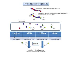
Protein detoxification is the process by which proteins containing methylated arginine are broken down and removed from the body.

Protein methylation is a type of post-translational modification featuring the addition of methyl groups to proteins. It can occur on the nitrogen-containing side-chains of arginine and lysine, but also at the amino- and carboxy-termini of a number of different proteins. In biology, methyltransferases catalyze the methylation process, activated primarily by S-adenosylmethionine. Protein methylation has been most studied in histones, where the transfer of methyl groups from S-adenosyl methionine is catalyzed by histone methyltransferases. Histones that are methylated on certain residues can act epigenetically to repress or activate gene expression.

References

  1. Vallance, P.; Leone, A.; Calver, A.; Collier, J.; Moncada, S. (1992). "Endogenous dimethylarginine as an inhibitor of nitric oxide synthesis". Journal of Cardiovascular Pharmacology. 20 (Suppl 12): S60–S62. doi: 10.1097/00005344-199204002-00018 . PMID   1282988. S2CID   38113918.
  2. Rawal N, Rajpurohit R, Lischwe MA, Williams KR, Paik WK, Kim S (1995). "Structural specificity of substrate for S-adenosylmethionine:protein arginine N-methyltransferases". Biochim Biophys Acta. 1248 (1): 11–8. doi: 10.1016/0167-4838(94)00213-Z . PMID   7536038.
  3. Boger RH, Sydow K, Borlak J, Thum T, Lenzen H, Schubert B, Tsikas D, Bode-Boger SM (21 July 2000). "LDL cholesterol upregulates synthesis of asymmetrical dimethylarginine in human endothelial cells: involvement of S-adenosylmethionine-dependent methyltransferases". Circ Res. 87 (2): 99–105. doi: 10.1161/01.RES.87.2.99 . PMID   10903992.
  4. Eid HM, Arnesen H, Hjerkinn EM, Lyberg T, Ellingsen I, Seljeflot I (2006). "Effect of diet and omega-3 fatty acid intervention on asymmetric dimethylarginine". Nutr Metab (Lond). 3 (1): 4. doi: 10.1186/1743-7075-3-4 . PMC   1343562 . PMID   16396682.
  5. Namiranian K, Mittermayer F, Artwohl M, Pleiner J, Schaller G, Mayer BX, Bayerle-Eder M, Roden M, Baumgartner-Parzer S, Wolzt M (2005). "Free fatty acids do not acutely increase asymmetrical dimethylarginine concentrations". Horm Metab Res. 37 (12): 768–72. doi:10.1055/s-2005-921100. PMID   16372232. S2CID   260169813.
  6. Krzyzanowska K, Mittermayer F, Krugluger W, Schnack C, Hofer M, Wolzt M, Schernthaner G (2006). "Asymmetric dimethylarginine is associated with macrovascular disease and total homocysteine in patients with type 2 diabetes". Atherosclerosis. 189 (1): 236–40. doi:10.1016/j.atherosclerosis.2005.12.007. PMID   16414052.
  7. Dayal S, Lentz SR (2005). "ADMA and hyperhomocysteinemia". Vasc Med. 10 Suppl 1 (2): S27–33. doi:10.1191/1358863x05vm599oa. PMID   16444866. S2CID   24645709.
  8. Stuhlinger MC, Stanger O (2005). "Asymmetric dimethyl-L-arginine (ADMA): a possible link between homocyst(e)ine and endothelial dysfunction". Curr Drug Metab. 6 (1): 3–14. doi:10.2174/1389200052997393. PMID   15720202.
  9. Zoungas S, McGrath BP, Branley P, Kerr PG, Muske C, Wolfe R, Atkins RC, Nicholls K, Fraenkel M, Hutchison BG, Walker R, McNeil JJ (2006). "Cardiovascular morbidity and mortality in the Atherosclerosis and Folic Acid Supplementation Trial (ASFAST) in chronic renal failure: a multicenter, randomized, controlled trial". J Am Coll Cardiol. 47 (6): 1108–16. doi:10.1016/j.jacc.2005.10.064. PMID   16545638. S2CID   37275349.
  10. Lonn, E; Yusuf, S; Arnold, MJ; Sheridan, P; Pogue, J; Micks, M; McQueen, MJ; Probstfield, J; et al. (2006). "Homocysteine Lowering with Folic Acid and B Vitamins in Vascular Disease". N Engl J Med. 354 (15): 1567–77. doi: 10.1056/NEJMoa060900 . PMID   16531613. S2CID   21790155.{{cite journal}}: CS1 maint: numeric names: authors list (link)
  11. Bonaa KH, Njolstad I, Ueland PM, Schirmer H, Tverdal A, Steigen T, Wang H, Nordrehaug JE, Arnesen E, Rasmussen K (2006). "Homocysteine Lowering and Cardiovascular Events after Acute Myocardial Infarction". N Engl J Med. 354 (15): 1578–88. doi: 10.1056/NEJMoa055227 . PMID   16531614.
  12. Bode-Boger SM, Muke J, Surdacki A, Brabant G, Boger RH, Frolich JC (2003). "Oral L-arginine improves endothelial function in healthy individuals older than 70 years". Vasc Med. 8 (2): 77–81. doi: 10.1191/1358863x03vm474oa . PMID   14518608.
  13. John P. Cooke (2002). The Cardiovascular Cure . Random House. ISBN   0-7679-0881-3.
  14. Rector TS, Bank AJ, Mullen KA, Tschumperlin LK, Sih R, Pillai K, Kubo SH (15 June 1996). "Randomized, double-blind, placebo-controlled study of supplemental oral L-arginine in patients with heart failure". Circulation. 93 (12): 2135–41. doi:10.1161/01.CIR.93.12.2135. PMID   8925582.
  15. Ceremuzynski L, Chamiec T, Herbaczynska-Cedro K (1997). "Effect of supplemental oral L-arginine on exercise capacity in patients with stable angina pectoris". Am J Cardiol. 80 (3): 331–3. doi:10.1016/S0002-9149(97)00354-8. PMID   9264427.
  16. Loscalzo J (2004). "L-arginine and atherothrombosis". J Nutr. 134 (10 Suppl): 2798S–2800S, discussion 2818S–2819S. doi: 10.1093/jn/134.10.2798S . PMID   15465788.
  17. Janatuinen T, Laakso J, Laaksonen R, Vesalainen R, Nuutila P, Lehtimaki T, Raitakari OT, Knuuti J (2003). "Plasma asymmetric dimethylarginine modifies the effect of pravastatin on myocardial blood flow in young adults". Vasc Med. 8 (3): 185–9. doi:10.1191/1358863x03vm490oa. PMID   14989559. S2CID   36938530.
  18. Vanaveski, Taavi; Narvik, Jane; Innos, Jürgen; Philips, Mari-Anne; Ottas, Aigar; Plaas, Mario; Haring, Liina; Zilmer, Mihkel; Vasar, Eero (2018-06-12). "Repeated Administration of D-Amphetamine Induces Distinct Alterations in Behavior and Metabolite Levels in 129Sv and Bl6 Mouse Strains". Frontiers in Neuroscience. 12: 399. doi: 10.3389/fnins.2018.00399 . ISSN   1662-4548. PMC   6005828 . PMID   29946233.