Barrel cortex

Last updated
Barrel cortex
Identifiers
NeuroLex ID nlx_81
Anatomical terms of neuroanatomy
Pictomicrograph shows the barrel field in layer IV of the rat somatosensory cortex. Each barrel receives input from one whisker. The tissue in the image has been stained with cytochrome oxidase and is 50mm thick. RatBarrelFieldCOstain.jpg
Pictomicrograph shows the barrel field in layer IV of the rat somatosensory cortex. Each barrel receives input from one whisker. The tissue in the image has been stained with cytochrome oxidase and is 50μm thick.

The barrel cortex is a region of the somatosensory cortex that is identifiable in some species of rodents and species of at least two other orders [1] and contains the barrel field. The 'barrels' of the barrel field are regions within cortical layer IV that are visibly darker when stained to reveal the presence of cytochrome c oxidase and are separated from each other by lighter areas called septa. These dark-staining regions are a major target for somatosensory inputs from the thalamus, and each barrel corresponds to a region of the body. Due to this distinctive cellular structure, organisation, and functional significance, the barrel cortex is a useful tool to understand cortical processing and has played an important role in neuroscience. [2] The majority of what is known about corticothalamic processing comes from studying the barrel cortex, and researchers have intensively studied the barrel cortex as a model of neocortical column.

Contents

The most distinctive aspect of the barrel field are the whisker barrels. These structures were first discovered by Woolsey and Van der Loos in 1970. [3] Staining in the whisker barrels is more distinct than that in other areas of the somatosensory cortex. Recognizing that the array was similar to that of the vibrissae (whiskers) on the mystacial pad (region where whiskers grow from) of certain mammals, they hypothesized that the barrels were the "cortical correlates of the mystacial vibrissae" and that "one barrel represents one vibrissa". Whereas small non-whisker areas of barrel cortex correspond to large and sometimes overlapping areas of the body, each much larger whisker barrel corresponds to a single whisker. As a result, the whisker barrels are the focus of the majority of barrel cortex research, and 'barrel cortex' is often used to refer primarily to the whisker barrels. Consequently, much of this article focuses on rodent whisker barrel cortex.

Organisation of the barrel fields

Pictomicrograph shows the posteromedial barrel subfield in layer IV of the rat somatosensory cortex. Barrels in the PMBSF are particularly large and distinct. The tissue in the image has been stained with cytochrome oxidase and is 50mm thick. Labeled40xVibrissae.jpg
Pictomicrograph shows the posteromedial barrel subfield in layer IV of the rat somatosensory cortex. Barrels in the PMBSF are particularly large and distinct. The tissue in the image has been stained with cytochrome oxidase and is 50μm thick.

The barrel field, like many regions of cortex, is organised into a topographic map. In the case of the barrel field, the map is somatotopic - based on the arrangement of body parts. Areas corresponding to the nose and mouth are more rostral and lateral in the map, the forelimb, hindlimb and trunk are more medial, with the forelimb rostral of the hindlimb, and the whisker barrel subfields - the posteromedial barrel subfield, which corresponds to the major facial whiskers (the mystacial vibrissae), and the anteriolateral barrel subfield, which corresponds to the smaller whiskers of the face - are caudal and lateral. Although the whiskers make up a relatively small portion of the animal, they dominate the somatotopic map. [4] [5]

Barrels of the major facial whiskers

The barrels that correspond to the major facial whiskers (mystacial vibrissae) are contained within the posteromedial barrel subfield (PMBSF). The barrels here are the largest and most elliptical in shape and have a striking topographical organization that is identical to that of the whiskers; they are organized into 5 rows of 4-7 large whiskers that run close to parallel with the bridge of the nose. [6] The organisation of the mystacial vibrissae and corresponding barrels is so consistent that there is a naming convention to identify each whisker in rats and mice. Rows are designated A to E from top to bottom, and columns of whiskers within each row are numbered from back to front. The first four rows also have an additional whisker behind column 1, which is designated with a lower case letter or a Greek letter (α, β, γ, or δ). These four whiskers are also called straddlers.

Anatomy and connectivity of the barrels

Sensory information flows in parallel pathways from whiskers to cortex. Barrel cortex pathways.jpg
Sensory information flows in parallel pathways from whiskers to cortex.

The barrels of the barrel cortex were named because the densities of cells resembled barrels, that is, they are collected into cylindrical shapes that are narrowed at the top and bottom. The centre of the barrel is designated the hollow, and the spaces between the barrels are the septa (singular: septum) [6]

Sensory information flows from whisker follicles to barrel cortex via the trigeminal nerve nuclei and the thalamus. Barrel like divisions can be seen in some, but not all parts of the trigeminal nuclei (where they are called barrelets) and the thalamus (where they are called barreloids). The trigeminal nerve carries afferent fibres from the follicles into the brainstem where they connect to neurons in four different trigeminal nerve nuclei: principal, interpolar, oral, and caudal. Projections from the trigeminal nuclei to the thalamus are split into pathways designated lemniscal, extralemniscal, and paralemniscal. In the lemniscal pathway, axons from the principal trigeminal nucleus cross over the midline and project to “barreloids” in the thalamus, specifically in the dorsomedial section of the ventroposterior medial nucleus (VPMdm). Neurons in VPMdm project mainly to barrels in layer 4 of primary somatosensory cortex (S1). In the extralemniscal pathway, neurons of the interpolar nucleus project to the ventrolateral section of the ventroposterior medial nucleus (VPMvl). Neurons in VPMvl project to septa between the barrels and to secondary somatosensory cortex (S2). The paralemniscal pathway runs from the interpolar trigeminal nucleus via posterior nucleus (POm) of the thalamus to S2 and to diffuse targets in barrel cortex especially layer 5. Each pathway also has secondary projections to other layers within barrel cortex and other regions of cortex, including motor cortex. [7] These different pathways are thought to transmit different modalities of sensory information from the whisker. [2] [8]

Whisker barrel neurophysiology

The whisker barrel cortex contains different types of neurons that receive input from a range of sources that themselves receive and process an array of different types of information. As a result, neurons of the whisker barrel cortex respond to whisker-related input, but in a way that is specific to the neurons type and location. This can manifest in different ways. The simplest way is whether the cortical neuron responds only to the deflection of one whisker, or to the deflection of many whiskers. Neurons in layer 4 barrels tend to strongly or exclusively respond to one whisker, while neurons in other layers are less strongly tuned and can respond to multiple whiskers. Neurons that respond to the deflection of multiple whiskers typically have a primary whisker, to which they respond the most. The difference in response magnitude between deflection of the primary whisker and secondary whiskers can also vary between neurons. Stimulation of multiple whiskers may produce a response that is equal to the sum of the responses if each whisker was stimulated independently, or it may be different. Some neurons show greater responses when multiple neurons are stimulated in sequence, and the sequence may be direction specific. [9]

As well as combinations of which whiskers have been stimulated, neurons may also respond to specific types of whisker stimulation. The simplest response, seen in neurons within the layer IV barrel cortex, directly code for whisker displacement. That is to say, that the neuron within a given barrel will fire when the whisker that barrel represents is moved at a rate that is roughly proportional to the angular displacement of the neuron. These neurons also show directional sensitivity; certain neurons will only fire when the whisker is moved in a specific direction. [10] [11] Deflection-based firing neurons can sustain their response throughout the deflection of the whisker. Other neurons respond to the initial deflection, but then quickly return to their previous level of activity. Much of this activity is also modulated by the behaviour of the animal - rats and mice actively move their whiskers to explore their environment, and the response of a neuron to a particular stimulus can vary depending on what the animal is doing.

Experience-dependent plasticity

Because the barrel cortex has a well-organised structure that relates clearly to the whisker pad, it has been used extensively as a tool to study sensory processing and development, and the phenomenon of experience-dependent plasticity - changes in the activity, connectivity, and structure of neural circuits in response to experience. Neurons in the barrel cortex exhibit the property of synaptic plasticity that allows them to alter the vibrissae to which they respond depending on the rodent's history of tactile experience. [12] Experience-dependent plasticity is commonly studied in the barrel cortex by partially depriving it of sensory input, either by lesioning elements of the afferent pathway (e.g. the trigeminal nerve) or by ablating, plucking, or trimming some of the facial whiskers. The anatomical structure of the barrels is only affected by lesioning elements of the pathway, but innocuous forms of deprivation can induce rapid changes in the cortical map into adulthood, without any corresponding changes in the barrel structures. [13] Because of their different effects, it seems these two paradigms work by different mechanisms.

Some forms of plasticity in the barrel cortex display a critical period. Plucking whiskers in neonatal rats causes a long-lasting expansion of the representation of the spared whisker in layer 4. [14] However, layer 4 plasticity rapidly diminishes if sensory deprivation begins after day 4 of life (P4) whereas representations in layer 2/3 remain highly plastic into adulthood. [15] [16]

Two cortical processes run alongside each other when barrel cortex is deprived of sensory input from some whiskers to produce representational plasticity. In deprived cortex, neuronal responses to spared whiskers are enhanced and responses to deprived whiskers are weakened. These two processes have different time courses, with the weakening of deprived response preceding the strengthening of spared response, implying that they have different underlying mechanisms. These two effects combine to produce an expansion of the cortical representation of spared whiskers into the representation of adjacent deprived whiskers. [15] [17]

It is likely that several different mechanisms are involved in producing experience-dependent plasticity in a whisker deprivation protocol (adapted from Feldman and Brecht, 2005 [17] ):

  1. Almost immediately, loss of input to a deprived barrel column leads to a loss of inhibitory firing in that column. This unmasks horizontal excitatory connections from adjacent spared columns. [18] This does not explain longer-lasting plastic changes as the unmasking would disappear immediately if the deprived input was reinstated (for example by allowing the whisker to regrow).
  2. LTP- and LTD-like processes also seem to be involved. This can be inferred by using transgenic mice where there are changes in the expression of enzymes related to LTP and LTD e.g. calmodulin-dependent protein kinase II (CaMKII) or cyclic-AMP response element binding protein (CREB). In these mice, plasticity is compromised [19] [20] Spike timing rather than frequency may be an important factor. Associative LTP has been demonstrated at layer 4 to layer 2/3 synapses when the layer 4 neuron fires 0-15 ms before the layer 2/3 neuron, and LTD is observed when this timing order is reversed. [21] Such mechanisms could act rapidly to produce plastic changes within hours or days.
  3. Sensory deprivation has been demonstrated to cause changes in synaptic dynamics such as EPSP amplitude and frequency. The net effect of these changes is to increase the proportion of synaptic input which layer 2/3 neurons in deprived barrels receive from spared barrels. [22] These observations suggest that other, more specific, mechanisms besides LTP/LTD are at play in experience-dependent plasticity.
  4. It seems intuitively likely that structural changes at the level of axons, dendrite branches, and dendrite spines underlie some of the long-term plastic changes in the cortex. Changes in axon structure have been reported in plasticity following lesions [23] and more recently by studies using whisker trimming. [24] Dendritic branching is important during prenatal and neonatal development, is involved in plasticity induced by lesions, but is not involved in experience-dependent plasticity. [25] In vivo two-photon microscopy reveals that dendritic spines in mouse barrel cortex are highly dynamic and subject to continuous turnover, and may be associated with formation or deletion of synapses. [25] It is likely that spine turnover is necessary but not sufficient to produce experience-dependent plasticity, and other mechanisms such as axonal remodelling are also needed to explain features such as savings from prior experience. [24]

Plasticity and remodelling of barrel cortex has also been studied in the context of traumatic brain injury, [26] where environmental enrichment of stimuli has been shown to induce plasticity/recovery [27] and patterns of temporal coding have been altered via plasticity and recovery mechanisms. [28]

Notes

  1. Woolsey et al., 1975
  2. 1 2 Fox, 2008
  3. Woolsey & Van der Loos, 1970
  4. Hoover et al., 2003
  5. Enriquez-Barreto et al., 2012
  6. 1 2 Woolsey & Van der Loos, 1970
  7. Bosman et al., 2011
  8. Diamond et al., 2008
  9. Bosman et al., 2011
  10. Swadlow, 1989
  11. Swadlow HA (1991). "Efferent neurons and suspected interneurons in second somatosensory cortex of the awake rabbit: receptive fields and axonal properties". J Neurophysiol. 66 (4): 1392–1409. doi:10.1152/jn.1991.66.4.1392. PMID   1761989.
  12. Hardingham N, Glazewski S, Pakhotin P, Mizuno K, Chapman PF, Giese KP, Fox K. Neocortical long-term potentiation and experience-dependent synaptic plasticity require alpha-calcium/calmodulin-dependent protein kinase II auto-phosphorylation. J Neurosci. 2003 Jun 1;23(11):4428-36.
  13. Fox K (2002). "Anatomical pathways and molecular mechanisms for plasticity in the barrel cortex". Neuroscience. 111 (4): 799–814. doi:10.1016/s0306-4522(02)00027-1. PMID   12031405. S2CID   39423181.
  14. Fox K (1992). "A critical period for experience-dependent synaptic plasticity in rat barrel cortex". J Neurosci. 12 (5): 1826–1838. doi: 10.1523/JNEUROSCI.12-05-01826.1992 . PMC   6575898 . PMID   1578273.
  15. 1 2 Glazewski S, Fox K (1996). "Time course of experience-dependent synaptic potentiation anddepression in barrel cortex of adolescent rats". J Neurophysiol. 75 (4): 1714–1729. doi:10.1152/jn.1996.75.4.1714. PMID   8727408.
  16. Stern EA, Maravall M, Svoboda K (2001). "Rapid development and plasticity of layer 2/3 maps in rat barrel cortex in vivo". Neuron. 31 (2): 305–315. doi: 10.1016/s0896-6273(01)00360-9 . PMID   11502260. S2CID   2819415.
  17. 1 2 Feldman DE, Brecht M (2005). "Map plasticity in somatosensory cortex". Science. 310 (5749): 810–815. doi:10.1126/science.1115807. PMID   16272113. S2CID   2892382.
  18. Kelly MK, Carvell GE, Kodger JM, Simons DJ (1999). "Sensory loss by selected whisker removal produces immediate disinhibition in the somatosensory cortex of behaving rats". J. Neurosci. 19 (20): 9117–25. doi:10.1523/JNEUROSCI.19-20-09117.1999. PMC   6782760 . PMID   10516329.
  19. Glazewski S, Chen CM, Silva A, Fox K (1996). "Requirement for alpha-CaMKII in experience dependent plasticity of the barrel cortex". Science. 272 (5260): 421–423. Bibcode:1996Sci...272..421G. doi:10.1126/science.272.5260.421. PMID   8602534. S2CID   84433995.
  20. Glazewski S, Barth AL, Wallace H, McKenna M, Silva A, Fox K (1999). "Impaired experiencedependent plasticity in barrel cortex of mice lacking the alpha and delta isoforms of CREB". Cereb Cortex. 9 (3): 249–256. doi: 10.1093/cercor/9.3.249 . PMID   10355905.
  21. Feldman DE (2000). "Timing-based LTP and LTD at vertical inputs to layer II/III pyramidal cells in rat barrel cortex". Neuron. 27 (1): 45–56. doi: 10.1016/s0896-6273(00)00008-8 . PMID   10939330. S2CID   17650728.
  22. Finnerty GT, Roberts LS, Connors BW (1999). "Sensory experience modifies the short-term dynamics of neocortical synapses". Nature. 400 (6742): 367–371. Bibcode:1999Natur.400..367F. doi:10.1038/22553. PMID   10432115. S2CID   4413560.
  23. Chklovskii DB, Mel BW, Svoboda K (2004). "Cortical rewiring and information storage". Nature. 431 (7010): 782–788. Bibcode:2004Natur.431..782C. doi:10.1038/nature03012. PMID   15483599. S2CID   4430167.
  24. 1 2 Cheetham CE, Hammond MS, MacFarlane R, Finnerty GT (2008) Altered sensory experience induces targeted rewiring of local excitatory connections in mature neocortex. J Neurosci (in press).
  25. 1 2 Trachtenberg JT, Chen BE, Knott GW, Feng G, Sanes JR, Welker E, Svoboda K (2002). "Long-term in vivo imaging of experience-dependent synaptic plasticity in adult cortex". Nature. 420 (6917): 788–794. Bibcode:2002Natur.420..788T. doi:10.1038/nature01273. PMID   12490942. S2CID   4341820.
  26. Carron, Simone F.; Alwis, Dasuni S.; Rajan, Ramesh (2016). "Carron SF, Alwis DS, Rajan R. Traumatic Brain Injury and Neuronal Functionality Changes in Sensory Cortex. Front Syst Neurosci. 2016;10(June):47. doi:10.3389/fnsys.2016.00047". Frontiers in Systems Neuroscience. 10: 47. doi: 10.3389/fnsys.2016.00047 . PMC   4889613 . PMID   27313514.
  27. Alwis, D. S.; Yan, E. B.; Johnstone, V.; Carron, S.; Hellewell, S.; Morganti-Kossmann, M. C.; Rajan, R. (2016). "Alwis DS, Yan EB, Johnstone V, et al. Environmental enrichment attenuates traumatic brain injury: Induced neuronal hyperexcitability in supragranular layers of sensory cortex. J Neurotrauma. 2016;33(11). doi:10.1089/neu.2014.3774". Journal of Neurotrauma. 33 (11): 1084–1101. doi:10.1089/neu.2014.3774. PMID   26715144.
  28. THOMAS FRANCIS BURNS (2019). Burns (2019) Temporal neuronal activity patterns in barrel cortex to simple and complex stimuli and the effects of traumatic brain injury. Monash University. Thesis. 10.26180/5b7166ad13e47 (thesis). Monash University. doi:10.26180/5b7166ad13e47.

Related Research Articles

<span class="mw-page-title-main">Cerebral cortex</span> Outer layer of the cerebrum of the mammalian brain

The cerebral cortex, also known as the cerebral mantle, is the outer layer of neural tissue of the cerebrum of the brain in humans and other mammals. The cerebral cortex mostly consists of the six-layered neocortex, with just 10% consisting of the allocortex. It is separated into two cortices, by the longitudinal fissure that divides the cerebrum into the left and right cerebral hemispheres. The two hemispheres are joined beneath the cortex by the corpus callosum. The cerebral cortex is the largest site of neural integration in the central nervous system. It plays a key role in attention, perception, awareness, thought, memory, language, and consciousness. The cerebral cortex is part of the brain responsible for cognition.

<span class="mw-page-title-main">Whiskers</span> Type of animal hair used for sensing

Vibrissae, more generally called whiskers, are a type of stiff, functional hair used by mammals to sense their environment. These hairs are finely specialised for this purpose, whereas other types of hair are coarser as tactile sensors. Although whiskers are specifically those found around the face, vibrissae are known to grow in clusters at various places around the body. Most mammals have them, including all non-human primates and especially nocturnal mammals.

In neurophysiology, long-term depression (LTD) is an activity-dependent reduction in the efficacy of neuronal synapses lasting hours or longer following a long patterned stimulus. LTD occurs in many areas of the CNS with varying mechanisms depending upon brain region and developmental progress.

<span class="mw-page-title-main">Claustrum</span> Structure in the brain

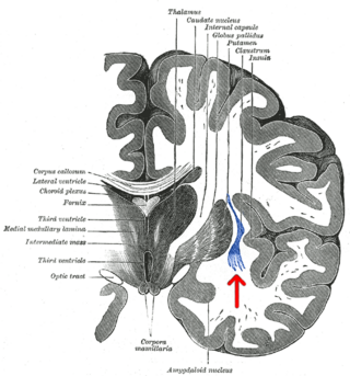
The claustrum is a thin sheet of neurons and supporting glial cells, that connects to the cerebral cortex and subcortical regions including the amygdala, hippocampus and thalamus of the brain. It is located between the insular cortex laterally and the putamen medially, encased by the extreme and external capsules respectively. Blood to the claustrum is supplied by the middle cerebral artery. It is considered to be the most densely connected structure in the brain, and thus hypothesized to allow for the integration of various cortical inputs such as vision, sound and touch, into one experience. Other hypotheses suggest that the claustrum plays a role in salience processing, to direct attention towards the most behaviorally relevant stimuli amongst the background noise. The claustrum is difficult to study given the limited number of individuals with claustral lesions and the poor resolution of neuroimaging.

Cortical maps are collections (areas) of minicolumns in the brain cortex that have been identified as performing a specific information processing function.

Multisensory integration, also known as multimodal integration, is the study of how information from the different sensory modalities may be integrated by the nervous system. A coherent representation of objects combining modalities enables animals to have meaningful perceptual experiences. Indeed, multisensory integration is central to adaptive behavior because it allows animals to perceive a world of coherent perceptual entities. Multisensory integration also deals with how different sensory modalities interact with one another and alter each other's processing.

Neuroplasticity, also known as neural plasticity, or brain plasticity, is the ability of neural networks in the brain to change through growth and reorganization. It is when the brain is rewired to function in some way that differs from how it previously functioned. These changes range from individual neuron pathways making new connections, to systematic adjustments like cortical remapping or neural oscillation. Other forms of neuroplasticity include homologous area adaptation, cross modal reassignment, map expansion, and compensatory masquerade. Examples of neuroplasticity include circuit and network changes that result from learning a new ability, information acquisition, environmental influences, practice, and psychological stress.

<span class="mw-page-title-main">Thalamocortical radiations</span> Neural pathways between the thalamus and cerebral cortex

In neuroanatomy, thalamocortical radiations also known as thalamocortical fibres, are the efferent fibres that project from the thalamus to distinct areas of the cerebral cortex. They form fibre bundles that emerge from the lateral surface of the thalamus.

<span class="mw-page-title-main">Stellate cell</span>

Stellate cells are neurons in the central nervous system, named for their star-like shape formed by dendritic processes radiating from the cell body. Many stellate cells are GABAergic and are located in the molecular layer of the cerebellum. Stellate cells are derived from dividing progenitor cells in the white matter of postnatal cerebellum. Dendritic trees can vary between neurons. There are two types of dendritic trees in the cerebral cortex, which include pyramidal cells, which are pyramid shaped and stellate cells which are star shaped. Dendrites can also aid neuron classification. Dendrites with spines are classified as spiny, those without spines are classified as aspinous. Stellate cells can be spiny or aspinous, while pyramidal cells are always spiny. Most common stellate cells are the inhibitory interneurons found within the upper half of the molecular layer in the cerebellum. Cerebellar stellate cells synapse onto the dendritic trees of Purkinje cells and send inhibitory signals. Stellate neurons are sometimes found in other locations in the central nervous system; cortical spiny stellate cells are found in layer IVC of the primary visual cortex. In the somatosensory barrel cortex of mice and rats, glutamatergic (excitatory) spiny stellate cells are organized in the barrels of layer 4. They receive excitatory synaptic fibres from the thalamus and process feed forward excitation to 2/3 layer of the primary visual cortex to pyramidal cells. Cortical spiny stellate cells have a 'regular' firing pattern. Stellate cells are chromophobes, that is cells that does not stain readily, and thus appears relatively pale under the microscope.

The zona incerta (ZI) is a horizontally elongated region of gray matter in the subthalamus below the thalamus. Its connections project extensively over the brain from the cerebral cortex down into the spinal cord.

A topographic map is the ordered projection of a sensory surface, like the retina or the skin, or an effector system, like the musculature, to one or more structures of the central nervous system. Topographic maps can be found in all sensory systems and in many motor systems.

<span class="mw-page-title-main">Perineuronal net</span> Structures of the brain

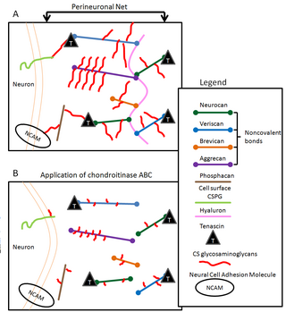
Perineuronal nets (PNNs) are specialized extracellular matrix structures responsible for synaptic stabilization in the adult brain. PNNs are found around certain neuron cell bodies and proximal neurites in the central nervous system. PNNs play a critical role in the closure of the childhood critical period, and their digestion can cause restored critical period-like synaptic plasticity in the adult brain. They are largely negatively charged and composed of chondroitin sulfate proteoglycans, molecules that play a key role in development and plasticity during postnatal development and in the adult.

Activity-dependent plasticity is a form of functional and structural neuroplasticity that arises from the use of cognitive functions and personal experience; hence, it is the biological basis for learning and the formation of new memories. Activity-dependent plasticity is a form of neuroplasticity that arises from intrinsic or endogenous activity, as opposed to forms of neuroplasticity that arise from extrinsic or exogenous factors, such as electrical brain stimulation- or drug-induced neuroplasticity. The brain's ability to remodel itself forms the basis of the brain's capacity to retain memories, improve motor function, and enhance comprehension and speech amongst other things. It is this trait to retain and form memories that is associated with neural plasticity and therefore many of the functions individuals perform on a daily basis. This plasticity occurs as a result of changes in gene expression which are triggered by signaling cascades that are activated by various signaling molecules during increased neuronal activity.

<span class="mw-page-title-main">Somatosensory system</span> Nerve system for sensing touch, temperature, body position, and pain

In physiology, the somatosensory system is the network of neural structures in the brain and body that produce the perception of touch, as well as temperature (thermoception), body position (proprioception), and pain. It is a subset of the sensory nervous system, which also represents visual, auditory, olfactory, gustatory and vestibular stimuli.

<span class="mw-page-title-main">Cross modal plasticity</span> Reorganization of neurons in the brain to integrate the function of two or more sensory systems

Cross modal plasticity is the adaptive reorganization of neurons to integrate the function of two or more sensory systems. Cross modal plasticity is a type of neuroplasticity and often occurs after sensory deprivation due to disease or brain damage. The reorganization of the neural network is greatest following long-term sensory deprivation, such as congenital blindness or pre-lingual deafness. In these instances, cross modal plasticity can strengthen other sensory systems to compensate for the lack of vision or hearing. This strengthening is due to new connections that are formed to brain cortices that no longer receive sensory input.

<span class="mw-page-title-main">Hydrodynamic reception</span> Ability of an organism to sense water movements

In animal physiology, hydrodynamic reception refers to the ability of some animals to sense water movements generated by biotic or abiotic sources. This form of mechanoreception is useful for orientation, hunting, predator avoidance, and schooling. Frequent encounters with conditions of low visibility can prevent vision from being a reliable information source for navigation and sensing objects or organisms in the environment. Sensing water movements is one resolution to this problem.

Sensory maps and brain development is a concept in neuroethology that links the development of the brain over an animal’s lifetime with the fact that there is spatial organization and pattern to an animal’s sensory processing. Sensory maps are the representations of sense organs as organized maps in the brain, and it is the fundamental organization of processing. Sensory maps are not always close to an exact topographic projection of the senses. The fact that the brain is organized into sensory maps has wide implications for processing, such as that lateral inhibition and coding for space are byproducts of mapping. The developmental process of an organism guides sensory map formation; the details are yet unknown. The development of sensory maps requires learning, long term potentiation, experience-dependent plasticity, and innate characteristics. There is significant evidence for experience-dependent development and maintenance of sensory maps, and there is growing evidence on the molecular basis, synaptic basis and computational basis of experience-dependent development.

<span class="mw-page-title-main">Cortical remapping</span>

Cortical remapping, also referred to as cortical reorganization, is the process by which an existing cortical map is affected by a stimulus resulting in the creating of a 'new' cortical map. Every part of the body is connected to a corresponding area in the brain which creates a cortical map. When something happens to disrupt the cortical maps such as an amputation or a change in neuronal characteristics, the map is no longer relevant. The part of the brain that is in charge of the amputated limb or neuronal change will be dominated by adjacent cortical regions that are still receiving input, thus creating a remapped area. Remapping can occur in the sensory or motor system. The mechanism for each system may be quite different. Cortical remapping in the somatosensory system happens when there has been a decrease in sensory input to the brain due to deafferentation or amputation, as well as a sensory input increase to an area of the brain. Motor system remapping receives more limited feedback that can be difficult to interpret.

Jessica Cardin is an American neuroscientist who is an associate professor of neuroscience at Yale University School of Medicine. Cardin's lab studies local circuits within the primary visual cortex to understand how cellular and synaptic interactions flexibly adapt to different behavioral states and contexts to give rise to visual perceptions and drive motivated behaviors. Cardin's lab applies their knowledge of adaptive cortical circuit regulation to probe how circuit dysfunction manifests in disease models.

Corey C. Harwell is an American neuroscientist who is an assistant professor in the Department of Neurobiology at Harvard Medical School.

References

Research groups working on barrel cortex:

Books on barrel cortex