
Gadolinium is a chemical element with the symbol Gd and atomic number 64. Gadolinium is a silvery-white metal when oxidation is removed. It is only slightly malleable and is a ductile rare-earth element. Gadolinium reacts with atmospheric oxygen or moisture slowly to form a black coating. Gadolinium below its Curie point of 20 °C (68 °F) is ferromagnetic, with an attraction to a magnetic field higher than that of nickel. Above this temperature it is the most paramagnetic element. It is found in nature only in an oxidized form. When separated, it usually has impurities of the other rare-earths because of their similar chemical properties.

Magnetic resonance imaging (MRI) is a medical imaging technique used in radiology to form pictures of the anatomy and the physiological processes of the body. MRI scanners use strong magnetic fields, magnetic field gradients, and radio waves to generate images of the organs in the body. MRI does not involve X-rays or the use of ionizing radiation, which distinguishes it from CT and PET scans. MRI is a medical application of nuclear magnetic resonance (NMR) which can also be used for imaging in other NMR applications, such as NMR spectroscopy.

Breast cancer is cancer that develops from breast tissue. Signs of breast cancer may include a lump in the breast, a change in breast shape, dimpling of the skin, milk rejection, fluid coming from the nipple, a newly inverted nipple, or a red or scaly patch of skin. In those with distant spread of the disease, there may be bone pain, swollen lymph nodes, shortness of breath, or yellow skin.

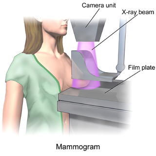
Mammography is the process of using low-energy X-rays to examine the human breast for diagnosis and screening. The goal of mammography is the early detection of breast cancer, typically through detection of characteristic masses or microcalcifications.

Virtual colonoscopy is the use of CT scanning or magnetic resonance imaging (MRI) to produce two- and three-dimensional images of the colon, from the lowest part, the rectum, to the lower end of the small intestine, and to display the images on an electronic display device. The procedure is used to screen for colon cancer and polyps, and may detect diverticulosis. A virtual colonoscopy can provide 3D reconstructed endoluminal views of the bowel. VC provides a secondary benefit of revealing diseases or abnormalities outside the colon.
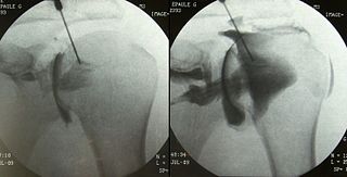
An arthrogram is a series of images of a joint after injection of a contrast medium, usually done by fluoroscopy or MRI. The injection is normally done under a local anesthetic such as Novocain or lidocaine. The radiologist or radiographer performs the study using fluoroscopy or x-ray to guide the placement of the needle into the joint and then injects around 10 ml of contrast based on age. There is some burning pain from the anesthetic and a painful bubbling feeling in the joint after the contrast is injected. This only lasts 20 – 30 hours until the Contrast is absorbed. During this time, while it is allowed, it is painful to use the limb for around 10 hours. After that the radiologist can more clearly see what is going on under your skin and can get results out within 24 to 48 hours.

Gadopentetic acid, sold under the brand name Magnevist, is a gadolinium-based MRI contrast agent.

Gadodiamide, sold under the brand name Omniscan, is a gadolinium-based MRI contrast agent (GBCA), used in magnetic resonance imaging (MRI) procedures to assist in the visualization of blood vessels.
Nephrogenic systemic fibrosis is a rare syndrome that involves fibrosis of skin, joints, eyes, and internal organs. NSF is caused by exposure to gadolinium in gadolinium-based MRI contrast agents (GBCAs) in patients with impaired kidney function. Epidemiological studies suggest that the incidence of NSF is unrelated to gender or ethnicity and it is not thought to have a genetic basis. After GBCAs were identified as a cause of the disorder in 2006, and screening and prevention measures put in place, it is now considered rare.
Breast cancer screening is the medical screening of asymptomatic, apparently healthy women for breast cancer in an attempt to achieve an earlier diagnosis. The assumption is that early detection will improve outcomes. A number of screening tests have been employed, including clinical and self breast exams, mammography, genetic screening, ultrasound, and magnetic resonance imaging.
MRI contrast agents are contrast agents used to improve the visibility of internal body structures in magnetic resonance imaging (MRI). The most commonly used compounds for contrast enhancement are gadolinium-based. Such MRI contrast agents shorten the relaxation times of nuclei within body tissues following oral or intravenous administration.

Gadobutrol (INN) (Gd-DO3A-butrol) is a gadolinium-based MRI contrast agent (GBCA).

Gadoteric acid, sold under the brand name Dotarem among others, is a macrocycle-structured gadolinium-based MRI contrast agent (GBCA). It consists of the organic acid DOTA as a chelating agent, and gadolinium (Gd3+), and is used in form of the meglumine salt (gadoterate meglumine). The paramagnetic property of gadoteric acid reduces the T1 relaxation time (and to some extent the T2 and T2* relaxation times) in MRI, which is the source of its clinical utility. Because it has magnetic properties, gadoteric acid develops a magnetic moment when put under a magnetic field, which increases the signal intensity (brightness) of tissues during MRI imaging.
Dynamic angiothermography (DATG) is a technique for the diagnosis of breast cancer. This technique, though springing from the thermography of old conception, is based on a completely different principle. DATG records the temperature variations linked to the vascular changes in the breast due to angiogenesis. The presence, change, and growth of tumors and lesions in breast tissue change the vascular network in the breast. Consequently, measuring the vascular structure over time, DATG effectively monitors the change in breast tissue due to tumors and lesions. It is currently used in combination with other techniques for diagnosis of breast cancer. This diagnostic method is a low cost one compared with other techniques.
Breast cancer diagnosis and treatment is influenced by different cultural backgrounds. Factors include differences in beliefs, attitudes, and treatment options that impact diverse populations throughout the world.
In medicine, breast imaging is a sub-speciality of diagnostic radiology that involves imaging of the breasts for screening or diagnostic purposes. There are various methods of breast imaging using a variety of technologies as described in detail below. Traditional screening and diagnostic mammography uses x-ray technology and has been the mainstay of breast imaging for many decades. Breast tomosynthesis is a relatively new digital x-ray mammography technique that produces multiple image slices of the breast similar to, but distinct from, Computed Tomography (CT). Xeromammography and Galactography are somewhat outdated technologies that also use x-ray technology and are now used infrequently in the detection of breast cancer. Breast ultrasound is another technology employed in diagnosis and screening that can help differentiate between fluid filled and solid lesions, an important factor to determine if a lesion may be cancerous. Breast MRI is a technology typically reserved for high-risk patients and patients recently diagnosed with breast cancer. Lastly, scintimammography is used in a subgroup of patients who have abnormal mammograms or whose screening is not reliable on the basis of using traditional mammography or ultrasound.
Magnetic resonance enterography is a magnetic resonance imaging technique used to evaluate bowel wall features of both upper and lower gastro-intestinal tract, although it is usually used for small bowel evaluation. It is a less invasive technique with the advantages of no ionizing radiation exposure, multiplanarity and high contrast resolution for soft tissue.

Medical imaging in pregnancy may be indicated because of pregnancy complications, intercurrent diseases or routine prenatal care.
Nola M. Hylton is an American oncologist who is Professor of Radiology and Director of the Breast Imaging Research Group at the University of California, San Francisco. She pioneered the usage of magnetic resonance imaging for the detection, diagnosis, and staging of breast cancer by using MRIs to locate tumors and characterize the surrounding tissue.
Sylvia Katina Plevritis is Professor and Chair of the Department of Biomedical Data Science at Stanford University.
