Brefeldin A

Last updated
Brefeldin A
Brefeldin A Structural Formula V1.svg
Names
Preferred IUPAC name
(1R,2E,6S,10E,11aS,13S,14aR)-1,13-Dihydroxy-6-methyl-1,6,7,8,9,11a,12,13,14,14a-decahydro-4H-cyclopenta[f][1]oxacyclotridecin-4-one
Other names
γ,4-Dihydroxy-2-(6-hydroxy-1-heptenyl)-4-cyclopentanecrotonic acid λ-lactone[ citation needed ]
Identifiers
3D model (JSmol)
ChEBI
ChEMBL
ChemSpider
DrugBank
ECHA InfoCard 100.127.053 OOjs UI icon edit-ltr-progressive.svg
PubChem CID
UNII
  • InChI=1S/C16H24O4/c1-11-5-3-2-4-6-12-9-13(17)10-14(12)15(18)7-8-16(19)20-11/h4,6-8,11-15,17-18H,2-3,5,9-10H2,1H3/b6-4+,8-7+/t11-,12+,13-,14+,15+/m0/s1 Yes check.svgY
    Key: KQNZDYYTLMIZCT-KQPMLPITSA-N Yes check.svgY
  • InChI=1/C16H24O4/c1-11-5-3-2-4-6-12-9-13(17)10-14(12)15(18)7-8-16(19)20-11/h4,6-8,11-15,17-18H,2-3,5,9-10H2,1H3/b6-4+,8-7+/t11-,12+,13-,14+,15+/m0/s1
    Key: KQNZDYYTLMIZCT-KQPMLPITBH
  • O=C/1O[C@H](CCC/C=C/[C@H]2[C@H]([C@H](O)/C=C\1)C[C@@H](O)C2)C
Properties
C16H24O4
Molar mass 280.36 g/mol
AppearanceWhite to off-white crystalline powder
Melting point 204 to 205 °C (399 to 401 °F; 477 to 478 K)
Except where otherwise noted, data are given for materials in their standard state (at 25 °C [77 °F], 100 kPa).
Yes check.svgY  verify  (what is  Yes check.svgYX mark.svgN ?)

Brefeldin A is a lactone antiviral produced by the fungus Penicillium brefeldianum . [1] Brefeldin A inhibits protein transport from the endoplasmic reticulum to the golgi complex indirectly by preventing association of COP-I coat [2] to the Golgi membrane. Brefeldin A was initially isolated with hopes to become an antiviral drug [3] but is now primarily used in research to study protein transport.

Contents

History

The compound gets its name from a species of anamorph fungus of the Penicillium genus known as Eupenicillium brefeldianum, though it is found in a variety of species that span several genera. [4] It was first isolated from Penicillium decumbens in 1958 by V.L. Singleton who initially called it Decumbin. [5] It was later identified as a metabolite by H.P. Siggs who then went on to identify the chemical structure of the compound in 1971. [5] Since then several successful total synthesis methods have been described. [5] Interest in researching brefeldin A was initially lacking due to poor antiviral activity. [5] However, upon discovery of its mechanism involving disruption of protein transport by Takatsuki and Tamura in 1985 and the cytotoxic effects observed in certain cancer cell lines, research efforts were revitalized. [5] It is currently used solely in research mainly as an assay tool for studying membrane traffic and vesicle transport dynamics between the endoplasmic reticulum and Golgi apparatus.[ citation needed ]

Physical properties and storage information

Brefeldin A is found naturally as a white to off-white crystalline solid. It forms a clear colorless solution when dissolved. It is soluble in methanol (10 mg/mL), ethanol (5 mg/mL), DMSO (20 mg/mL), acetone, and ethyl acetate (1 mg/mL) without the aid of heating. [6] It is poorly soluble in water (slightly miscible). [6] It is sold commercially with a purity of 98% or greater. [6] It is recommended that it be stored desiccate at -20 °C away from direct sunlight. Its suggested shelf life for use is 6 months as a solid and 1 month as a solution with tightly sealed storage at -20 °C. Since the compound is combustible, contamination with oxidizing agents should be avoided to prevent the risk of fire. Direct contact should be avoided as well.[ citation needed ]

Mechanism of action

Brefeldin A inhibits vesicle formation and transport between the endoplasmic reticulum and the Golgi apparatus which ultimately results in collapse of the Golgi apparatus into the endoplasmic reticulum via membrane fusion. Brefeldin A Inhibition of Intracellular Vesicle Transport.png
Brefeldin A inhibits vesicle formation and transport between the endoplasmic reticulum and the Golgi apparatus which ultimately results in collapse of the Golgi apparatus into the endoplasmic reticulum via membrane fusion.

In mammalian and yeast cells, the main target of brefeldin A appears to be a guanine nucleotide exchange factor (GEF) called GBF1. [7] GBF1 is a member of the Arf family of GEFs which are recruited to membranes of the Golgi. [8] It is responsible for the regulation of Arf1p GTPase. [8] It does this through converting the inactive GDP-bound form of Arf1p to the active GTP-bound form. [8] The nucleotide exchange occurs at the catalytic Sec7 domain of GBF1. Activated Arf1p then recruits coat protein β-COP, a subunit of the COP-I complex, to cargo-bound receptors on the membrane. [8] Coat protein recruitment is necessary for proper vesicle formation and transport. Brefeldin A reversibly inhibits the function of GBF1 uncompetitively by binding to the complex it forms with GDP-bound Arf1p and preventing conversion to the GTP-bound form. [8] The lack of active Arf1p prevents coat protein recruitment, which then ultimately induces the fusion of neighboring ER and Golgi membranes due to lack of vesicle formation. This is because lack of vesicle formation results in a buildup of SNARE proteins in the Golgi which would otherwise be bound to coat protein-coated vesicles and removed with the vesicles once they bud off. [9] SNARE proteins mediate membrane fusion and it is postulated that the described SNARE build up in the Golgi increases the chances of aberrant fusion of the Golgi membrane with that of the ER. [9] The collapse of the Golgi into the ER triggers activation of unfolded protein response (UPR) (or ER stress) [10] [11] which can result in apoptosis.

Toxicity

The toxological effects of brefeldin A have not been studied extensively yet. [12] Some animal LD50 values have been reported including 250 mg/kg in mice (interperitoneal) and 275 mg/kg in rats (oral). [12] Generally, antibiotic macrolides that share a similar macrocyclic lactone ring to that of brefeldin A have been shown to produce gastrointestinal discomfort as the most common side effect. [13] Some macrolides have been shown to produce allergic reactions and though uncommon this possibility in the case of brefeldin A cannot be disregarded as of yet. [13] The compound may bind to hemoglobin and inhibit oxygen uptake resulting in methemoglobinemia, a form of oxygen starvation, though this is not confirmed. [13] Brefeldin A is not considered to be harmful from direct skin or eye exposure other than transient irritation. [13] It may cause irritation of the respiratory system if inhaled. [13]

See also

Related Research Articles

<span class="mw-page-title-main">Endoplasmic reticulum</span> Cell organelle that synthesizes, folds and processes proteins

The endoplasmic reticulum (ER) is, in essence, the transportation system of the eukaryotic cell, and has many other important functions such as protein folding. It is a type of organelle made up of two subunits – rough endoplasmic reticulum (RER), and smooth endoplasmic reticulum (SER). The endoplasmic reticulum is found in most eukaryotic cells and forms an interconnected network of flattened, membrane-enclosed sacs known as cisternae, and tubular structures in the SER. The membranes of the ER are continuous with the outer nuclear membrane. The endoplasmic reticulum is not found in red blood cells, or spermatozoa.

<span class="mw-page-title-main">Endomembrane system</span> Membranes in the cytoplasm of a eukaryotic cell

The endomembrane system is composed of the different membranes (endomembranes) that are suspended in the cytoplasm within a eukaryotic cell. These membranes divide the cell into functional and structural compartments, or organelles. In eukaryotes the organelles of the endomembrane system include: the nuclear membrane, the endoplasmic reticulum, the Golgi apparatus, lysosomes, vesicles, endosomes, and plasma (cell) membrane among others. The system is defined more accurately as the set of membranes that forms a single functional and developmental unit, either being connected directly, or exchanging material through vesicle transport. Importantly, the endomembrane system does not include the membranes of plastids or mitochondria, but might have evolved partially from the actions of the latter.

<span class="mw-page-title-main">Golgi apparatus</span> Cell organelle that packages proteins for export

The Golgi apparatus, also known as the Golgi complex, Golgi body, or simply the Golgi, is an organelle found in most eukaryotic cells. Part of the endomembrane system in the cytoplasm, it packages proteins into membrane-bound vesicles inside the cell before the vesicles are sent to their destination. It resides at the intersection of the secretory, lysosomal, and endocytic pathways. It is of particular importance in processing proteins for secretion, containing a set of glycosylation enzymes that attach various sugar monomers to proteins as the proteins move through the apparatus.

<span class="mw-page-title-main">Vesicle (biology and chemistry)</span> Any small, fluid-filled, spherical organelle enclosed by a membrane

In cell biology, a vesicle is a structure within or outside a cell, consisting of liquid or cytoplasm enclosed by a lipid bilayer. Vesicles form naturally during the processes of secretion (exocytosis), uptake (endocytosis), and the transport of materials within the plasma membrane. Alternatively, they may be prepared artificially, in which case they are called liposomes. If there is only one phospholipid bilayer, the vesicles are called unilamellar liposomes; otherwise they are called multilamellar liposomes. The membrane enclosing the vesicle is also a lamellar phase, similar to that of the plasma membrane, and intracellular vesicles can fuse with the plasma membrane to release their contents outside the cell. Vesicles can also fuse with other organelles within the cell. A vesicle released from the cell is known as an extracellular vesicle.

The Coat Protein Complex II, or COPII, is a group of proteins that facilitate the formation of vesicles to transport proteins from the endoplasmic reticulum to the Golgi apparatus or endoplasmic-reticulum–Golgi intermediate compartment. This process is termed anterograde transport, in contrast to the retrograde transport associated with the COPI complex. COPII is assembled in two parts: first an inner layer of Sar1, Sec23, and Sec24 forms; then the inner coat is surrounded by an outer lattice of Sec13 and Sec31.

<span class="mw-page-title-main">COPI</span> Protein complex

COPI is a coatomer, a protein complex that coats vesicles transporting proteins from the cis end of the Golgi complex back to the rough endoplasmic reticulum (ER), where they were originally synthesized, and between Golgi compartments. This type of transport is retrograde transport, in contrast to the anterograde transport associated with the COPII protein. The name "COPI" refers to the specific coat protein complex that initiates the budding process on the cis-Golgi membrane. The coat consists of large protein subcomplexes that are made of seven different protein subunits, namely α, β, β', γ, δ, ε and ζ.

<span class="mw-page-title-main">Vesicular-tubular cluster</span>

The vesicular-tubular cluster (VTC), also referred to as the endoplasmic-reticulum–Golgi intermediate compartment (ERGIC), is an organelle in eukaryotic cells. This compartment mediates trafficking between the endoplasmic reticulum (ER) and Golgi complex, facilitating the sorting of cargo. The cluster was first identified in 1988 using an antibody to the protein that has since been named ERGIC-53.

The coatomer is a protein complex that coats membrane-bound transport vesicles. Two types of coatomers are known:

<span class="mw-page-title-main">USO1</span> Protein-coding gene in the species Homo sapiens

General vesicular transport factor p115 is a protein that in humans is encoded by the USO1 gene.

<span class="mw-page-title-main">STX5</span> Protein-coding gene in the species Homo sapiens

Syntaxin-5 is a protein that in humans is encoded by the STX5 gene.

<span class="mw-page-title-main">KDELR1</span> Protein-coding gene in the species Homo sapiens

KDEL (Lys-Asp-Glu-Leu) endoplasmic reticulum protein retention receptor 1, also known as KDELR1, is a protein which in humans is encoded by the KDELR1 gene.

<span class="mw-page-title-main">VAPA</span> Protein-coding gene in humans

VAMP-Associated Protein A is a protein that in humans is encoded by the VAPA gene. Together with VAPB and VAPC it forms the VAP protein family. They are integral endoplasmic reticulum membrane proteins of the type II and are ubiquitous among eukaryotes.
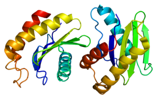
<span class="mw-page-title-main">SEC22B</span> Protein-coding gene in the species Homo sapiens

Vesicle-trafficking protein SEC22b is a protein that in humans is encoded by the SEC22B gene.

<span class="mw-page-title-main">BET1</span> Protein-coding gene in the species Homo sapiens

BET1 homolog is a protein that in humans is encoded by the BET1 gene.

<span class="mw-page-title-main">OSBP</span> Protein-coding gene in the species Homo sapiens

Oxysterol-binding protein 1 is a protein that in humans is encoded by the OSBP gene.

<span class="mw-page-title-main">GOSR2</span> Protein-coding gene in the species Homo sapiens

Golgi SNAP receptor complex member 2 is a protein that in humans is encoded by the GOSR2 gene.

KDEL is a target peptide sequence in mammals and plants located on the C-terminal end of the amino acid structure of a protein. The KDEL sequence prevents a protein from being secreted from the endoplasmic reticulum (ER) and facilitates its return if it is accidentally exported.

<span class="mw-page-title-main">Jennifer Lippincott-Schwartz</span> American biologist

Jennifer Lippincott-Schwartz is a Senior Group Leader at Howard Hughes Medical Institute's Janelia Research Campus and a founding member of the Neuronal Cell Biology Program at Janelia. Previously, she was the Chief of the Section on Organelle Biology in the Cell Biology and Metabolism Program, in the Division of Intramural Research in the Eunice Kennedy Shriver National Institute of Child Health and Human Development at the National Institutes of Health from 1993 to 2016. Lippincott-Schwartz received her PhD from Johns Hopkins University, and performed post-doctoral training with Richard Klausner at the NICHD, NIH in Bethesda, Maryland.

Membrane vesicle trafficking in eukaryotic animal cells involves movement of biochemical signal molecules from synthesis-and-packaging locations in the Golgi body to specific release locations on the inside of the plasma membrane of the secretory cell. It takes place in the form of Golgi membrane-bound micro-sized vesicles, termed membrane vesicles (MVs).

Modeccin is a toxic lectin, a group of glycoproteins capable of binding specifically to sugar moieties. Different toxic lectins are present in seeds of different origin. Modeccin is found in the roots of the African plant Adenia digitata. These roots are often mistaken for edible roots, which has led to some cases of intoxication. Sometimes the fruit is eaten, or a root extract is drunk as a manner of suicide.

References

  1. Hutchinson, C. R.; Shu-Wen, L.; McInnes, A. G.; Walter, J. A. (1983). "Comparative biochemistry of fatty acid and macrolide antibiotic (brefeldin a). Formation in penicillium brefeldianum". Tetrahedron. 39 (21): 3507. doi:10.1016/S0040-4020(01)88660-9.
  2. Helms, J. Bernd; Rothman, James E. (1992). "Inhibition by brefeldin A of a Golgi membrane enzyme that catalyses exchange of guanine nucleotide bound to ARF". Nature. 360 (6402): 352–354. Bibcode:1992Natur.360..352H. doi:10.1038/360352a0. PMID   1448152. S2CID   4306100.
  3. Tamura G, Ando K, Suzuki S, Takatsuki A, Arima K (February 1968). "Antiviral activity of brefeldin A and verrucarin A". J. Antibiot. 21 (2): 160–1. doi: 10.7164/antibiotics.21.160 . PMID   4299889.
  4. Wang, Jianfeng; Huang, Yaojian; Fang, Meijuan; Zhang, Yongjie; Zheng, Zhonghui; Zhao, Yufen; Su, Wenjin (2002-09-01). "Brefeldin A, a cytotoxin produced by Paecilomyces sp. and Aspergillus clavatus isolated from Taxus mairei and Torreya grandis". FEMS Immunology & Medical Microbiology. 34 (1): 51–57. doi: 10.1111/j.1574-695X.2002.tb00602.x . ISSN   0928-8244. PMID   12208606.
  5. 1 2 3 4 5 McCloud, T. G.; Burns, M. P.; Majadly, F. D.; Muschik, G. M.; Miller, D. A.; Poole, K. K.; Roach, J. M.; Ross, J. T.; Lebherz, W. B. (1995-07-01). "Production of brefeldin-A". Journal of Industrial Microbiology. 15 (1): 5–9. doi: 10.1007/BF01570006 . ISSN   0169-4146. PMID   7662298. S2CID   8511645.
  6. 1 2 3 "Brefeldin A (CAS 20350-15-6)". Santa Cruz Biotechnology. 8 May 2017.
  7. "GBF1 Gene - GeneCards | GBF1 Protein | GBF1 Antibody". www.genecards.org.
  8. 1 2 3 4 5 Niu, Ting-Kuang; Pfeifer, Andrea C.; Lippincott-Schwartz, Jennifer; Jackson, Catherine L. (2005-03-01). "Dynamics of GBF1, a Brefeldin A-Sensitive Arf1 Exchange Factor at the Golgi". Molecular Biology of the Cell. 16 (3): 1213–1222. doi:10.1091/mbc.E04-07-0599. ISSN   1059-1524. PMC   551486 . PMID   15616190.
  9. 1 2 Nebenführ, Andreas; Ritzenthaler, Christophe; Robinson, David G. (2002-11-01). "Brefeldin A: Deciphering an Enigmatic Inhibitor of Secretion". Plant Physiology. 130 (3): 1102–1108. doi:10.1104/pp.011569. ISSN   1532-2548. PMC   1540261 . PMID   12427977.
  10. Pahl HL, Baeuerle (Jun 1995). "A novel signal transduction pathway from the endoplasmic reticulum to the nucleus is mediated by transcription factor NF-kappa B". EMBO J. 14 (11): 2580–8. doi:10.1002/j.1460-2075.1995.tb07256.x. PMC   398372 . PMID   7781611.
  11. Kober L, Zehe C, Bode J (October 2012). "Development of a novel ER stress based selection system for the isolation of highly productive clones". Biotechnol. Bioeng. 109 (10): 2599–611. doi:10.1002/bit.24527. PMID   22510960. S2CID   25858120.
  12. 1 2 "SAFETY DATA SHEET Brefeldin A" (PDF). Cayman Chemical. 6 February 2015.
  13. 1 2 3 4 5 "Material Safety Data Sheet. Brefeldin A (BFA) sc-200861" (PDF). Santa Cruz Biotechnology. 20 January 2009.