Cancer immunology

Last updated
Tumor-associated immune cells in the tumor microenvironment (TME) of breast cancer models Tumor-associated immune cells in the tumor microenvironment (TME) of breast cancer models.svg
Tumor-associated immune cells in the tumor microenvironment (TME) of breast cancer models

Cancer immunology (immuno-oncology) is an interdisciplinary branch of biology and a sub-discipline of immunology that is concerned with understanding the role of the immune system in the progression and development of cancer; the most well known application is cancer immunotherapy, which utilises the immune system as a treatment for cancer. Cancer immunosurveillance and immunoediting are based on protection against development of tumors in animal systems and (ii) identification of targets for immune recognition of human cancer.

Contents

Definition

Cancer immunology is an interdisciplinary branch of biology concerned with the role of the immune system in the progression and development of cancer; the most well known application is cancer immunotherapy, where the immune system is used to treat cancer. [1] [2] Cancer immunosurveillance is a theory formulated in 1957 by Burnet and Thomas, who proposed that lymphocytes act as sentinels in recognizing and eliminating continuously arising, nascent transformed cells. [3] [4] Cancer immunosurveillance appears to be an important host protection process that decreases cancer rates through inhibition of carcinogenesis and maintaining of regular cellular homeostasis. [5] It has also been suggested that immunosurveillance primarily functions as a component of a more general process of cancer immunoediting. [3]

Tumor antigens

Tumors may express tumor antigens that are recognized by the immune system and may induce an immune response. [6] These tumor antigens are either TSA (Tumor-specific antigen) or TAA (Tumor-associated antigen). [7]

Tumor-specific

Tumor-specific antigens (TSA) are antigens that only occur in tumor cells. [7] TSAs can be products of oncoviruses like E6 and E7 proteins of human papillomavirus, occurring in cervical carcinoma, or EBNA-1 protein of EBV, occurring in Burkitt's lymphoma cells. [8] [9] Another example of TSAs are abnormal products of mutated oncogenes (e.g. Ras protein) and anti-oncogenes (e.g. p53). [10]

Tumor-associated antigens

Tumor-associated antigens (TAA) are present in healthy cells, but for some reason they also occur in tumor cells. [7] However, they differ in quantity, place or time period of expression. [11] Oncofetal antigens are tumor-associated antigens expressed by embryonic cells and by tumors. [12] Examples of oncofetal antigens are AFP (α-fetoprotein), produced by hepatocellular carcinoma, or CEA (carcinoembryonic antigen), occurring in ovarian and colon cancer. [13] [14] More tumor-associated antigens are HER2/neu, EGFR or MAGE-1. [15] [16] [17]

Immunoediting

Cancer immunoediting is a process in which immune system interacts with tumor cells. It consists of three phases: elimination, equilibrium and escape. These phases are often referred to as "the three Es" of cancer immunoediting. Both adaptive and innate immune system participate in immunoediting. [18]

In the elimination phase, the immune response leads to destruction of tumor cells and therefore to tumor suppression. However, some tumor cells may gain more mutations, change their characteristics and evade the immune system. These cells might enter the equilibrium phase, in which the immune system does not recognise all tumor cells, but at the same time the tumor does not grow. This condition may lead to the phase of escape, in which the tumor gains dominance over immune system, starts growing and establishes immunosuppressive environment. [19]

As a consequence of immunoediting, tumor cell clones less responsive to the immune system gain dominance in the tumor through time, as the recognized cells are eliminated. This process may be considered akin to Darwinian evolution, where cells containing pro-oncogenic or immunosuppressive mutations survive to pass on their mutations to daughter cells, which may themselves mutate and undergo further selective pressure. This results in the tumor consisting of cells with decreased immunogenicity and can hardly be eliminated. [19] This phenomenon was proven to happen as a result of immunotherapies of cancer patients. [20]

Tumor evasion mechanisms

Multiple factors determine whether tumor cells will be eliminated by the immune system or will escape detection. During the elimination phase immune effector cells such as CTL's and NK cells with the help of dendritic and CD4+ T-cells are able to recognize and eliminate tumor cells. Tumor microenvironment.jpg
Multiple factors determine whether tumor cells will be eliminated by the immune system or will escape detection. During the elimination phase immune effector cells such as CTL's and NK cells with the help of dendritic and CD4+ T-cells are able to recognize and eliminate tumor cells.

Tumor microenvironment

Immune checkpoints of immunosuppressive actions associated with breast cancer Immune checkpoints of immunosuppressive actions associated with breast cancer.svg
Immune checkpoints of immunosuppressive actions associated with breast cancer

Immunomodulation methods

Immune system is the key player in fighting cancer. As described above in mechanisms of tumor evasion, the tumor cells are modulating the immune response in their profit. It is possible to improve the immune response in order to boost the immunity against tumor cells.

Relationship to chemotherapy

Obeid et al. [42] investigated how inducing immunogenic cancer cell death ought to become a priority of cancer chemotherapy. He reasoned, the immune system would be able to play a factor via a 'bystander effect' in eradicating chemotherapy-resistant cancer cells. [43] [44] [45] [2] However, extensive research is still needed on how the immune response is triggered against dying tumour cells. [2] [46]

Professionals in the field have hypothesized that 'apoptotic cell death is poorly immunogenic whereas necrotic cell death is truly immunogenic'. [47] [48] [49] This is perhaps because cancer cells being eradicated via a necrotic cell death pathway induce an immune response by triggering dendritic cells to mature, due to inflammatory response stimulation. [50] [51] On the other hand, apoptosis is connected to slight alterations within the plasma membrane causing the dying cells to be attractive to phagocytic cells. [52] However, numerous animal studies have shown the superiority of vaccination with apoptotic cells, compared to necrotic cells, in eliciting anti-tumor immune responses. [53] [54] [55] [56] [57]

Thus Obeid et al. [42] propose that the way in which cancer cells die during chemotherapy is vital. Anthracyclins produce a beneficial immunogenic environment. The researchers report that when killing cancer cells with this agent uptake and presentation by antigen presenting dendritic cells is encouraged, thus allowing a T-cell response which can shrink tumours. Therefore, activating tumour-killing T-cells is crucial for immunotherapy success. [2] [58]

However, advanced cancer patients with immunosuppression have left researchers in a dilemma as to how to activate their T-cells. The way the host dendritic cells react and uptake tumour antigens to present to CD4+ and CD8+ T-cells is the key to success of the treatment. [2] [59]

See also

Related Research Articles

<span class="mw-page-title-main">Antigen</span> Molecule triggering an immune response (antibody production) in the host

In immunology, an antigen (Ag) is a molecule, moiety, foreign particulate matter, or an allergen, such as pollen, that can bind to a specific antibody or T-cell receptor. The presence of antigens in the body may trigger an immune response.

<span class="mw-page-title-main">Cytotoxic T cell</span> T cell that kills infected, damaged or cancerous cells

A cytotoxic T cell (also known as TC, cytotoxic T lymphocyte, CTL, T-killer cell, cytolytic T cell, CD8+ T-cell or killer T cell) is a T lymphocyte (a type of white blood cell) that kills cancer cells, cells that are infected by intracellular pathogens (such as viruses or bacteria), or cells that are damaged in other ways.

<span class="mw-page-title-main">Natural killer cell</span> Type of cytotoxic lymphocyte

Natural killer cells, also known as NK cells or large granular lymphocytes (LGL), are a type of cytotoxic lymphocyte critical to the innate immune system. They belong to the rapidly expanding family of known innate lymphoid cells (ILC) and represent 5–20% of all circulating lymphocytes in humans. The role of NK cells is analogous to that of cytotoxic T cells in the vertebrate adaptive immune response. NK cells provide rapid responses to virus-infected cells, stressed cells, tumor cells, and other intracellular pathogens based on signals from several activating and inhibitory receptors. Most immune cells detect the antigen presented on major histocompatibility complex I (MHC-I) on infected cell surfaces, but NK cells can recognize and kill stressed cells in the absence of antibodies and MHC, allowing for a much faster immune reaction. They were named "natural killers" because of the notion that they do not require activation to kill cells that are missing "self" markers of MHC class I. This role is especially important because harmful cells that are missing MHC I markers cannot be detected and destroyed by other immune cells, such as T lymphocyte cells.

Immunotherapy or biological therapy is the treatment of disease by activating or suppressing the immune system. Immunotherapies designed to elicit or amplify an immune response are classified as activation immunotherapies, while immunotherapies that reduce or suppress are classified as suppression immunotherapies. Immunotherapy is under preliminary research for its potential to treat various forms of cancer.

A cancer vaccine, or oncovaccine, is a vaccine that either treats existing cancer or prevents development of cancer. Vaccines that treat existing cancer are known as therapeutic cancer vaccines or tumor antigen vaccines. Some of the vaccines are "autologous", being prepared from samples taken from the patient, and are specific to that patient.

The regulatory T cells (Tregs or Treg cells), formerly known as suppressor T cells, are a subpopulation of T cells that modulate the immune system, maintain tolerance to self-antigens, and prevent autoimmune disease. Treg cells are immunosuppressive and generally suppress or downregulate induction and proliferation of effector T cells. Treg cells express the biomarkers CD4, FOXP3, and CD25 and are thought to be derived from the same lineage as naïve CD4+ cells. Because effector T cells also express CD4 and CD25, Treg cells are very difficult to effectively discern from effector CD4+, making them difficult to study. Research has found that the cytokine transforming growth factor beta (TGF-β) is essential for Treg cells to differentiate from naïve CD4+ cells and is important in maintaining Treg cell homeostasis.

<span class="mw-page-title-main">Cancer immunotherapy</span> Artificial stimulation of the immune system to treat cancer

Cancer immunotherapy (immuno-oncotherapy) is the stimulation of the immune system to treat cancer, improving the immune system's natural ability to fight the disease. It is an application of the fundamental research of cancer immunology (immuno-oncology) and a growing subspecialty of oncology.

<span class="mw-page-title-main">Antigen-presenting cell</span> Cell that displays antigen bound by MHC proteins on its surface

An antigen-presenting cell (APC) or accessory cell is a cell that displays an antigen bound by major histocompatibility complex (MHC) proteins on its surface; this process is known as antigen presentation. T cells may recognize these complexes using their T cell receptors (TCRs). APCs process antigens and present them to T cells.

Cross-presentation is the ability of certain professional antigen-presenting cells (mostly dendritic cells) to take up, process and present extracellular antigens with MHC class I molecules to CD8 T cells (cytotoxic T cells). Cross-priming, the result of this process, describes the stimulation of naive cytotoxic CD8+ T cells into activated cytotoxic CD8+ T cells. This process is necessary for immunity against most tumors and against viruses that infect dendritic cells and sabotage their presentation of virus antigens. Cross presentation is also required for the induction of cytotoxic immunity by vaccination with protein antigens, for example, tumour vaccination.

<span class="mw-page-title-main">CD40 (protein)</span> Mammalian protein found in humans

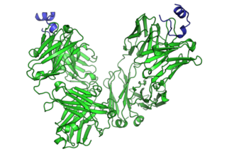
Cluster of differentiation 40, CD40 is a type I transmembrane protein found on antigen-presenting cells and is required for their activation. The binding of CD154 (CD40L) on TH cells to CD40 activates antigen presenting cells and induces a variety of downstream effects.

Immune tolerance, also known as immunological tolerance or immunotolerance, refers to the immune system's state of unresponsiveness to substances or tissues that would otherwise trigger an immune response. It arises from prior exposure to a specific antigen and contrasts the immune system's conventional role in eliminating foreign antigens. Depending on the site of induction, tolerance is categorized as either central tolerance, occurring in the thymus and bone marrow, or peripheral tolerance, taking place in other tissues and lymph nodes. Although the mechanisms establishing central and peripheral tolerance differ, their outcomes are analogous, ensuring immune system modulation.

<span class="mw-page-title-main">CD80</span> Mammalian protein found in Homo sapiens

The Cluster of differentiation 80 is a B7, type I membrane protein in the immunoglobulin superfamily, with an extracellular immunoglobulin constant-like domain and a variable-like domain required for receptor binding. It is closely related to CD86, another B7 protein (B7-2), and often works in tandem. Both CD80 and CD86 interact with costimulatory receptors CD28, CTLA-4 (CD152) and the p75 neurotrophin receptor.

Understanding of the antitumor immunity role of CD4+ T cells has grown substantially since the late 1990s. CD4+ T cells (mature T-helper cells) play an important role in modulating immune responses to pathogens and tumor cells, and are important in orchestrating overall immune responses.

Gamma delta T cells are T cells that have a γδ T-cell receptor (TCR) on their surface. Most T cells are αβ T cells with TCR composed of two glycoprotein chains called α (alpha) and β (beta) TCR chains. In contrast, γδ T cells have a TCR that is made up of one γ (gamma) chain and one δ (delta) chain. This group of T cells is usually less common than αβ T cells. Their highest abundance is in the gut mucosa, within a population of lymphocytes known as intraepithelial lymphocytes (IELs).

<span class="mw-page-title-main">HAVCR2</span> Protein-coding gene in the species Homo sapiens

Hepatitis A virus cellular receptor 2 (HAVCR2), also known as T-cell immunoglobulin and mucin-domain containing-3 (TIM-3), is a protein that in humans is encoded by the HAVCR2 (TIM-3)gene. HAVCR2 was first described in 2002 as a cell surface molecule expressed on IFNγ producing CD4+ Th1 and CD8+ Tc1 cells. Later, the expression was detected in Th17 cells, regulatory T-cells, and innate immune cells. HAVCR2 receptor is a regulator of the immune response.

ALECSAT technology is a novel method of epigenetic cancer immunotherapy being used by the company CytoVac. It uses a patient's own immune system to target tumor cells in prostate cancer, glioblastomas, and potentially pancreatic cancer. ALECSAT research, directed by Alexei Kirken and Karine Dzhandzhugazyan, has led to several clinical trials.

Immunogenic cell death is any type of cell death eliciting an immune response. Both accidental cell death and regulated cell death can result in immune response. Immunogenic cell death contrasts to forms of cell death that do not elicit any response or even mediate immune tolerance.

Immunoediting is a dynamic process that consists of immunosurveillance and tumor progression. It describes the relation between the tumor cells and the immune system. It is made up of three phases: elimination, equilibrium, and escape.

<span class="mw-page-title-main">Immune checkpoint</span> Regulators of the immune system

Immune checkpoints are regulators of the immune system. These pathways are crucial for self-tolerance, which prevents the immune system from attacking cells indiscriminately. However, some cancers can protect themselves from attack by stimulating immune checkpoint targets.

Whole-cell vaccines are a type of vaccine that has been prepared in the laboratory from entire cells. Such vaccines simultaneously contain multiple antigens to activate the immune system. They induce antigen-specific T-cell responses.

References

  1. Miller JF, Sadelain M (April 2015). "The journey from discoveries in fundamental immunology to cancer immunotherapy". Cancer Cell. 27 (4): 439–49. doi: 10.1016/j.ccell.2015.03.007 . PMID   25858803.
  2. 1 2 3 4 5 Syn NL, Teng MW, Mok TS, Soo RA (December 2017). "De-novo and acquired resistance to immune checkpoint targeting". The Lancet. Oncology. 18 (12): e731–e741. doi:10.1016/s1470-2045(17)30607-1. PMID   29208439.
  3. 1 2 Dunn GP, Bruce AT, Ikeda H, Old LJ, Schreiber RD (November 2002). "Cancer immunoediting: from immunosurveillance to tumor escape". Nature Immunology. 3 (11): 991–8. doi:10.1038/ni1102-991. PMID   12407406. S2CID   3355084.
  4. Burnet M (April 1957). "Cancer; a biological approach. I. The processes of control". British Medical Journal. 1 (5022): 779–86. doi:10.1136/bmj.1.3356.779. JSTOR   25382096. PMC   1973174 . PMID   13404306.
  5. Kim R, Emi M, Tanabe K (May 2007). "Cancer immunoediting from immune surveillance to immune escape". Immunology. 121 (1): 1–14. doi:10.1111/j.1365-2567.2007.02587.x. PMC   2265921 . PMID   17386080.
  6. Pandolfi F, Cianci R, Pagliari D, Casciano F, Bagalà C, Astone A, et al. (2011). "The immune response to tumors as a tool toward immunotherapy". Clinical & Developmental Immunology. 2011: 894704. doi: 10.1155/2011/894704 . PMC   3235449 . PMID   22190975.
  7. 1 2 3 Storkus WJ, Finn OJ, DeLeo A, Zarour HM (2003). "Categories of Tumor Antigens". In Kufe DW, Pollock RE, Weichselbaum RR, Bast Jr RC, Gansler TS, Holland JF, Frei III E (eds.). Holland-Frei Cancer Medicine (6th ed.). BC Decker.
  8. Ramos CA, Narala N, Vyas GM, Leen AM, Gerdemann U, Sturgis EM, et al. (January 2013). "Human papillomavirus type 16 E6/E7-specific cytotoxic T lymphocytes for adoptive immunotherapy of HPV-associated malignancies". Journal of Immunotherapy. 36 (1): 66–76. doi:10.1097/CJI.0b013e318279652e. PMC   3521877 . PMID   23211628.
  9. Kelly GL, Stylianou J, Rasaiyaah J, Wei W, Thomas W, Croom-Carter D, et al. (March 2013). "Different patterns of Epstein-Barr virus latency in endemic Burkitt lymphoma (BL) lead to distinct variants within the BL-associated gene expression signature". Journal of Virology. 87 (5): 2882–94. doi:10.1128/JVI.03003-12. PMC   3571367 . PMID   23269792.
  10. Disis ML, Cheever MA (October 1996). "Oncogenic proteins as tumor antigens". Current Opinion in Immunology. 8 (5): 637–42. doi:10.1016/s0952-7915(96)80079-3. PMID   8902388.
  11. Finn OJ (May 2017). "Human Tumor Antigens Yesterday, Today, and Tomorrow". Cancer Immunology Research. 5 (5): 347–354. doi:10.1158/2326-6066.CIR-17-0112. PMC   5490447 . PMID   28465452.
  12. Orell SR, Dowling KD (November 1983). "Oncofetal antigens as tumor markers in the cytologic diagnosis of effusions". Acta Cytologica. 27 (6): 625–9. PMID   6196931.
  13. Hsieh MY, Lu SN, Wang LY, Liu TY, Su WP, Lin ZY, et al. (November 1992). "Alpha-fetoprotein in patients with hepatocellular carcinoma after transcatheter arterial embolization". Journal of Gastroenterology and Hepatology. 7 (6): 614–7. doi:10.1111/j.1440-1746.1992.tb01495.x. PMID   1283085. S2CID   7112149.
  14. Khoo SK, MacKay EV (October 1976). "Carcinoembryonic antigen (CEA) in ovarian cancer: factors influencing its incidence and changes which occur in response to cytotoxic drugs". British Journal of Obstetrics and Gynaecology. 83 (10): 753–9. doi:10.1111/j.1471-0528.1976.tb00739.x. PMID   990213. S2CID   6945964.
  15. Wang B, Zaidi N, He LZ, Zhang L, Kuroiwa JM, Keler T, et al. (March 2012). "Targeting of the non-mutated tumor antigen HER2/neu to mature dendritic cells induces an integrated immune response that protects against breast cancer in mice". Breast Cancer Research. 14 (2): R39. doi: 10.1186/bcr3135 . PMC   3446373 . PMID   22397502.
  16. Li G, Wong AJ (September 2008). "EGF receptor variant III as a target antigen for tumor immunotherapy". Expert Review of Vaccines. 7 (7): 977–85. doi:10.1586/14760584.7.7.977. PMID   18767947. S2CID   207196758.
  17. Weon JL, Potts PR (December 2015). "The MAGE protein family and cancer". Current Opinion in Cell Biology. 37: 1–8. doi:10.1016/j.ceb.2015.08.002. PMC   4688208 . PMID   26342994.
  18. Dunn GP, Old LJ, Schreiber RD (2004-03-19). "The three Es of cancer immunoediting". Annual Review of Immunology. 22 (1): 329–60. CiteSeerX   10.1.1.459.1918 . doi:10.1146/annurev.immunol.22.012703.104803. PMID   15032581.
  19. 1 2 Mittal D, Gubin MM, Schreiber RD, Smyth MJ (April 2014). "New insights into cancer immunoediting and its three component phases--elimination, equilibrium and escape". Current Opinion in Immunology. 27: 16–25. doi:10.1016/j.coi.2014.01.004. PMC   4388310 . PMID   24531241.
  20. von Boehmer L, Mattle M, Bode P, Landshammer A, Schäfer C, Nuber N, et al. (2013-07-15). "NY-ESO-1-specific immunological pressure and escape in a patient with metastatic melanoma". Cancer Immunity. 13: 12. PMC   3718732 . PMID   23882157.
  21. 1 2 3 Daniyan AF, Brentjens RJ (June 2017). "Immunotherapy: Hiding in plain sight: immune escape in the era of targeted T-cell-based immunotherapies". Nature Reviews. Clinical Oncology. 14 (6): 333–334. doi:10.1038/nrclinonc.2017.49. PMC   5536112 . PMID   28397826.
  22. Cai L, Michelakos T, Yamada T, Fan S, Wang X, Schwab JH, et al. (June 2018). "Defective HLA class I antigen processing machinery in cancer". Cancer Immunology, Immunotherapy. 67 (6): 999–1009. doi:10.1007/s00262-018-2131-2. PMC   8697037 . PMID   29487978. S2CID   3580894.
  23. Mojic M, Takeda K, Hayakawa Y (December 2017). "The Dark Side of IFN-γ: Its Role in Promoting Cancer Immunoevasion". International Journal of Molecular Sciences. 19 (1): 89. doi: 10.3390/ijms19010089 . PMC   5796039 . PMID   29283429.
  24. Vinay DS, Ryan EP, Pawelec G, Talib WH, Stagg J, Elkord E, et al. (December 2015). "Immune evasion in cancer: Mechanistic basis and therapeutic strategies". Seminars in Cancer Biology. A broad-spectrum integrative design for cancer prevention and therapy. 35 Suppl: S185–S198. doi: 10.1016/j.semcancer.2015.03.004 . PMID   25818339.
  25. 1 2 Wagner M, Koyasu S (May 2019). "Cancer Immunoediting by Innate Lymphoid Cells". Trends in Immunology. 40 (5): 415–430. doi:10.1016/j.it.2019.03.004. PMID   30992189. S2CID   119093972.
  26. Tirapu I, Huarte E, Guiducci C, Arina A, Zaratiegui M, Murillo O, et al. (February 2006). "Low surface expression of B7-1 (CD80) is an immunoescape mechanism of colon carcinoma". Cancer Research. 66 (4): 2442–50. doi: 10.1158/0008-5472.CAN-05-1681 . hdl: 10171/21800 . PMID   16489051.
  27. Pettit SJ, Ali S, O'Flaherty E, Griffiths TR, Neal DE, Kirby JA (April 1999). "Bladder cancer immunogenicity: expression of CD80 and CD86 is insufficient to allow primary CD4+ T cell activation in vitro". Clinical and Experimental Immunology. 116 (1): 48–56. doi:10.1046/j.1365-2249.1999.00857.x. PMC   1905215 . PMID   10209504.
  28. Peter ME, Hadji A, Murmann AE, Brockway S, Putzbach W, Pattanayak A, et al. (April 2015). "The role of CD95 and CD95 ligand in cancer". Cell Death and Differentiation. 22 (4): 549–59. doi:10.1038/cdd.2015.3. PMC   4356349 . PMID   25656654.
  29. Buchbinder EI, Desai A (February 2016). "CTLA-4 and PD-1 Pathways: Similarities, Differences, and Implications of Their Inhibition". American Journal of Clinical Oncology. 39 (1): 98–106. doi:10.1097/COC.0000000000000239. PMC   4892769 . PMID   26558876.
  30. Kim R, Kin T, Beck WT (February 2024). "Impact of Complex Apoptotic Signaling Pathways on Cancer Cell Sensitivity to Therapy". Cancers. 16 (5): 984. doi: 10.3390/cancers16050984 . PMC   10930821 . PMID   38473345.
  31. Sordo-Bahamonde C, Lorenzo-Herrero S, Payer ÁR, Gonzalez S, López-Soto A (May 2020). "Mechanisms of Apoptosis Resistance to NK Cell-Mediated Cytotoxicity in Cancer". International Journal of Molecular Sciences. 21 (10): 3726. doi: 10.3390/ijms21103726 . PMC   7279491 . PMID   32466293.
  32. Frenzel A, Grespi F, Chmelewskij W, Villunger A (April 2009). "Bcl2 family proteins in carcinogenesis and the treatment of cancer". Apoptosis. 14 (4): 584–96. doi:10.1007/s10495-008-0300-z. PMC   3272401 . PMID   19156528.
  33. Obexer P, Ausserlechner MJ (2014-07-28). "X-linked inhibitor of apoptosis protein - a critical death resistance regulator and therapeutic target for personalized cancer therapy". Frontiers in Oncology. 4: 197. doi: 10.3389/fonc.2014.00197 . PMC   4112792 . PMID   25120954.
  34. Polanczyk MJ, Walker E, Haley D, Guerrouahen BS, Akporiaye ET (July 2019). "+ T cells". Journal of Translational Medicine. 17 (1): 219. doi: 10.1186/s12967-019-1967-3 . PMC   6617864 . PMID   31288845.
  35. Ha TY (December 2009). "The role of regulatory T cells in cancer". Immune Network. 9 (6): 209–35. doi:10.4110/in.2009.9.6.209. PMC   2816955 . PMID   20157609.
  36. Mantovani A (December 2010). "The growing diversity and spectrum of action of myeloid-derived suppressor cells". European Journal of Immunology. 40 (12): 3317–20. doi: 10.1002/eji.201041170 . PMID   21110315.
  37. Quaranta V, Schmid MC (July 2019). "Macrophage-Mediated Subversion of Anti-Tumour Immunity". Cells. 8 (7): 747. doi: 10.3390/cells8070747 . PMC   6678757 . PMID   31331034.
  38. Lin A, Yan WH (2018). "Heterogeneity of HLA-G Expression in Cancers: Facing the Challenges". Frontiers in Immunology. 9: 2164. doi: 10.3389/fimmu.2018.02164 . PMC   6170620 . PMID   30319626.
  39. Brunner-Weinzierl MC, Rudd CE (2018-11-27). "CTLA-4 and PD-1 Control of T-Cell Motility and Migration: Implications for Tumor Immunotherapy". Frontiers in Immunology. 9: 2737. doi: 10.3389/fimmu.2018.02737 . PMC   6277866 . PMID   30542345.
  40. Feins S, Kong W, Williams EF, Milone MC, Fraietta JA (May 2019). "An introduction to chimeric antigen receptor (CAR) T-cell immunotherapy for human cancer". American Journal of Hematology. 94 (S1): S3–S9. doi: 10.1002/ajh.25418 . PMID   30680780.
  41. Abbas AK (2018). Cellular and molecular immunology. Elsevier. p. 409. ISBN   978-0-323-47978-3.
  42. 1 2 Obeid M, Tesniere A, Ghiringhelli F, Fimia GM, Apetoh L, Perfettini JL, et al. (Jan 2007). "Calreticulin exposure dictates the immunogenicity of cancer cell death". Nature Medicine. 13 (1): 54–61. doi:10.1038/nm1523. PMID   17187072. S2CID   12641252.
  43. Steinman RM, Mellman I (Jul 2004). "Immunotherapy: bewitched, bothered, and bewildered no more". Science. 305 (5681): 197–200. Bibcode:2004Sci...305..197S. doi:10.1126/science.1099688. PMID   15247468. S2CID   5169245.
  44. Lake RA, van der Most RG (Jun 2006). "A better way for a cancer cell to die". The New England Journal of Medicine. 354 (23): 2503–4. doi:10.1056/NEJMcibr061443. PMID   16760453.
  45. Zitvogel L, Tesniere A, Kroemer G (Oct 2006). "Cancer despite immunosurveillance: immunoselection and immunosubversion". Nature Reviews. Immunology. 6 (10): 715–27. doi: 10.1038/nri1936 . PMID   16977338.
  46. Zitvogel L, Casares N, Péquignot MO, Chaput N, Albert ML, Kroemer G (2004). Immune Response Against Dying Tumor Cells. Advances in Immunology. Vol. 84. pp. 131–79. doi:10.1016/S0065-2776(04)84004-5. ISBN   978-0-12-022484-5. PMID   15246252.
  47. Bellamy CO, Malcomson RD, Harrison DJ, Wyllie AH (Feb 1995). "Cell death in health and disease: the biology and regulation of apoptosis". Seminars in Cancer Biology. 6 (1): 3–16. doi:10.1006/scbi.1995.0002. PMID   7548839.
  48. Thompson CB (Mar 1995). "Apoptosis in the pathogenesis and treatment of disease". Science. 267 (5203): 1456–62. Bibcode:1995Sci...267.1456T. doi:10.1126/science.7878464. PMID   7878464. S2CID   12991980.
  49. Igney FH, Krammer PH (Apr 2002). "Death and anti-death: tumour resistance to apoptosis". Nature Reviews. Cancer. 2 (4): 277–88. doi:10.1038/nrc776. PMID   12001989. S2CID   205470264.
  50. Steinman RM, Turley S, Mellman I, Inaba K (Feb 2000). "The induction of tolerance by dendritic cells that have captured apoptotic cells". The Journal of Experimental Medicine. 191 (3): 411–6. doi:10.1084/jem.191.3.411. PMC   2195815 . PMID   10662786.
  51. Liu K, Iyoda T, Saternus M, Kimura Y, Inaba K, Steinman RM (Oct 2002). "Immune tolerance after delivery of dying cells to dendritic cells in situ". The Journal of Experimental Medicine. 196 (8): 1091–7. doi:10.1084/jem.20021215. PMC   2194037 . PMID   12391020.
  52. Kroemer G, El-Deiry WS, Golstein P, Peter ME, Vaux D, Vandenabeele P, et al. (Nov 2005). "Classification of cell death: recommendations of the Nomenclature Committee on Cell Death". Cell Death and Differentiation. 12 (Suppl 2): 1463–7. doi:10.1038/sj.cdd.4401724. PMC   2744427 . PMID   16247491.
  53. Buckwalter MR, Srivastava PK (2013). "Mechanism of dichotomy between CD8+ responses elicited by apoptotic and necrotic cells". Cancer Immunity. 13: 2. PMC   3559190 . PMID   23390373.
  54. Gamrekelashvili J, Ormandy LA, Heimesaat MM, Kirschning CJ, Manns MP, Korangy F, et al. (Oct 2012). "Primary sterile necrotic cells fail to cross-prime CD8(+) T cells". Oncoimmunology. 1 (7): 1017–1026. doi:10.4161/onci.21098. PMC   3494616 . PMID   23170250.
  55. Janssen E, Tabeta K, Barnes MJ, Rutschmann S, McBride S, Bahjat KS, et al. (Jun 2006). "Efficient T cell activation via a Toll-Interleukin 1 Receptor-independent pathway". Immunity. 24 (6): 787–99. doi: 10.1016/j.immuni.2006.03.024 . PMID   16782034.
  56. Ronchetti A, Rovere P, Iezzi G, Galati G, Heltai S, Protti MP, et al. (Jul 1999). "Immunogenicity of apoptotic cells in vivo: role of antigen load, antigen-presenting cells, and cytokines". Journal of Immunology. 163 (1): 130–6. doi: 10.4049/jimmunol.163.1.130 . PMID   10384108. S2CID   27286647.
  57. Scheffer SR, Nave H, Korangy F, Schlote K, Pabst R, Jaffee EM, et al. (Jan 2003). "Apoptotic, but not necrotic, tumor cell vaccines induce a potent immune response in vivo". International Journal of Cancer. 103 (2): 205–11. doi: 10.1002/ijc.10777 . PMID   12455034.
  58. Storkus WJ, Falo LD (Jan 2007). "A 'good death' for tumor immunology". Nature Medicine. 13 (1): 28–30. doi:10.1038/nm0107-28. PMID   17206130. S2CID   28596435.
  59. Dunn GP, Koebel CM, Schreiber RD (Nov 2006). "Interferons, immunity and cancer immunoediting". Nature Reviews. Immunology. 6 (11): 836–48. doi:10.1038/nri1961. PMID   17063185. S2CID   223082.