
In vertebrates, the gallbladder, also known as the cholecyst, is a small hollow organ where bile is stored and concentrated before it is released into the small intestine. In humans, the pear-shaped gallbladder lies beneath the liver, although the structure and position of the gallbladder can vary significantly among animal species. It receives and stores bile, produced by the liver, via the common hepatic duct, and releases it via the common bile duct into the duodenum, where the bile helps in the digestion of fats.

The common bile duct, sometimes abbreviated as CBD, is a duct in the gastrointestinal tract of organisms that have a gallbladder. It is formed by the confluence of the common hepatic duct and cystic duct and terminates by uniting with pancreatic duct, forming the ampulla of Vater. The flow of bile from the ampulla of Vater into the duodenum is under the control of the sphincter of Oddi.
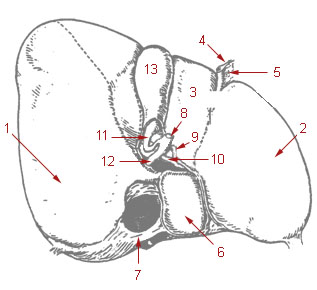
The cystic duct is the short duct that joins the gallbladder to the common hepatic duct. It usually lies next to the cystic artery. It is of variable length. It contains 'spiral valves of Heister', which do not provide much resistance to the flow of bile.
The common hepatic duct is the first part of the biliary tract. It joins the cystic duct coming from the gallbladder to form the common bile duct.
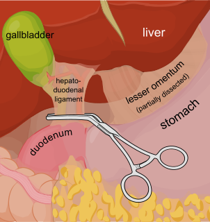
The Pringle manoeuvre is a surgical technique used in some abdominal operations and in liver trauma. The hepatoduodenal ligament is clamped either with a surgical tool called a haemostat, an umbilical tape or by hand. This limits blood inflow through the hepatic artery and the portal vein, controlling bleeding from the liver.

The lesser omentum is the double layer of peritoneum that extends from the liver to the lesser curvature of the stomach, and to the first part of the duodenum. The lesser omentum is usually divided into these two connecting parts: the hepatogastric ligament, and the hepatoduodenal ligament.

The hepatic artery proper is the artery that supplies the liver and gallbladder. It raises from the common hepatic artery, a branch of the celiac artery.

The cystic artery supplies oxygenated blood to the gallbladder and cystic duct.

Dr. Wu Mengchao, was a Chinese surgeon and a medical scientist who specialized in hepatobiliary surgery. He was also known as the "Father of Chinese Hepatobiliary Surgery".

The porta hepatis or transverse fissure of the liver is a short but deep fissure, about 5 cm long, extending transversely beneath the left portion of the right lobe of the liver, nearer its posterior surface than its anterior border.

The hepatoduodenal ligament is the portion of the lesser omentum extending between the porta hepatis of the liver and the superior part of the duodenum.

In histology, the lobules of liver, or hepatic lobules, are small divisions of the liver defined at the microscopic scale. The hepatic lobule is a building block of the liver tissue, consisting of a portal triad, hepatocytes arranged in linear cords between a capillary network, and a central vein.
The biliary tract, refers to the liver, gall bladder and bile ducts, and how they work together to make, store and secrete bile. Bile consists of water, electrolytes, bile acids, cholesterol, phospholipids and conjugated bilirubin. Some components are synthesised by hepatocytes, the rest are extracted from the blood by the liver.

A hepatoportoenterostomy or Kasai portoenterostomy is a surgical treatment performed on infants with Type IVb choledochal cyst and biliary atresia to allow for bile drainage. In these infants, the bile is not able to drain normally from the small bile ducts within the liver into the larger bile ducts that connect to the gall bladder and small intestine.
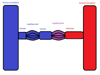
In the circulatory system of animals, a portal venous system occurs when a capillary bed pools into another capillary bed through veins, without first going through the heart. Both capillary beds and the blood vessels that connect them are considered part of the portal venous system.

Recurrent pyogenic cholangitis (RPC), also known as Hong Kong disease, Oriental cholangitis, and Oriental infestational cholangitis, is a chronic infection characterized by recurrent bouts of bacterial cholangitis with primary hepatolithiasis. It is exclusive to people who live or have lived in southeast Asia.

The liver is a major organ only found in vertebrates which performs many essential biological functions such as detoxification of the organism, and the synthesis of proteins and biochemicals necessary for digestion and growth. In humans, it is located in the right upper quadrant of the abdomen, below the diaphragm. Its other roles in metabolism include the regulation of glycogen storage, decomposition of red blood cells, and the production of hormones.

A liver segment is one of eight segments of the liver as described in the widely used Couinaud classification in the anatomy of the liver. This system divides the lobes of the liver into eight segments based on a transverse plane through the bifurcation of the main portal vein, arranged in a clockwise manner starting from the caudate lobe.

In human anatomy, the liver is divided grossly into four parts or lobes: the right lobe, the left lobe, the caudate lobe, and the quadrate lobe. Seen from the front – the diaphragmatic surface - the liver is divided into two lobes: the right lobe and the left lobe. Viewed from the underside – the visceral surface, the other two smaller lobes the caudate lobe, and the quadrate lobe are also visible. The two smaller lobes, the caudate lobe and the quadrate lobe, are known as superficial or accessory lobes, and both are located on the underside of the right lobe.
