Jaundice, also known as icterus, is a yellowish or greenish pigmentation of the skin and whites of the eyes due to high bilirubin levels. Jaundice in adults is typically a sign indicating the presence of underlying diseases involving abnormal heme metabolism, liver dysfunction, or biliary-tract obstruction. The prevalence of jaundice in adults is rare, while jaundice in babies is common, with an estimated 80% affected during their first week of life. The most commonly associated symptoms of jaundice are itchiness, pale feces, and dark urine.

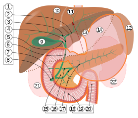
In vertebrates, the gallbladder, also known as the cholecyst, is a small hollow organ where bile is stored and concentrated before it is released into the small intestine. In humans, the pear-shaped gallbladder lies beneath the liver, although the structure and position of the gallbladder can vary significantly among animal species. It receives and stores bile, produced by the liver, via the common hepatic duct, and releases it via the common bile duct into the duodenum, where the bile helps in the digestion of fats.

A bile duct is any of a number of long tube-like structures that carry bile, and is present in most vertebrates.

Epithelium is one of the four basic types of animal tissue, along with connective tissue, muscle tissue and nervous tissue. It is a thin, continuous, protective layer of compactly packed cells with little intercellular matrix. Epithelial tissues line the outer surfaces of organs and blood vessels throughout the body, as well as the inner surfaces of cavities in many internal organs. An example is the epidermis, the outermost layer of the skin.

The common bile duct, sometimes abbreviated as CBD, is a duct in the gastrointestinal tract of organisms that have a gallbladder. It is formed by the confluence of the common hepatic duct and cystic duct and terminates by uniting with pancreatic duct, forming the ampulla of Vater. The flow of bile from the ampulla of Vater into the duodenum is under the control of the sphincter of Oddi.
The cystic duct is the short duct that joins the gallbladder to the common hepatic duct. It usually lies next to the cystic artery. It is of variable length. It contains 'spiral valves of Heister', which do not provide much resistance to the flow of bile.
The common hepatic duct is the first part of the biliary tract. It joins the cystic duct coming from the gallbladder to form the common bile duct.
Cholestasis is a condition where bile cannot flow from the liver to the duodenum. The two basic distinctions are an obstructive type of cholestasis where there is a mechanical blockage in the duct system that can occur from a gallstone or malignancy, and metabolic types of cholestasis which are disturbances in bile formation that can occur because of genetic defects or acquired as a side effect of many medications. Classification is further divided into acute or chronic and extrahepatic or intrahepatic.
Cholangiocytes are the epithelial cells of the bile duct. They are cuboidal epithelium in the small interlobular bile ducts, but become columnar and mucus secreting in larger bile ducts approaching the porta hepatis and the extrahepatic ducts. They contribute to hepatocyte survival by transporting bile acids.

Bile canaliculus is a thin tube that collects bile secreted by hepatocytes. The bile canaliculi empty into a series of progressively larger bile ductules and ducts, which eventually become common hepatic duct. The bile canaliculi empty directly into the Canals of Hering.

The lobules of liver, or hepatic lobules, are small divisions of the liver defined at the microscopic (histological) scale. The hepatic lobule is a building block of the liver tissue, consisting of a portal triad, hepatocytes arranged in linear cords between a capillary network, and a central vein.
The biliary tract, refers to the liver, gall bladder and bile ducts, and how they work together to make, store and secrete bile. Bile consists of water, electrolytes, bile acids, cholesterol, phospholipids and conjugated bilirubin. Some components are synthesised by hepatocytes, the rest are extracted from the blood by the liver.

Congenital hepatic fibrosis is an inherited fibrocystic liver disease associated with proliferation of interlobular bile ducts within the portal areas and fibrosis that do not alter hepatic lobular architecture. The fibrosis would affect resistance in portal veins leading to portal hypertension.

The canals of Hering, or intrahepatic bile ductules, are part of the outflow system of exocrine bile product from the liver. Liver stem cells are located in the canals of Hering.
The interlobular bile ducts carry bile in the liver between the Canals of Hering and the interlobar bile ducts. They are part of the interlobular portal triad and can be easily localized by looking for the much larger portal vein. The cells of the ducts are described as cuboidal epithelium with increasing amounts of connective tissue around it.
Ductopenia refers to a reduction in the number of ducts in an organ, in particular the absence of bile ducts of the expected size in the portal tract of the liver. It is the histological hallmark of vanishing bile duct syndrome. The most common cause of ductopenia is primary biliary cholangitis.

The liver is a major organ only found in vertebrates which performs many essential biological functions such as detoxification of the organism, and the synthesis of proteins and biochemicals necessary for digestion and growth. In humans, it is located in the right upper quadrant of the abdomen, below the diaphragm. Its other roles in metabolism include the regulation of glycogen storage, decomposition of red blood cells, and the production of hormones.

Technetium (99mTc) mebrofenin is a diagnostic radiopharmaceutical used for imaging of the liver and the gallbladder. Under the brand name Choletec it is available from Bracco Diagnostic. Supplied as a sterile kit of mebrofenin and dehydrated stannous fluoride. The vial is reconstituted with 1 to 5 mL up to 3.7 gigabecquerels (100 mCi) of sodium pertechnetate solution to form the final radio labeled 99mTc mebrofenin.
Liver cytology is the branch of cytology that studies the liver cells and its functions. The liver is a vital organ, in charge of almost all the body’s metabolism. Main liver cells are hepatocytes, Kupffer cells, and hepatic stellate cells; each one with a specific function.

Bilirubin glucuronide is a water-soluble reaction intermediate over the process of conjugation of indirect bilirubin. Bilirubin glucuronide itself belongs to the category of conjugated bilirubin along with bilirubin di-glucuronide. However, only the latter one is primarily excreted into the bile in the normal setting.