Cell sorting

Last updated

Cell sorting is the process through which a particular cell type is separated from others contained in a sample on the basis of its physical or biological properties, such as size, morphological parameters, viability and both extracellular and intracellular protein expression. The homogeneous cell population obtained after sorting can be used for a variety of applications including research, diagnosis, and therapy. [1]

Contents

Methods

Methods of cell sorting fall into two major categories: fluorescence activated cell sorting (FACS) and immunomagnetic cell sorting. [2] Due to many years of refinement and increased demand for cell separation however, researchers are working to develop microfluidic sorting devices that have many benefits in comparison to the main types of fluorescence-activated cell sorting and immunomagnetic cell sorting methods.

Fluorescence-activated

Diagram A: Fluorescence assisted cell sorting for negative selection. Fluorescence Assisted Cell Sorting (FACS) A.jpg
Diagram A: Fluorescence assisted cell sorting for negative selection.

Fluorescence-Activated Cell Sorting, is also known as flow cytometry cell sorting, or commonly known by the acronym FACS, which is a trademark of Becton Dickinson and Company. Fluorescence activated cell sorting utilizes flow cytometry to separate cells based on morphological parameters and the expression of multiple extracellular and intracellular proteins. This method allows multiparameter cell sorting and involves encapsulating cells into small liquid droplets which are selectively given electric charges and sorted by an external electric field. Fluorescence activated cell sorting has several systems that work together to achieve successful sorting of events of interest. These include fluidic, optical, and electrostatic systems. The fluidic system has to establish a precisely-timedbreak off from the liquid stream in small uniform droplets, so that droplets containing individual cells can then be deflected electrostatically [2] Based on the invention of Richard Sweet, [3] droplet formation of the liquid jet of a cell sorter is stabilized by vibrations of an ultrasonic transducer at the exit of the nozzle orifice. The disturbances grow exponentially and lead to break up of the jet in droplets with precise timing. A cell of interest that should be sorted is measured at the sensing zone and moves down the stream to the breakoff point. During the separation of the droplet with the cell in it from the intact liquid jet, a voltage pulse is given to the liquid jet so that droplets containing the cells of interest can be deflected in an electric field between two deflection plates for sorting. The droplets are then caught by collection tubes or vessels placed below the deflection plates. [2] Flow cytometry cell sorting yields very high specificity according to one or several surface markers, but one limitation is constituted by the number of cells that can be processed during a work-day. For this reason pre-enrichment of the population of interest by immunomagnetic cell sorting is often considered, especially when the target cells are comparatively rare and a large batch of cells must be processed. Moreover, flow cytometry cell sorters are complex instruments that are generally used only by well-trained staff in flow cytometry facilities or well-equipped laboratories and, since they are normally big in size, it is not always possible to place them inside a biological safety cabinet. Therefore, it is not always possible to ensure sample sterility and, since the fluidic systems can be cleaned but it is not single-use, there is the possibility of cross-contamination among samples. Another aspect to be considered is that droplet generation inside the instrument could lead to aerosol formation that are hazardous for the operator when using infectious samples. These last considerations are of particular importance when cell sorting is used for clinical applications, for example cell therapy and should therefore be performed under Good Manufacturing Practice (GMP) conditions. Researchers can use a variety of fluorescent dyes to design multi-color panels to achieve successful, simultaneous sorting of multiple, precisely defined cell-types. Diagram A shows fluorescence-activated cell sorting of negative cell selection (undesired group) and diagram B shows FACS of positive cell selection (desired group).

Diagram B: Fluorescence assisted cell sorting for positive selection. Fluorescence Assisted Cell Sorting (FACS) B.jpg
Diagram B: Fluorescence assisted cell sorting for positive selection.

Fluorescent Dyes in Cell Sorting

Fluorescent dyes can act very differently. Generally, a fluorescent dye will be excited by a light source (a laser) at a particular wavelength and emit light at a lower energy and longer wavelength. The most common dyes act by binding to antigens presented on cells. Common antigens targeted are clusters of differentiation (CDs). [4] These are specific to a certain type of cell. If you can identify which CD is presented on your cells of interest, then you can stain your sample with a fluorescent dye specific to it and use fluorescence-activated cell sorting to separate the population of interest. However, there are many other mechanisms by which fluorescent dyes can act.

Some dyes are able to diffuse across membranes. By taking advantage of this property of the dye, users can characterize intracellular activity as well as surface-expression of proteins. For example, in dead cells, propidium iodide (PI) can penetrate the nucleus where it binds to DNA. The fluorescent signal of PI can be used to quantify DNA content for cell cycle analysis or to identify dead cells in a sample.

Certain fluorescent dyes can be used to characterize kinetic intracellular activity rather than fixing cells in formaldehyde and losing viable cells. The table below outlines dyes that can be used to measure several parameters of cytotoxicity caused by oxidative stress.

DyeParameterMechanism of ActionExcitation/ Emission
DCFH-DAReactive Oxygen Species

(ROS)

Deacetylated to

2’7’ dichlorofluorescin which reacts with ROS under radical conditions to 2’7 dichlorofluorescein (DCF)

488 nm/525 nm
Rh123Mitochondria Membrane Potential

(MMP)

Sequestered by active mitochondria488 nm/525 nm
Indo-1 AMCalcium LevelsEmits at two different wavelengths depending on presence of calcium ions350 nm/[400 nm/485 nm]
PILive/DeadPermeates dead cells only and binds to DNA488 nm/675 nm

This experimental setup is just one example of the capability of flow cytometry. In FACS systems, these characterized cells can then be sorted and purified for further experiments.

Immunomagnetic cell sorting

MACS

Immunomagnetic cell sorting is also known as immunomagnetic cell separation, immunomagnetic cell enrichment, or magnetic-activated cell sorting, and commonly known by the acronym MACS which is a trademark of Miltenyi GmbH. Immunomagnetic cell sorting is based on separation of beads passing a magnetic field. A variety of companies offer different solutions for enrichment or depletion of cell populations. Immunomagnetic cell sorting provides a method for enriching a heterogeneous mixture of cells based on cell-surface protein expression (antigens). This technology is based on the attachment of small, inert, supra-magnetic particles to mAbs specific for antigens on the target cell population. Cells labelled to these antibody-bead conjugates are then separated via a column containing a ferromagnetic matrix. By applying a magnetic field to the matrix, the beads stick to the matrix inside the column and the bead-carrying cells are held back from passing through. Unlabelled cells can pass through the matrix and are collected in the flow-through. To elute the trapped cells from the column, the magnetic field is simply removed. Immunomagnetic cell sorting therefore enables different strategies for positive enrichment or depletion of cells. [2] Immunomagnetic beads are small and usually do not interfere with downstream assays, however for some applications it may be necessary to remove them . Using this separation method up-scaling the cell numbers does not significantly increase processing times and the sterility of the sample is guaranteed if the cell sorting is performed inside a biosafety cabinet. On the other hand, this technique allows to separate the cells based only on a single marker and it is not able to discriminate between different levels of protein expression (quantitative analysis). Immunomagnetic cell sorting has shown to be beneficial when used with NPC (neural progenitor cell) cultures in particular, as it is easier to manage and causes minimal damage to live cells. [5]

NPC cultures are especially difficult to work with because live brain cells are sensitive and tend to contaminate each other. [5] In order to get clearer results, labs need cleaner materials, meaning more pure NPC lines. [5] A study done in 2019 (with the funding support of New York Stem Cell Foundation and the Association for Frontotemporal Degeneration) found immunomagnetic cell sorting to be a cheap, simple way to yield such purity with minimal damage to the cell lines, therefore maintaining better quality cells, collecting more homogeneous NPCs, and increasing their chances of finding effective treatments for neurological disorders. [5] They used both the immunomagnetic and fluorescence-activated methods to filter out CD271- (useful markers for mesenchymal stem cells) and CD133+ (markers for cancer stem cells) to compare viability of each method. [5]

The immunomagnetic cell sorting has also been used in assistance with reproduction (artificial insemination) and retinal transplant treatment. [6] [7] In the case of assisting reproduction, apoptotic sperm cells (dead or damaged cells) are separated out so more non-apoptotic sperm (non-fragmented) cells can be collected and used to increase the subject's chances of fertility. [6] This type of treatment has shown to be more effective when done repeatedly, increasing the amount of non-apoptotic cells present during insemination. [6]

A 2018 study done in France (with the support of multiple individuals and agencies including: the Institut de la Vision in Paris and the Retina France Association) used rats and the immunomagnetic cell sorting method to show that photoreceptors (cells in the retina which respond to light) may be transplanted to cure blindness. [7] In this process, the microbeads were attached to the CD73 enzyme to assist in the separation of PRs (photoreceptors) from retinal organoids. [7] When a CD73+ antigen expressed itself with RCVRN+ cells (calcium-binding proteins in the eye), it showed researchers that this combination of CD73+ and RCVRN+ could be used with post-mitotic PR precursors for repair. [7] Although the study could not verify success in humans, they have the foundation for further research based on the success of pairing non-damaged photoreceptors with a CD73 antigen and the transplantation in rats. [7] This success in cell separation and pairing through transplantation shows promise for a potential cure for retinal diseases including total blindness. So far, only partial vision repair has been reported. [7]

Microfluidic devices

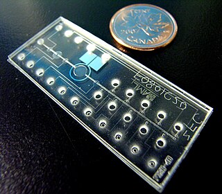
Due to various limitations of fluorescence-activated and immunomagnetic cell sorting devices, a wide range of microfluidic cell-sorting devices have emerged. A few of these are now commercially available or in commercial development. Research into microfluidic cell sorter designs often employ soft lithography techniques utilizing materials such as polydimethylsiloxane (PDMS).

A key benefit of microfluidic sorters is the potential to perform fluorescence-activated cell sorting in a closed single-use sterile cartridge. Such a closed cartridge would prevent the exposure of an operator to biohazards through the droplets that are emitted by FACS systems. Other benefits include reduced impact on cell viability due to reduced hydrodynamic stress on the cells; Some published devices show the potential for multi-way sorting, decreased cost of a cartridge due to low-cost manufacturing methods, lower power consumption, and smaller-sized footprints, with some devices being the size of a credit card. Some have achieved high purity outputs and rates of up to around 50,000 cells/s. [8] [9] [10] [11]

Microfluidic cell sorters can be divided into two categories: active and passive. Active devices deflect individual cells by the cytometric measurements of the cells, made in real time. Passive devices exploit physical differences between cells in how they interact with the fluid flow or surfaces.

Active

Active microfluidic cell sorters involve the deflection of individual cells following their measurement using cytometric methods, including fluorescent labelling, light scatter and image analysis. Individual cells are deflected by either a force directly on the cell or a force on the fluid surrounding the cells, so that they flow into separate output vessels.

Methods of cell deflection employ several kinds of macroscopic, optical or MEMS (micro-electro-mechanical systems) actuators to deflect a particle or liquid volume within a microchannel. Notable recent examples are based on surface acoustic wave actuators, [12] [13] [14] [15] [16] [17] macroscopic actuators (such as piezoelectric actuators) coupled to microchannels, [18] [19] [20] [21] dielectrophoresis of droplets, [22] thermal vapor bubble actuators, [23] [24] [25] [26] [27] transient micro-vortices generated by thermal vapour bubble actuators, [28] optical manipulation, [29] and micro-mechanical valves. The fastest of these have demonstrated sort rates in excess of 1000/s and potential maximum throughput rates in some cases approaching those of FACS. [30] [31] [32] [33] [34] [35] Active microfluidic cell sorting requires similar cytometry instrumentation as fluorescence-activated cell sorting described above. [36]

Active microfluidic cells sorters have the potential of throughput scaling by parallelisation on chip. [37] The fastest published active microfluidic sorting device has demonstrated a 160,000/s throughput [38]

Passive

Passive cell sorting uses the behavior of the fluid within the microchannels to alter and separate cells based on size and morphology. [39] The fluid in a colloidal solution is subject to a velocity profile due to the interactions of the fluid with the walls of the channel; the cells in the solution are subject to various drag and inertial forces that are dependent on the size of the cell and balance accordingly at different locations along the velocity profile. [40] In curved microfluidic channels, vortices are formed due to the Dean force which locate different sized particles in different cross sectional locations due to the Reynolds number and curve radius of curvature. [41]

For example, in a straight channel, larger cells in colloidal solution are found closer to the center of the microchannel than smaller cells due to the larger drag forces from the wall that pushes the cell away from the wall and the shear gradient force from the velocity profile that balances this wall drag force to set the cell into equilibrium. [42]

Other antibody-based methods of cell separation

Several methods of cell separation and enrichment using antibodies were employed before fluorescence-based and immunomagnetic cell sorting gained popularity. [43] These included antibody- and complement-mediated cell separation, polystyrene immunoaffinity devices, and the CellPro CEPRATE® SC System which employed immobilised antibodies in a porous column. The latter was the first FDA-approved medical device for the separation of hematopoietic stem cells.

A newer cell separation technique employing antibodies is buoyancy-activated cell sorting (BACS) is a separation technique in which microbubbles bind to cells through antibodies binding to the surface of cells. The targeted cells are then removed from a biological sample through flotation. [44]

Single-cell sorting

Single-cell sorting provides a method for sorting a heterogeneous mixture of cells based on intracellular and extracellular properties. Single-cell methods enable understanding of cellular properties that may be obscured or non-evident. There are several methods for sorting single cells:

The microraft array provides a rapid, cost-effective method for isolating cells, analyzing cells over time, and generating clonal populations with the unique ability to monitor all intra- and extracellular properties. [45] This system is ideal for both adherent and non-adherent cell types.

A single-cell method to observe the response to an external stimulus (in this case, cellular response to a ligand) was studied using a microfluidic device with micro-channel valves to trap a single cell in a chamber. A 23-valve system was used for actuation, and fluorescent dye was used in stimulus-response imaging. [46]

Single-cell clustering methods are a series of statistical methods designed by data scientists based on intracellular properties. The process includes Single Cell RNA-Seq data gathering; data preprocessing for clustering; clustering; and evaluations of clustering. Scientists apply machine-learning methods (mainly clustering analysis) on the single-cell RNA-Seq data to divide the cells into different categories. All the methods are modified to resolve the problems in RNA-Seq data such as dropout of low-expression genes and ambiguous cell markers in the presence of technical biases. State-of-the-art methods include SC3., [47] CIDR., [48] Seurat and for more detailed information, please refer to the Wiki page: Single Cell RNA-Seq Clustering.

Related Research Articles

Microfluidics refers to a system that manipulates a small amount of fluids using small channels with sizes ten to hundreds micrometres. It is a multidisciplinary field that involves molecular analysis, molecular biology, and microelectronics. It has practical applications in the design of systems that process low volumes of fluids to achieve multiplexing, automation, and high-throughput screening. Microfluidics emerged in the beginning of the 1980s and is used in the development of inkjet printheads, DNA chips, lab-on-a-chip technology, micro-propulsion, and micro-thermal technologies.

<span class="mw-page-title-main">Digital microfluidics</span>

Digital microfluidics (DMF) is a platform for lab-on-a-chip systems that is based upon the manipulation of microdroplets. Droplets are dispensed, moved, stored, mixed, reacted, or analyzed on a platform with a set of insulated electrodes. Digital microfluidics can be used together with analytical analysis procedures such as mass spectrometry, colorimetry, electrochemical, and electrochemiluminescense.

<span class="mw-page-title-main">Surface acoustic wave</span> Sound wave which travels along the surface of an elastic material

A surface acoustic wave (SAW) is an acoustic wave traveling along the surface of a material exhibiting elasticity, with an amplitude that typically decays exponentially with depth into the material, such that they are confined to a depth of about one wavelength.

<span class="mw-page-title-main">Flow cytometry</span> Lab technique in biology and chemistry

Flow cytometry (FC) is a technique used to detect and measure physical and chemical characteristics of a population of cells or particles.

A lab-on-a-chip (LOC) is a device that integrates one or several laboratory functions on a single integrated circuit of only millimeters to a few square centimeters to achieve automation and high-throughput screening. LOCs can handle extremely small fluid volumes down to less than pico-liters. Lab-on-a-chip devices are a subset of microelectromechanical systems (MEMS) devices and sometimes called "micro total analysis systems" (µTAS). LOCs may use microfluidics, the physics, manipulation and study of minute amounts of fluids. However, strictly regarded "lab-on-a-chip" indicates generally the scaling of single or multiple lab processes down to chip-format, whereas "µTAS" is dedicated to the integration of the total sequence of lab processes to perform chemical analysis.

Chemical biology is a scientific discipline between the fields of chemistry and biology. The discipline involves the application of chemical techniques, analysis, and often small molecules produced through synthetic chemistry, to the study and manipulation of biological systems. In contrast to biochemistry, which involves the study of the chemistry of biomolecules and regulation of biochemical pathways within and between cells, chemical biology deals with chemistry applied to biology.

<span class="mw-page-title-main">Plasma cleaning</span>

Plasma cleaning is the removal of impurities and contaminants from surfaces through the use of an energetic plasma or dielectric barrier discharge (DBD) plasma created from gaseous species. Gases such as argon and oxygen, as well as mixtures such as air and hydrogen/nitrogen are used. The plasma is created by using high frequency voltages to ionise the low pressure gas, although atmospheric pressure plasmas are now also common.

<span class="mw-page-title-main">Bio-MEMS</span>

Bio-MEMS is an abbreviation for biomedical microelectromechanical systems. Bio-MEMS have considerable overlap, and is sometimes considered synonymous, with lab-on-a-chip (LOC) and micro total analysis systems (μTAS). Bio-MEMS is typically more focused on mechanical parts and microfabrication technologies made suitable for biological applications. On the other hand, lab-on-a-chip is concerned with miniaturization and integration of laboratory processes and experiments into single chips. In this definition, lab-on-a-chip devices do not strictly have biological applications, although most do or are amenable to be adapted for biological purposes. Similarly, micro total analysis systems may not have biological applications in mind, and are usually dedicated to chemical analysis. A broad definition for bio-MEMS can be used to refer to the science and technology of operating at the microscale for biological and biomedical applications, which may or may not include any electronic or mechanical functions. The interdisciplinary nature of bio-MEMS combines material sciences, clinical sciences, medicine, surgery, electrical engineering, mechanical engineering, optical engineering, chemical engineering, and biomedical engineering. Some of its major applications include genomics, proteomics, molecular diagnostics, point-of-care diagnostics, tissue engineering, single cell analysis and implantable microdevices.

<span class="mw-page-title-main">Cytometry</span> Measurement of number and characteristics of cells

Cytometry is the measurement of number and characteristics of cells. Variables that can be measured by cytometric methods include cell size, cell count, cell morphology, cell cycle phase, DNA content, and the existence or absence of specific proteins on the cell surface or in the cytoplasm. Cytometry is used to characterize and count blood cells in common blood tests such as the complete blood count. In a similar fashion, cytometry is also used in cell biology research and in medical diagnostics to characterize cells in a wide range of applications associated with diseases such as cancer and AIDS.

<span class="mw-page-title-main">Centrifugal micro-fluidic biochip</span>

The centrifugal micro-fluidic biochip or centrifugal micro-fluidic biodisk is a type of lab-on-a-chip technology, also known as lab-on-a-disc, that can be used to integrate processes such as separating, mixing, reaction and detecting molecules of nano-size in a single piece of platform, including a compact disk or DVD. This type of micro-fluidic biochip is based upon the principle of microfluidics; to take advantage of noninertial pumping for lab-on-a-chip devices using noninertial valves and switches under centrifugal force and Coriolis effect to distribute fluids about the disks in a highly parallel order.

<span class="mw-page-title-main">Single-cell analysis</span> Testbg biochemical processes and reactions in an individual cell

In the field of cellular biology, single-cell analysis and subcellular analysis is the study of genomics, transcriptomics, proteomics, metabolomics and cell–cell interactions at the single cell level. The concept of single-cell analysis originated in the 1970s. Before the discovery of heterogeneity, single-cell analysis mainly referred to the analysis or manipulation of an individual cell in a bulk population of cells at a particular condition using optical or electronic microscope. To date, due to the heterogeneity seen in both eukaryotic and prokaryotic cell populations, analyzing a single cell makes it possible to discover mechanisms not seen when studying a bulk population of cells. Technologies such as fluorescence-activated cell sorting (FACS) allow the precise isolation of selected single cells from complex samples, while high throughput single cell partitioning technologies, enable the simultaneous molecular analysis of hundreds or thousands of single unsorted cells; this is particularly useful for the analysis of transcriptome variation in genotypically identical cells, allowing the definition of otherwise undetectable cell subtypes. The development of new technologies is increasing our ability to analyze the genome and transcriptome of single cells, as well as to quantify their proteome and metabolome. Mass spectrometry techniques have become important analytical tools for proteomic and metabolomic analysis of single cells. Recent advances have enabled quantifying thousands of protein across hundreds of single cells, and thus make possible new types of analysis. In situ sequencing and fluorescence in situ hybridization (FISH) do not require that cells be isolated and are increasingly being used for analysis of tissues.

Acoustic tweezers are a set of tools that use sound waves to manipulate the position and movement of very small objects. Strictly speaking, only a single-beam based configuration can be called acoustical tweezers. However, the broad concept of acoustical tweezers involves two configurations of beams: single beam and standing waves. The technology works by controlling the position of acoustic pressure nodes that draw objects to specific locations of a standing acoustic field. The target object must be considerably smaller than the wavelength of sound used, and the technology is typically used to manipulate microscopic particles.

Optoelectrowetting (OEW) is a method of liquid droplet manipulation used in microfluidics applications. This technique builds on the principle of electrowetting, which has proven useful in liquid actuation due to fast switching response times and low power consumption. Where traditional electrowetting runs into challenges, however, such as in the simultaneous manipulation of multiple droplets, OEW presents a lucrative alternative that is both simpler and cheaper to produce. OEW surfaces are easy to fabricate, since they require no lithography, and have real-time, reconfigurable, large-scale manipulation control, due to its reaction to light intensity.

Microfluidic cell culture integrates knowledge from biology, biochemistry, engineering, and physics to develop devices and techniques for culturing, maintaining, analyzing, and experimenting with cells at the microscale. It merges microfluidics, a set of technologies used for the manipulation of small fluid volumes within artificially fabricated microsystems, and cell culture, which involves the maintenance and growth of cells in a controlled laboratory environment. Microfluidics has been used for cell biology studies as the dimensions of the microfluidic channels are well suited for the physical scale of cells. For example, eukaryotic cells have linear dimensions between 10 and 100 μm which falls within the range of microfluidic dimensions. A key component of microfluidic cell culture is being able to mimic the cell microenvironment which includes soluble factors that regulate cell structure, function, behavior, and growth. Another important component for the devices is the ability to produce stable gradients that are present in vivo as these gradients play a significant role in understanding chemotactic, durotactic, and haptotactic effects on cells.

Droplet-based microfluidics manipulate discrete volumes of fluids in immiscible phases with low Reynolds number and laminar flow regimes. Interest in droplet-based microfluidics systems has been growing substantially in past decades. Microdroplets offer the feasibility of handling miniature volumes of fluids conveniently, provide better mixing, encapsulation, sorting, sensing and are suitable for high throughput experiments. Two immiscible phases used for the droplet based systems are referred to as the continuous phase and dispersed phase.

Paper-based microfluidics are microfluidic devices that consist of a series of hydrophilic cellulose or nitrocellulose fibers that transport fluid from an inlet through the porous medium to a desired outlet or region of the device, by means of capillary action. This technology builds on the conventional lateral flow test which is capable of detecting many infectious agents and chemical contaminants. The main advantage of this is that it is largely a passively controlled device unlike more complex microfluidic devices. Development of paper-based microfluidic devices began in the early 21st century to meet a need for inexpensive and portable medical diagnostic systems.

Open microfluidics can be employed in the multidimensional culturing of cell types for various applications including organ-on-a-chip studies, oxygen-driven reactions, neurodegeneration, cell migration, and other cellular pathways.

FAST is a small, genetically-encoded, protein tag which allows for fluorescence reporting of proteins of interest. Unlike natural fluorescent proteins and derivates such as GFP or mCherry, FAST is not fluorescent by itself. It can bind selectively a fluorogenic chromophore derived from 4-hydroxybenzylidene rhodanine (HBR), which is itself non fluorescent unless bound. Once bound, the pair of molecules goes through a unique fluorogen activation mechanism based on two spectroscopic changes, increase of fluorescence quantum yield and absorption red shift, hence providing high labeling selectivity. The FAST-fluorogen reporting system can be used in fluorescence microscopy, flow cytometry and any other fluorometric method to explore the living world: biosensors, protein trafficking.

Jonathan Cooper is Professor of Engineering in the College of Science & Engineering at the University of Glasgow. Professor Cooper has held the Wolfson Chair in Bioengineering at the school since 2009.

Cell biomechanics a branch of biomechanics that involves single molecules, molecular interactions, or cells as the system of interest. Cells generate and maintain mechanical forces within their environment as a part of their physiology. Cell biomechanics deals with how mRNA, protein production, and gene expression is affected by said environment and with mechanical properties of isolated molecules or interaction of proteins that make up molecular motors.

References

  1. Steven A Soper, Malgorzata A Witek (7 January 2020). "Cell Separations and Sorting". Anal Chem. 92 (1): 105–131. doi:10.1021/acs.analchem.9b05357. PMC   7469080 . PMID   31808677.
  2. 1 2 3 4 Arturo Zychlinsky, Andrea Cossarizza (October 2019). "Guidelines for the use of flow cytometry and cell sorting in immunological studies (second edition)". Eur J Immunol. 49 (10): 1457–1973. doi:10.1002/eji.201970107. PMC   7350392 . PMID   31633216.
  3. Richard G Sweet, Richard G Sweet. "Fluid droplet recorder".
  4. "Human CD & Other Cellular Antigens - US". www.thermofisher.com. Retrieved 2018-12-11.
  5. 1 2 3 4 5 Alison M Goate, Kathryn R Bowles (27 March 2019). "Reduced variability of neural progenitor cells and improved purity of neuronal cultures using magnetic activated cell sorting". PLOS ONE. 14 (3): e0213374. doi: 10.1371/journal.pone.0213374 . PMC   6436701 . PMID   30917153.
  6. 1 2 3 Dirican, Enver Kerem (2012), "Magnetic-Activated Cell Sorting of Human Spermatozoa", Practical Manual of in Vitro Fertilization, Springer New York, pp. 265–272, doi:10.1007/978-1-4419-1780-5_29, ISBN   9781441917799
  7. 1 2 3 4 5 6 Gagliardi, Giuliana; Ben M'Barek, Karim; Chaffiol, Antoine; Slembrouck-Brec, Amélie; Conart, Jean-Baptiste; Nanteau, Céline; Rabesandratana, Oriane; Sahel, José-Alain; Duebel, Jens; Orieux, Gael; Reichman, Sacha (September 2018). "Characterization and Transplantation of CD73-Positive Photoreceptors Isolated from Human iPSC-Derived Retinal Organoids". Stem Cell Reports. 11 (3): 665–680. doi:10.1016/j.stemcr.2018.07.005. PMC   6135113 . PMID   30100409.
  8. Gossett DR, Weaver WM, Mach AJ, Hur SC, Tse HT, Lee W, Amini H, Di Carlo D (August 2010). "Label-free cell separation and sorting in microfluidic systems". Analytical and Bioanalytical Chemistry. 397 (8): 3249–67. doi:10.1007/s00216-010-3721-9. PMC   2911537 . PMID   20419490.
  9. Hulspas R, Villa-Komaroff L, Koksal E, Etienne K, Rogers P, Tuttle M, Korsgren O, Sharpe JC, Berglund D (October 2014). "Purification of regulatory T cells with the use of a fully enclosed high-speed microfluidic system". Cytotherapy. 16 (10): 1384–9. doi:10.1016/j.jcyt.2014.05.016. PMID   25065635.
  10. Shields CW, Reyes CD, López GP (March 2015). "Microfluidic cell sorting: a review of the advances in the separation of cells from debulking to rare cell isolation". Lab on a Chip. 15 (5): 1230–49. doi:10.1039/C4LC01246A. PMC   4331226 . PMID   25598308.
  11. Ding X, Lin SC, Lapsley MI, Li S, Guo X, Chan CY, Chiang IK, Wang L, McCoy JP, Huang TJ (November 2012). "Standing surface acoustic wave (SSAW) based multichannel cell sorting". Lab on a Chip. 12 (21): 4228–31. doi:10.1039/C2LC40751E. PMC   3956451 . PMID   22992833.
  12. Ding X, Lin SC, Lapsley MI, Li S, Guo X, Chan CY, Chiang IK, Wang L, McCoy JP, Huang TJ (November 2012). "Standing surface acoustic wave (SSAW) based multichannel cell sorting". Lab on a Chip. 12 (21): 4228–31. doi:10.1039/c2lc40751e. PMC   3956451 . PMID   22992833.
  13. Schmid, L., Weitz, D. A., Franke, T. (7 October 2014). "Sorting drops and cells with acoustics: acoustic microfluidic fluorescence-activated cell sorter". Lab on a Chip. 14 (19): 3710–3718. doi:10.1039/c4lc00588k. ISSN   1473-0189. PMID   25031157.
  14. Ma, Z., Zhou, Y., Collins, D. J., Ai, Y. (12 September 2017). "Fluorescence activated cell sorting via a focused traveling surface acoustic beam". Lab on a Chip. 17 (18): 3176–3185. doi:10.1039/C7LC00678K. ISSN   1473-0189. PMID   28815231.
  15. Ren, L., Chen, Y., Li, P., Mao, Z., Huang, P.-H., Rufo, J., Guo, F., Wang, L., McCoy, J. P., Levine, S. J., Huang, T. J. (7 October 2015). "A high-throughput acoustic cell sorter". Lab on a Chip. 15 (19): 3870–3879. doi:10.1039/c5lc00706b. ISSN   1473-0189. PMC   4641751 . PMID   26289231.
  16. Schmid L, Weitz DA, Franke T (October 2014). "Sorting drops and cells with acoustics: acoustic microfluidic fluorescence-activated cell sorter". Lab on a Chip. 14 (19): 3710–8. doi:10.1039/c4lc00588k. PMID   25031157. S2CID   9893236.
  17. Ren L, Chen Y, Li P, Mao Z, Huang PH, Rufo J, Guo F, Wang L, McCoy JP, Levine SJ, Huang TJ (October 2015). "A high-throughput acoustic cell sorter". Lab on a Chip. 15 (19): 3870–3879. doi:10.1039/c5lc00706b. PMC   4641751 . PMID   26289231.
  18. Cho, S. H., Chen, C. H., Tsai, F. S., Godin, J. M., Lo, Y.-H. (21 June 2010). "Human mammalian cell sorting using a highly integrated micro-fabricated fluorescence-activated cell sorter (μFACS)". Lab on a Chip. 10 (12): 1567–1573. doi:10.1039/c000136h. ISSN   1473-0197. PMC   3118392 . PMID   20379604.
  19. Lee, C., Lee, J., Kim, H. H., Teh, S.-Y., Lee, A., Chung, I.-Y., Park, J. Y., Shung, K. K. (7 August 2012). "Microfluidic droplet sorting with a high frequency ultrasound beam". Lab on a Chip. 12 (15): 2736–2742. doi:10.1039/c2lc21123h. ISSN   1473-0189. PMC   3400154 . PMID   22643737.
  20. Hulspas, R., Villa-Komaroff, L., Koksal, E., Etienne, K., Rogers, P., Tuttle, M., Korsgren, O., Sharpe, J. C., Berglund, D. (October 2014). "Purification of regulatory T cells with the use of a fully enclosed high-speed microfluidic system". Cytotherapy. 16 (10): 1384–1389. doi:10.1016/j.jcyt.2014.05.016. ISSN   1465-3249. PMID   25065635.
  21. Cho SH, Chen CH, Tsai FS, Godin JM, Lo YH (June 2010). "Human mammalian cell sorting using a highly integrated micro-fabricated fluorescence-activated cell sorter (microFACS)". Lab on a Chip. 10 (12): 1567–73. doi:10.1039/c000136h. PMC   3118392 . PMID   20379604.
  22. Agresti JJ, Antipov E, Abate AR, Ahn K, Rowat AC, Baret JC, Marquez M, Klibanov AM, Griffiths AD, Weitz DA (March 2010). "Ultrahigh-throughput screening in drop-based microfluidics for directed evolution". Proceedings of the National Academy of Sciences of the United States of America. 107 (9): 4004–9. Bibcode:2010PNAS..107.4004A. doi: 10.1073/pnas.0910781107 . PMC   2840095 . PMID   20142500.
  23. Chen Y, Chung AJ, Wu TH, Teitell MA, Di Carlo D, Chiou PY (May 2014). "Pulsed laser activated cell sorting with three dimensional sheathless inertial focusing". Small. 10 (9): 1746–51. doi:10.1002/smll.201302885. PMC   4324602 . PMID   24536017.
  24. Wu, T.-H., Chen, Y., Park, S.-Y., Hong, J., Teslaa, T., Zhong, J. F., Di Carlo, D., Teitell, M. A., Chiou, P.-Y. (2012). "Pulsed laser triggered high speed microfluidic fluorescence activated cell sorter". Lab on a Chip. 12 (7): 1378–1383. doi:10.1039/c2lc21084c. ISSN   1473-0197. PMC   3965373 . PMID   22361780.
  25. Hoefemann, H., Wadle, S., Bakhtina, N., Kondrashov, V., Wangler, N., Zengerle, R. (June 2012). "Sorting and lysis of single cells by BubbleJet technology". Sensors and Actuators B: Chemical. 168: 442–445. doi:10.1016/j.snb.2012.04.005. ISSN   0925-4005.
  26. Wijs, K. de, Liu, C., Dusa, A., Vercruysse, D., Majeed, B., Tezcan, D. S., Blaszkiewicz, K., Loo, J., Lagae, L. (2017). "Micro vapor bubble jet flow for safe and high-rate fluorescence-activated cell sorting". Lab Chip. 17 (7): 1287–1296. doi:10.1039/C6LC01560C. ISSN   1473-0197. PMID   28252674 . Retrieved 14 March 2017.
  27. Chen, C. C., Wang, J. S., Solgaard, O. (October 2006). "Micromachined bubble-jet cell sorter with multiple operation modes". Sensors and Actuators B: Chemical. 117 (2): 523–529. doi:10.1016/j.snb.2006.05.011. ISSN   0925-4005.
  28. Pritchard, R. H., Zhukov, A. A., Fullerton, J. N., Want, A. J., Hussain, F., Cour, M. F. la, Bashtanov, M. E., Gold, R. D., Hailes, A., Banham-Hall, E., Rogers, S. S. (18 June 2019). "Cell sorting actuated by a microfluidic inertial vortex". Lab on a Chip. 19 (14): 2456–2465. doi: 10.1039/C9LC00120D . ISSN   1473-0189. PMID   31210196. S2CID   190538792 . Retrieved 20 June 2019.
  29. Chen Y, Chung AJ, Wu TH, Teitell MA, Di Carlo D, Chiou PY (May 2014). "Pulsed laser activated cell sorting with three dimensional sheathless inertial focusing". Small. 10 (9): 1746–51. doi:10.1002/smll.201302885. PMC   4324602 . PMID   24536017.
  30. Wijs, K. de, Liu, C., Dusa, A., Vercruysse, D., Majeed, B., Tezcan, D. S., Blaszkiewicz, K., Loo, J., Lagae, L. (2017). "Micro vapor bubble jet flow for safe and high-rate fluorescence-activated cell sorting". Lab Chip. 17 (7): 1287–1296. doi:10.1039/C6LC01560C. ISSN   1473-0197. PMID   28252674 . Retrieved 14 March 2017.
  31. Ma, Z., Zhou, Y., Collins, D. J., Ai, Y. (12 September 2017). "Fluorescence activated cell sorting via a focused traveling surface acoustic beam". Lab on a Chip. 17 (18): 3176–3185. doi:10.1039/C7LC00678K. ISSN   1473-0189. PMID   28815231.
  32. Wu, T.-H., Chen, Y., Park, S.-Y., Hong, J., Teslaa, T., Zhong, J. F., Di Carlo, D., Teitell, M. A., Chiou, P.-Y. (2012). "Pulsed laser triggered high speed microfluidic fluorescence activated cell sorter". Lab on a Chip. 12 (7): 1378–1383. doi:10.1039/c2lc21084c. ISSN   1473-0197. PMC   3965373 . PMID   22361780.
  33. Cho, S. H., Chen, C. H., Tsai, F. S., Godin, J. M., Lo, Y.-H. (21 June 2010). "Human mammalian cell sorting using a highly integrated micro-fabricated fluorescence-activated cell sorter (μFACS)". Lab on a Chip. 10 (12): 1567–1573. doi:10.1039/c000136h. ISSN   1473-0197. PMC   3118392 . PMID   20379604.
  34. Schmid, L., Weitz, D. A., Franke, T. (7 October 2014). "Sorting drops and cells with acoustics: acoustic microfluidic fluorescence-activated cell sorter". Lab on a Chip. 14 (19): 3710–3718. doi:10.1039/c4lc00588k. ISSN   1473-0189. PMID   25031157.
  35. Ren, L., Chen, Y., Li, P., Mao, Z., Huang, P.-H., Rufo, J., Guo, F., Wang, L., McCoy, J. P., Levine, S. J., Huang, T. J. (7 October 2015). "A high-throughput acoustic cell sorter". Lab on a Chip. 15 (19): 3870–3879. doi:10.1039/c5lc00706b. ISSN   1473-0189. PMC   4641751 . PMID   26289231.
  36. Shields CW, Ohiri KA, Szott LM, López GP (March 2017). "Translating microfluidics: Cell separation technologies and their barriers to commercialization". Cytometry Part B. 92 (2): 115–125. doi:10.1002/cyto.b.21388. PMC   5149119 . PMID   27282966.
  37. Hulspas, R., Villa-Komaroff, L., Koksal, E., Etienne, K., Rogers, P., Tuttle, M., Korsgren, O., Sharpe, J. C., Berglund, D. (October 2014). "Purification of regulatory T cells with the use of a fully enclosed high-speed microfluidic system". Cytotherapy. 16 (10): 1384–1389. doi:10.1016/j.jcyt.2014.05.016. ISSN   1465-3249. PMID   25065635.
  38. Zhukov, A. A., Pritchard, R. H., Withers, M. J., Hailes, T., Gold, R. D., Hayes, C., Cour, M. F. la, Hussein, F., Rogers, S. S. (April 2021). "Extremely High-Throughput Parallel Microfluidic Vortex-Actuated Cell Sorting". Micromachines. Multidisciplinary Digital Publishing Institute. 12 (4): 389. doi: 10.3390/mi12040389 . ISSN   2072-666X. PMC   8066247 . PMID   33918161.
  39. Zhou J, Kasper S, Papautsky I (November 2013). "Enhanced size-dependent trapping of particles using microvortices". Microfluidics and Nanofluidics. 15 (5): 611–623. doi:10.1007/s10404-013-1176-y. PMC   3810988 . PMID   24187531.
  40. Martel JM, Toner M (July 2014). "Inertial focusing in microfluidics". Annual Review of Biomedical Engineering. 16 (1): 371–96. doi:10.1146/annurev-bioeng-121813-120704. PMC   4467210 . PMID   24905880.
  41. Martel JM, Toner M (2013-11-26). "Particle Focusing in Curved Microfluidic Channels". Scientific Reports. 3: 3340. Bibcode:2013NatSR...3E3340M. doi: 10.1038/srep03340 .
  42. Geislinger TM, Franke T (June 2014). "Hydrodynamic lift of vesicles and red blood cells in flow--from Fåhræus & Lindqvist to microfluidic cell sorting". Advances in Colloid and Interface Science. 208: 161–76. doi:10.1016/j.cis.2014.03.002. PMID   24674656.
  43. Recktenwald, D. (4 November 1997). Cell Separation Methods and Applications. CRC Press. ISBN   978-0-8247-9864-2.
  44. "Cell Separation Terminology, Uses, Methods and Technologies". Akadeum Life Sciences. Retrieved 2022-05-04.
  45. "The Isoraft System" (website). Cell Microsystems. Retrieved 2013-03-19.
  46. Tan SJ, Kee MZ, Mathuru AS, Burkholder WF, Jesuthasan SJ (2013-11-08). "A microfluidic device to sort cells based on dynamic response to a stimulus". PLOS ONE. 8 (11): e78261. Bibcode:2013PLoSO...878261T. doi: 10.1371/journal.pone.0078261 . PMC   3826715 . PMID   24250795.
  47. Kiselev V, Kirschner K, Schaub M, et al. (2017). "SC3: consensus clustering of single-cell RNA-seq data". Nature Methods. 483 (486): 483–486. doi:10.1038/nmeth.4236. PMC   5410170 . PMID   28346451.
  48. Lin P, Troup M, Ho JW (2017). "CIDR: Ultrafast and accurate clustering through imputation for single-cell RNA-seq data". Genome Biol. 18 (59): 59. doi: 10.1186/s13059-017-1188-0 . PMC   5371246 . PMID   28351406.