Cerebrospinal fluid flow MRI

Last updated

Cerebrospinal fluid (CSF) flow MRI is used to assess pulsatile CSF flow both qualitatively and quantitatively. Time-resolved 2D phase-contrast MRI with velocity encoding is the most common method for CSF analysis. [1] CSF Fluid Flow MRI detects back and forth flow of Cerebrospinal fluid that corresponds to vascular pulsations from mostly the cardiac cycle of the choroid plexus. Bulk transport of CSF, characterized by CSF circulation through the Central Nervous System, is not used because it is too slow to assess clinically. [2] CSF would have to pass through the brain's lymphatic system and be absorbed by arachnoid granulations.

Contents

Cerebrospinal fluid (CSF)

CSF is a clear fluid that surrounds the brain and spinal cord. [3] The rate of CSF formation in humans is about 0.3–0.4 ml per minute and the total CSF volume is 90–150 ml in adults. [2]

Traditionally, CSF was evaluated mainly using invasive procedures such as lumbar puncture, myelographies, radioisotope studies, and intracranial pressure monitoring. Recently, rapid advances in imaging techniques have provided non-invasive methods for flow assessment. One of the best-known methods is Phase-Contrast MRI and it is the only imaging modality for both qualitative and quantitative evaluation. The constant progress of magnetic resonance sequences gives a new opportunity to develop new applications and enhance unknown mechanisms of CSF flow. [4]

Phase contrast MRI

The study of CSF flow became one of Phase-contrast MRI's major applications. The key to Phase-contrast MRI (PC-MRI) is the use of a bipolar gradient. [4] A bipolar gradient has equal positive and negative magnitudes that are applied for the same time duration. The bipolar gradient in PC-MRI is put in a sequence after RF excitation but before data collection during the echo time of the generic MRI modality. The bipolar lobe must be applied in all three axes to image flow in all three directions.

Bipolar gradient

The basis of the bipolar gradient in PC-MRI is that when using this gradient to change frequencies, there will be no phase shift for the stationary protons because they will experience equal positive and negative magnitudes. [4] However, the moving protons will undergo various degrees of phase shift because, along the gradient direction, their locations are constantly changing. This notion can be applied to monitor protons that are moving through a plane. From the phase contrast, the floating protons can be detected. In the equation for determining the phase, local susceptibility influence is not removed by this bipolar gradient. Thus, it is necessary to invert a second sequence with the bipolar gradient, and the signal must be subtracted from the original acquisition. The purpose of this step is to cancel out those static areas’ signals and produce the characteristic static appearance at phase-contrast imaging.

where = phase shift, = gyromagnetic ratio, is the proton velocity, and is the change in magnetic moment

Equation 1. This is used to calculate phase shift, which is directly proportional to the gradient strength according to the change in magnetic moment. [5]

In phase-contrast imaging, there is a direct correlation between the degree of phase shift and the proton velocity in the direction of the gradient. However, because of the limitation of angles above 360°, the angle will wrap back to 0°, and only a specific range of proton velocities can be measured. For example, if a certain velocity leads to a 361° phase shift, we cannot distinguish this one from a velocity that causes a 1° phase shift. This phenomenon is called aliasing. Because both the forward direction velocity and the backward direction velocity are important, phase angles are usually within the range from −180° to 180°. [5]

Using the bipolar gradient, it is possible to create a phase shift of spins that move with a specific velocity in the axis direction. Spins moving towards the bipolar gradient have a positive net phase shift, whereas spins moving away from the gradient have a negative net phase shift. Positive phase shifts are generally shown as white, while negative phase shifts are black. The net phase shift is directly proportional to both the time of bipolar gradient application and the flow velocity. This is why it is important to pick a velocity parameter that is similar in magnitude and width to that of the bipolar gradient - this is denoted as velocity encoding. [4]

Velocity encoding

Velocity encoding (VENC), measured in cm/s, is directly related to the properties of the bipolar gradient. The VENC is used as the highest estimated fluid velocity in PC-MRI. Underestimating VENC leads to aliasing artifacts, as any velocity slightly higher than the VENC value has the opposite sign phase shift. However, overestimating the VENC value leads to a lower acquired flow signal and a lower SNR. Typical CSF flow is 5–8 cm/s; however, patients with hyper-dynamic circulation often require higher VENCs of up to 25 cm/s. [2] An accurate VENC value helps generate the highest signal possible.

Equation 2. This is used to calculate VENC, which is inversely proportional to gradient strength. [5] The variables are equivalent to those defined in Equation 1.

Images

PC-MRI is made of a magnitude and phase image for each plane and VENC obtained. In the magnitude image, cerebrospinal fluid (CSF) that is flowing is a brighter signal and stationary tissues are suppressed and visualized as black background. The phase image is phase-shift encoded, where white high signals represent forward flowing CSF and black low signals represent backwards flow. Since the phase image is phase-dependent, the velocity can be quantitatively estimated from the image. The background is mid-grey in color. There is also a re-phased image, which is the magnitude of flow of the compensated signal. It includes bright high signal flow and a background that is visible. [1]

The phase-contrast velocity image has greater sensitivity to CSF flow than the magnitude image, since the velocity image reflects the phase shifts of the protons. [5] There are two sets of phase-contrast images used in evaluating CSF flow. The first is imaging of the axial plane, with through-plane velocity that shows the craniocaudal direction of flow (from cranial to caudal end of the structure). The second image is in the sagittal plane, where the velocity is shown in-plane and images the craniocaudal direction. The first technique allows for flow quantification, while the second allows for qualitative assessment. Through-plane analysis is usually done perpendicular to the aqueduct and is more accurate for quantitative evaluation because this minimizes the partial volume effect, a main limitation of PC-MRI. The partial volume effect occurs when a voxel includes a boundary of static and moving materials, this leads to an overestimate of phase which results in inaccurate velocities at material boundaries. These quantitative and qualitative CSF flow images can be acquired in about 8-10 additional minutes than a regular MRI. [4]

Choosing parameters

Factors that impact PC-MRI include VENC, repetition time (TR), and signal-to-noise ratio (SNR). To capture CSF flow of 5–8 cm/s, it is necessary to use a strong bipolar gradient. VENC is inversely proportional to magnitude and time of application. This means that a slower VENC value needs a higher magnitude bipolar gradient applied for a longer time. This results in a larger TR value; however, TR can only be increased to a certain extent, as a short repetition time is needed for higher temporal resolution since the data is plotted relative to a full cardiac cycle. Therefore, it is important to balance these parameters to maximize resolution.

Quantification

To quantify CSF flow, it is important to define the region of interest, which can be done using a cross-sectional area measurement, for example. Then, velocity versus time can be plotted. Velocity is typically pulsatile due to systole and diastole, and the area under the curve can yield the amount of flow. Systole produces forward flow, while diastole produces backwards flow. [1]

Applications

Clinical

CSF flow can be used in diagnosing and treating aqueduct stenosis, normal pressure hydrocephalus, and Chiari malformation. [6]

Aqueduct stenosis is the narrowing of the aqueduct of Sylvius which blocks the flow of CSF, causing fluid buildup in the brain called hydrocephalus. Decreased aqueduct stroke volume and peak systolic velocity could be detected through CSF flow to diagnose a patient with aqueduct stenosis.

Normal pressure hydrocephalus (NPH) looks at CSF flow values and velocities, which is important for diagnosis because NPH is idiopathic and has varying symptoms amongst patients including urinary incontinence, dementia, and gait disturbances. Increased aqueduct CSF stroke volume and velocity are indicators of NPH. [7] It is critically important to recognize and treat NPH because NPH is one of the few potentially treatable causes of dementia. The treatment of choice in NPH is ventriculoperitoneal shunt surgery (VPS). This treatment needs a VP shunt, which is a catheter with a valve aiming at implementing a one-way outflow of the excessive amount of CSF from the ventricles. It is obligatory to have patency control because of some possible complications such as infections and obstruction. Due to the development and widespread of PC-MRI, it superseded spin-echo(SE) images, which is the traditional way to choose patients who might benefit from a VPS. And PC-MRI gradually became the most often used sequence to evaluate the CSF flow pattern in patients with NPH in relation to the cardiac cycle. [4]

Figure 1. The MRI of a patient with normal pressure hydrocephalus (NPH) showing pulsations of CSF with heartbeat is shown in this moving image. NPH MRI 272 GILD.gif
Figure 1. The MRI of a patient with normal pressure hydrocephalus (NPH) showing pulsations of CSF with heartbeat is shown in this moving image.

Chiari malformation (CMI) is when the cerebellar tonsils push through the foramen magnum of the skull. CSF flow varies based on level of tonsil descent and type of Chiari malformation, so the MRI can also be helpful in deciding the type of surgery to be performed and monitoring progress. [8] CSF flow will be altered within different regions of the spinal cord and brain stem because of the changes in the morphology of the posterior fossa and craniocervical junction, which enables PC-MRI as a fundamental technique in CMI research studies and clinical evaluation.

Limitations

In PC-MRI, the quantitative analysis of stroke volume, mean peak velocity, and peak systolic velocity is possible only in the plane that is perpendicular to the unidirectional flow. Additionally, it is not possible to calculate multidirectional flow in multiaxial planes in 2D or 3D PC-MRI. This means that it is not a useful technique in clinical applications that have turbulent flow.

Future

Emerging 4D PC-MRI is showing promising results in the assessment of multidirectional flow. [4] The 4D imaging modality adds time as a dimension to the 3D image. There are many applications of 4D PC-MRI, including the ability to examine blood flow patterns. This is particularly helpful for cardiac and aortic imaging, but the major limitation remains the image acquisition time.

Related Research Articles

<span class="mw-page-title-main">Cerebrospinal fluid</span> Clear, colorless bodily fluid found in the brain and spinal cord

Cerebrospinal fluid (CSF) is a clear, colorless body fluid found within the tissue that surrounds the brain and spinal cord of all vertebrates.

<span class="mw-page-title-main">Magnetic resonance imaging</span> Medical imaging technique

Magnetic resonance imaging (MRI) is a medical imaging technique used in radiology to form pictures of the anatomy and the physiological processes of the body. MRI scanners use strong magnetic fields, magnetic field gradients, and radio waves to generate images of the organs in the body. MRI does not involve X-rays or the use of ionizing radiation, which distinguishes it from computed tomography (CT) and positron emission tomography (PET) scans. MRI is a medical application of nuclear magnetic resonance (NMR) which can also be used for imaging in other NMR applications, such as NMR spectroscopy.

<span class="mw-page-title-main">Syringomyelia</span> Disorder in which a cyst forms in the spinal cord

Syringomyelia is a generic term referring to a disorder in which a cyst or cavity forms within the spinal cord. Often, syringomyelia is used as a generic term before an etiology is determined. This cyst, called a syrinx, can expand and elongate over time, destroying the spinal cord. The damage may result in loss of feeling, paralysis, weakness, and stiffness in the back, shoulders, and extremities. Syringomyelia may also cause a loss of the ability to feel extremes of hot or cold, especially in the hands. It may also lead to a cape-like bilateral loss of pain and temperature sensation along the upper chest and arms. The combination of symptoms varies from one patient to another depending on the location of the syrinx within the spinal cord, as well as its extent.

<span class="mw-page-title-main">Hydrocephalus</span> Abnormal increase in cerebrospinal fluid in the ventricles of the brain

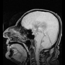
Hydrocephalus is a condition in which an accumulation of cerebrospinal fluid (CSF) occurs within the brain. This typically causes increased pressure inside the skull. Older people may have headaches, double vision, poor balance, urinary incontinence, personality changes, or mental impairment. In babies, it may be seen as a rapid increase in head size. Other symptoms may include vomiting, sleepiness, seizures, and downward pointing of the eyes.

<span class="mw-page-title-main">Ventricular system</span> Set of structures containing cerebrospinal fluid in the brain

The ventricular system is a set of four interconnected cavities known as cerebral ventricles in the brain. Within each ventricle is a region of choroid plexus which produces the circulating cerebrospinal fluid (CSF). The ventricular system is continuous with the central canal of the spinal cord from the fourth ventricle, allowing for the flow of CSF to circulate.

<span class="mw-page-title-main">Cerebral aqueduct</span> Conduit for CSF to the brain

The cerebral aqueduct is a narrow 15 mm conduit for cerebrospinal fluid (CSF) that connects the third ventricle to the fourth ventricle of the ventricular system of the brain. It is located in the midbrain dorsal to the pons and ventral to the cerebellum. The cerebral aqueduct is surrounded by an enclosing area of gray matter called the periaqueductal gray, or central gray. It was first named after Franciscus Sylvius.

Normal-pressure hydrocephalus (NPH), also called malresorptive hydrocephalus, is a form of communicating hydrocephalus in which excess cerebrospinal fluid (CSF) occurs in the ventricles, and with normal or slightly elevated cerebrospinal fluid pressure. As the fluid builds up, it causes the ventricles to enlarge and the pressure inside the head to increase, compressing surrounding brain tissue and leading to neurological complications. The disease presents in a classic triad of symptoms, which are memory impairment, urinary frequency, and balance problems/gait deviations. The disease was first described by Salomón Hakim and Adams in 1965.

<span class="mw-page-title-main">Magnetic resonance angiography</span> Group of techniques based on magnetic resonance imaging (MRI) to image blood vessels.

Magnetic resonance angiography (MRA) is a group of techniques based on magnetic resonance imaging (MRI) to image blood vessels. Magnetic resonance angiography is used to generate images of arteries in order to evaluate them for stenosis, occlusions, aneurysms or other abnormalities. MRA is often used to evaluate the arteries of the neck and brain, the thoracic and abdominal aorta, the renal arteries, and the legs.

A radionuclide cisternogram is a medical imaging study which involves injecting a radionuclide by lumbar puncture into a patient's cerebral spinal fluid (CSF) to determine if there is abnormal CSF flow within the brain and spinal canal which can be altered by hydrocephalus, Arnold–Chiari malformation, syringomyelia, or an arachnoid cyst. It may also evaluate a suspected leak from the CSF cavity into the nasal cavity. A leak can also be confirmed by the presence of beta-2 transferrin in fluid collected from the nose before this more invasive procedure is performed.

<span class="mw-page-title-main">Fluid-attenuated inversion recovery</span>

Fluid-attenuated inversion recovery (FLAIR) is an MRI sequence with an inversion recovery set to null fluids. For example, it can be used in brain imaging to suppress cerebrospinal fluid (CSF) effects on the image, so as to bring out the periventricular hyperintense lesions, such as multiple sclerosis (MS) plaques. It was invented by Graeme Bydder. FLAIR can be used with both three-dimensional imaging or two dimensional imaging.

<span class="mw-page-title-main">Spin–spin relaxation</span>

In physics, the spin–spin relaxation is the mechanism by which Mxy, the transverse component of the magnetization vector, exponentially decays towards its equilibrium value in nuclear magnetic resonance (NMR) and magnetic resonance imaging (MRI). It is characterized by the spin–spin relaxation time, known as T2, a time constant characterizing the signal decay. It is named in contrast to T1, the spin–lattice relaxation time. It is the time it takes for the magnetic resonance signal to irreversibly decay to 37% (1/e) of its initial value after its generation by tipping the longitudinal magnetization towards the magnetic transverse plane. Hence the relation

<i>k</i>-space (magnetic resonance imaging)

In magnetic resonance imaging (MRI), k-space is the 2D or 3D Fourier transform of the image measured. It was introduced in 1979 by Likes and in 1983 by Ljunggren and Twieg.

<span class="mw-page-title-main">Physics of magnetic resonance imaging</span> Overview article

The physics of magnetic resonance imaging (MRI) concerns fundamental physical considerations of MRI techniques and technological aspects of MRI devices. MRI is a medical imaging technique mostly used in radiology and nuclear medicine in order to investigate the anatomy and physiology of the body, and to detect pathologies including tumors, inflammation, neurological conditions such as stroke, disorders of muscles and joints, and abnormalities in the heart and blood vessels among others. Contrast agents may be injected intravenously or into a joint to enhance the image and facilitate diagnosis. Unlike CT and X-ray, MRI uses no ionizing radiation and is, therefore, a safe procedure suitable for diagnosis in children and repeated runs. Patients with specific non-ferromagnetic metal implants, cochlear implants, and cardiac pacemakers nowadays may also have an MRI in spite of effects of the strong magnetic fields. This does not apply on older devices, and details for medical professionals are provided by the device's manufacturer.

<span class="mw-page-title-main">Neurohydrodynamics</span>

Neurohydrodynamics is a division of neurophysics that focuses on the hydrodynamics of the neurological system. It applies physical principles and design concepts to neurophysics seeking to close the gap between fluid mechanics and neurosurgical and neurological medicine. It combines fluid mechanics principles with neuroscience to improve neurological disorder healthcare diagnosis, monitoring and therapy.

<span class="mw-page-title-main">ShuntCheck</span>

ShuntCheck is a non-invasive diagnostic medical device which detects flow in the cerebral shunts of hydrocephalus patients. Neurosurgeons can use ShuntCheck flow results along with other diagnostic tests to assess shunt function and malfunction.

<span class="mw-page-title-main">Aqueductal stenosis</span> Narrowing of the aqueduct of Sylvius

Aqueductal stenosis is a narrowing of the aqueduct of Sylvius which blocks the flow of cerebrospinal fluid (CSF) in the ventricular system. Blockage of the aqueduct can lead to hydrocephalus, specifically as a common cause of congenital and/or obstructive hydrocephalus.

<span class="mw-page-title-main">Phase contrast magnetic resonance imaging</span>

Phase contrast magnetic resonance imaging (PC-MRI) is a specific type of magnetic resonance imaging used primarily to determine flow velocities. PC-MRI can be considered a method of Magnetic Resonance Velocimetry. It also provides a method of magnetic resonance angiography. Since modern PC-MRI is typically time-resolved, it provides a means of 4D imaging.

<span class="mw-page-title-main">MRI sequence</span>

An MRI sequence in magnetic resonance imaging (MRI) is a particular setting of pulse sequences and pulsed field gradients, resulting in a particular image appearance.

An MRI artifact is a visual artifact in magnetic resonance imaging (MRI). It is a feature appearing in an image that is not present in the original object. Many different artifacts can occur during MRI, some affecting the diagnostic quality, while others may be confused with pathology. Artifacts can be classified as patient-related, signal processing-dependent and hardware (machine)-related.

Inversion recovery is an MRI sequence that provides high contrast between tissue and lesion. It can be used to provide high T1 weighted image, high T2 weighted image, and to suppress the signals from fat, blood, or cerebrospinal fluid (CSF).

References

  1. 1 2 3 Gaillard, Frank. "CSF flow studies | Radiology Reference Article | Radiopaedia.org". Radiopaedia. Retrieved 2021-11-24.
  2. 1 2 3 Battal, B; Kocaoglu, M; Bulakbasi, N; Husmen, G; Tuba Sanal, H; Tayfun, C (2011-08-01). "Cerebrospinal fluid flow imaging by using phase-contrast MR technique". The British Journal of Radiology. 84 (1004): 758–765. doi:10.1259/bjr/66206791. ISSN   0007-1285. PMC   3473435 . PMID   21586507.
  3. "Cerebrospinal Fluid (CSF) Leak: Causes, Symptoms, & Treatments". Cleveland Clinic. Retrieved 2021-11-24.
  4. 1 2 3 4 5 6 7 Korbecki, Adrian; Zimny, Anna; Podgórski, Przemysław; Sąsiadek, Marek; Bladowska, Joanna (2019-05-13). "Imaging of cerebrospinal fluid flow: fundamentals, techniques, and clinical applications of phase-contrast magnetic resonance imaging". Polish Journal of Radiology. 84: e240–e250. doi:10.5114/pjr.2019.86881. ISSN   1733-134X. PMC   6717940 . PMID   31481996.
  5. 1 2 3 4 Wymer, David T.; Patel, Kunal P.; Burke, William F.; Bhatia, Vinay K. (2020-01-01). "Phase-Contrast MRI: Physics, Techniques, and Clinical Applications". RadioGraphics. 40 (1): 122–140. doi:10.1148/rg.2020190039. ISSN   0271-5333. PMID   31917664. S2CID   210131904.
  6. Mbonane, Samukelisiwe Sithembile; Andronikou, Savvas (2013-03-05). "Interpretation and value of MR CSF flow studies for paediatric neurosurgery". South African Journal of Radiology. 17 (1): 26–29. doi:10.4102/sajr.v17i1.208. ISSN   2078-6778.
  7. Hurley, Robin A.; Bradley, William G.; Latifi, Haleema T.; Taber, Katherine H. (1999-08-01). "Normal Pressure Hydrocephalus: Significance of MRI in a Potentially Treatable Dementia". The Journal of Neuropsychiatry and Clinical Neurosciences. 11 (3): 297–300. doi:10.1176/jnp.11.3.297. ISSN   0895-0172. PMID   10440004.
  8. Panigrahi, M.; Reddy, B. Praveen; Reddy, A. K.; Reddy, J. J. M. (May 2004). "CSF flow study in Chiari I malformation". Child's Nervous System. 20 (5): 336–340. doi:10.1007/s00381-003-0881-3. ISSN   0256-7040. PMID   15085382. S2CID   36584252.