Phase contrast magnetic resonance imaging

Last updated
Phase contrast magnetic resonance imaging
Vastly undersampled Isotropic Projection Reconstruction (VIPR) Phase Contrast (PC) sequence MRI of arterial dissections.jpg
Vastly undersampled Isotropic Projection Reconstruction (VIPR) of a phase contrast (PC) MRI sequence of a 56-year-old male with dissections of the celiac artery (upper) and the superior mesenteric artery (lower). Laminar flow is present in the true lumen (closed arrow) and helical flow is present in the false lumen (open arrow). [1]
Purposemethod of magnetic resonance angiograph

Phase contrast magnetic resonance imaging (PC-MRI) is a specific type of magnetic resonance imaging used primarily to determine flow velocities. PC-MRI can be considered a method of Magnetic Resonance Velocimetry. It also provides a method of magnetic resonance angiography. Since modern PC-MRI is typically time-resolved, it provides a means of 4D imaging (three spatial dimensions plus time). [2]

Contents

How it Works

Atoms with an odd number of protons or neutrons have a randomly aligned angular spin momentum. When placed in a strong magnetic field, some of these spins align with the axis of the external field, which causes a net 'longitudinal' magnetization. These spins precess about the axis of the external field at a frequency proportional to the strength of that field. Then, energy is added to the system through a Radio frequency (RF) pulse to 'excite' the spins, changing the axis that the spins precess about. These spins can then be observed by receiver coils (Radiofrequency coils) using Faraday's law of induction. Different tissues respond to the added energy in different ways, and imaging parameters can be adjusted to highlight desired tissues.

All of these spins have a phase that is dependent on the atom's velocity. Phase shift of a spin is a function of the gradient field :

where is the Gyromagnetic ratio and is defined as:

,

is the initial position of the spin, is the spin velocity, and is the spin acceleration.

If we only consider static spins and spins in the x-direction, we can rewrite equation for phase shift as:

We then assume that acceleration and higher order terms are negligible to simplify the expression for phase to:

where is the zeroth moment of the x-gradient and is the first moment of the x gradient.

If we take two different acquisitions with applied magnetic gradients that are the opposite of each other (bipolar gradients), we can add the results of the two acquisitions together to calculate a change in phase that is dependent on gradient:

where . [3] [4]

The phase shift is measured and converted to a velocity according to the following equation:

where is the maximum velocity that can be recorded and is the recorded phase shift.

The choice of defines range of velocities visible, known as the ‘dynamic range’. A choice of below the maximum velocity in the slice will induce aliasing in the image where a velocity just greater than will be incorrectly calculated as moving in the opposite direction. However, there is a direct trade-off between the maximum velocity that can be encoded and the signal-to-noise ratio of the velocity measurements. This can be described by:

where is the signal-to-noise ratio of the image (which depends on the magnetic field of the scanner, the voxel volume, and the acquisition time of the scan).

For an example, setting a ‘low’ (below the maximum velocity expected in the scan) will allow for better visualization of slower velocities (better SNR), but any higher velocities will alias to an incorrect value. Setting a ‘high’ (above the maximum velocity expected in the scan) will allow for the proper velocity quantification, but the larger dynamic range will obscure the smaller velocity features as well as decrease SNR. Therefore, the setting of will be application dependent and care must be exercised in the selection. In order to further allow for proper velocity quantification, especially in clinical applications where the velocity dynamic range of flow is high (e.g. blood flow velocities in vessels across the thoracoabdominal cavity), a dual-echo PC-MRI (DEPC) method with dual velocity encoding in the same repetition time has been developed. [5] The DEPC method does not only allow for proper velocity quantification, but also reduces the total acquisition time (especially when applied to 4D flow imaging) compared to a single-echo single- PC-MRI acquisition carried out at two separate values.

To allow for more flexibility in selecting , instantaneous phase (phase unwrapping) can be used to increase both dynamic range and SNR. [6]

Encoding Methods

When each dimension of velocity is calculated based on acquisitions from oppositely applied gradients, this is known as a six-point method. However, more efficient methods are also used. Two are described here:

Simple Four-point Method

Four sets of encoding gradients are used. The first is a reference and applies a negative moment in ,, and . The next applies a positive moment in , and a negative moment in and . The third applies a positive moment in , and a negative moment in and . And the last applies a positive moment in , and a negative moment in and . [7] Then, the velocities can be solved based on the phase information from the corresponding phase encodes as follows:

Balanced Four-Point Method

The balanced four-point method also includes four sets of encoding gradients. The first is the same as in the simple four-point method with negative gradients applied in all directions. The second has a negative moment in , and a positive moment in and . The third has a negative moment in , and a positive moment in and . The last has a negative moment in and a positive moment in and . [8] This gives us the following system of equations:

Then, the velocities can be calculated:

Retrospective Cardiac and Respiratory Gating

For medical imaging, in order to get highly resolved scans in 3D space and time without motion artifacts from the heart or lungs, retrospective cardiac gating and respiratory compensation are employed. Beginning with cardiac gating, the patient's ECG signal is recorded throughout the imaging process. Similarly, the patient's respiratory patterns can be tracked throughout the scan. After the scan, the continuously collected data in k-space (temporary image space) can be assigned accordingly to match-up with the timing of the heart beat and lung motion of the patient. This means that these scans are cardiac-averaged so the measured blood velocities are an average over multiple cardiac cycles. [9]

Applications

Phase contrast MRI is one of the main techniques for magnetic resonance angiography (MRA). This is used to generate images of arteries (and less commonly veins) in order to evaluate them for stenosis (abnormal narrowing), occlusions, aneurysms (vessel wall dilatations, at risk of rupture) or other abnormalities. MRA is often used to evaluate the arteries of the neck and brain, the thoracic and abdominal aorta, the renal arteries, and the legs (the latter exam is often referred to as a "run-off").

Limitations

In particular, a few limitations of PC-MRI are of importance for the measured velocities:

Vastly undersampled Isotropic Projection Reconstruction (VIPR)

A Vastly undersampled Isotropic Projection Reconstruction (VIPR) is a radially acquired MRI sequence which results in high-resolution MRA with significantly reduced scan times, and without the need for breath-holding. [11]

Related Research Articles

<span class="mw-page-title-main">Lorentz force</span> Force acting on charged particles in electric and magnetic fields

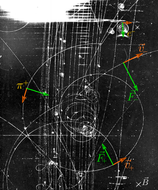
In physics, the Lorentz force is the combination of electric and magnetic force on a point charge due to electromagnetic fields. A particle of charge q moving with a velocity v in an electric field E and a magnetic field B experiences a force of

In special relativity, four-momentum (also called momentum–energy or momenergy) is the generalization of the classical three-dimensional momentum to four-dimensional spacetime. Momentum is a vector in three dimensions; similarly four-momentum is a four-vector in spacetime. The contravariant four-momentum of a particle with relativistic energy E and three-momentum p = (px, py, pz) = γmv, where v is the particle's three-velocity and γ the Lorentz factor, is

<span class="mw-page-title-main">Four-vector</span> 4-dimensional vector in relativity

In special relativity, a four-vector is an object with four components, which transform in a specific way under Lorentz transformations. Specifically, a four-vector is an element of a four-dimensional vector space considered as a representation space of the standard representation of the Lorentz group, the representation. It differs from a Euclidean vector in how its magnitude is determined. The transformations that preserve this magnitude are the Lorentz transformations, which include spatial rotations and boosts.

Hemorheology, also spelled haemorheology, or blood rheology, is the study of flow properties of blood and its elements of plasma and cells. Proper tissue perfusion can occur only when blood's rheological properties are within certain levels. Alterations of these properties play significant roles in disease processes. Blood viscosity is determined by plasma viscosity, hematocrit and mechanical properties of red blood cells. Red blood cells have unique mechanical behavior, which can be discussed under the terms erythrocyte deformability and erythrocyte aggregation. Because of that, blood behaves as a non-Newtonian fluid. As such, the viscosity of blood varies with shear rate. Blood becomes less viscous at high shear rates like those experienced with increased flow such as during exercise or in peak-systole. Therefore, blood is a shear-thinning fluid. Contrarily, blood viscosity increases when shear rate goes down with increased vessel diameters or with low flow, such as downstream from an obstruction or in diastole. Blood viscosity also increases with increases in red cell aggregability.

In physics, the Hamilton–Jacobi equation, named after William Rowan Hamilton and Carl Gustav Jacob Jacobi, is an alternative formulation of classical mechanics, equivalent to other formulations such as Newton's laws of motion, Lagrangian mechanics and Hamiltonian mechanics.

In physics, the gyromagnetic ratio of a particle or system is the ratio of its magnetic moment to its angular momentum, and it is often denoted by the symbol γ, gamma. Its SI unit is the radian per second per tesla (rad⋅s−1⋅T−1) or, equivalently, the coulomb per kilogram (C⋅kg−1).

In differential geometry, the four-gradient is the four-vector analogue of the gradient from vector calculus.

A theoretical motivation for general relativity, including the motivation for the geodesic equation and the Einstein field equation, can be obtained from special relativity by examining the dynamics of particles in circular orbits about the Earth. A key advantage in examining circular orbits is that it is possible to know the solution of the Einstein Field Equation a priori. This provides a means to inform and verify the formalism.

In MRI and NMR spectroscopy, an observable nuclear spin polarization (magnetization) is created by a homogeneous magnetic field. This field makes the magnetic dipole moments of the sample precess at the resonance (Larmor) frequency of the nuclei. At thermal equilibrium, nuclear spins precess randomly about the direction of the applied field. They become abruptly phase coherent when they are hit by radiofrequency (RF) pulses at the resonant frequency, created orthogonal to the field. The RF pulses cause the population of spin-states to be perturbed from their thermal equilibrium value. The generated transverse magnetization can then induce a signal in an RF coil that can be detected and amplified by an RF receiver. The return of the longitudinal component of the magnetization to its equilibrium value is termed spin-latticerelaxation while the loss of phase-coherence of the spins is termed spin-spin relaxation, which is manifest as an observed free induction decay (FID).

The Newman–Penrose (NP) formalism is a set of notation developed by Ezra T. Newman and Roger Penrose for general relativity (GR). Their notation is an effort to treat general relativity in terms of spinor notation, which introduces complex forms of the usual variables used in GR. The NP formalism is itself a special case of the tetrad formalism, where the tensors of the theory are projected onto a complete vector basis at each point in spacetime. Usually this vector basis is chosen to reflect some symmetry of the spacetime, leading to simplified expressions for physical observables. In the case of the NP formalism, the vector basis chosen is a null tetrad: a set of four null vectors—two real, and a complex-conjugate pair. The two real members often asymptotically point radially inward and radially outward, and the formalism is well adapted to treatment of the propagation of radiation in curved spacetime. The Weyl scalars, derived from the Weyl tensor, are often used. In particular, it can be shown that one of these scalars— in the appropriate frame—encodes the outgoing gravitational radiation of an asymptotically flat system.

The theoretical and experimental justification for the Schrödinger equation motivates the discovery of the Schrödinger equation, the equation that describes the dynamics of nonrelativistic particles. The motivation uses photons, which are relativistic particles with dynamics described by Maxwell's equations, as an analogue for all types of particles.

The intent of this article is to highlight the important points of the derivation of the Navier–Stokes equations as well as its application and formulation for different families of fluids.

The Cauchy momentum equation is a vector partial differential equation put forth by Cauchy that describes the non-relativistic momentum transport in any continuum.

In physics, magnetization dynamics is the branch of solid-state physics that describes the evolution of the magnetization of a material.

Nuclear magnetic resonance (NMR) in porous materials covers the application of using NMR as a tool to study the structure of porous media and various processes occurring in them. This technique allows the determination of characteristics such as the porosity and pore size distribution, the permeability, the water saturation, the wettability, etc.

<span class="mw-page-title-main">Relativistic Lagrangian mechanics</span> Mathematical formulation of special and general relativity

In theoretical physics, relativistic Lagrangian mechanics is Lagrangian mechanics applied in the context of special relativity and general relativity.

<span class="mw-page-title-main">Derivations of the Lorentz transformations</span>

There are many ways to derive the Lorentz transformations using a variety of physical principles, ranging from Maxwell's equations to Einstein's postulates of special relativity, and mathematical tools, spanning from elementary algebra and hyperbolic functions, to linear algebra and group theory.

In quantum mechanics, magnetic resonance is a resonant effect that can appear when a magnetic dipole is exposed to a static magnetic field and perturbed with another, oscillating electromagnetic field. Due to the static field, the dipole can assume a number of discrete energy eigenstates, depending on the value of its angular momentum (azimuthal) quantum number. The oscillating field can then make the dipole transit between its energy states with a certain probability and at a certain rate. The overall transition probability will depend on the field's frequency and the rate will depend on its amplitude. When the frequency of that field leads to the maximum possible transition probability between two states, a magnetic resonance has been achieved. In that case, the energy of the photons composing the oscillating field matches the energy difference between said states. If the dipole is tickled with a field oscillating far from resonance, it is unlikely to transition. That is analogous to other resonant effects, such as with the forced harmonic oscillator. The periodic transition between the different states is called Rabi cycle and the rate at which that happens is called Rabi frequency. The Rabi frequency should not be confused with the field's own frequency. Since many atomic nuclei species can behave as a magnetic dipole, this resonance technique is the basis of nuclear magnetic resonance, including nuclear magnetic resonance imaging and nuclear magnetic resonance spectroscopy.

Adiabatic radio frequency (RF) pulses are used in magnetic resonance imaging (MRI) to achieve excitation that is insensitive to spatial inhomogeneities in the excitation field or off-resonances in the sampled object.

Steered-response power (SRP) is a family of acoustic source localization algorithms that can be interpreted as a beamforming-based approach that searches for the candidate position or direction that maximizes the output of a steered delay-and-sum beamformer.

References

  1. Hartung, Michael P; Grist, Thomas M; François, Christopher J (2011). "Magnetic resonance angiography: current status and future directions". Journal of Cardiovascular Magnetic Resonance. 13 (1): 19. doi: 10.1186/1532-429X-13-19 . ISSN   1532-429X. PMC   3060856 . PMID   21388544. (CC-BY-2.0)
  2. Stankovic, Zoran; Allen, Bradley D.; Garcia, Julio; Jarvis, Kelly B.; Markl, Michael (2014). "4D flow imaging with MRI". Cardiovascular Diagnosis and Therapy . 4 (2): 173–192. doi:10.3978/j.issn.2223-3652.2014.01.02. PMC   3996243 . PMID   24834414.
  3. Elkins, C.; Alley, M.T. (2007). "Magnetic resonance velocimetry: applications of magnetic resonance imaging in the measurement of fluid motion". Experiments in Fluids . 43 (6): 823. Bibcode:2007ExFl...43..823E. doi:10.1007/s00348-007-0383-2. S2CID   121958168.
  4. Taylor, Charles A.; Draney, Mary T. (2004). "Experimental and computational methods in cardiovascular fluid mechanics". Annual Review of Fluid Mechanics . 36: 197–231. Bibcode:2004AnRFM..36..197T. doi:10.1146/annurev.fluid.36.050802.121944.
  5. Ajala, Afis; Zhang, Jiming; Pednekar, Amol; Buko, Erick; Wang, Luning; Cheong, Benjamin; Hor, Pei-Herng; Muthupillai, Raja (2020). "Mitral Valve Flow and Myocardial Motion Assessed with Dual-Echo Dual-Velocity Cardiac MRI". Radiology: Cardiothoracic Imaging. 3 (2): e190126. doi:10.1148/ryct.2020190126. PMC   7977974 . PMID   33778578.
  6. Salfitya, M.F.; Huntleya, J.M.; Gravesb, M.J.; Marklundc, O.; Cusackd, R.; Beauregardd, D.A. (2006). "Extending the dynamic range of phase contrast magnetic resonance velocity imaging using advanced higher-dimensional phase unwrapping algorithms". Journal of the Royal Society Interface . 3 (8): 415–427. doi:10.1098/rsif.2005.0096. PMC   1578755 . PMID   16849270.
  7. Pelc, Norbert J.; Bernstein, Matt A.; Shimakawa, Ann; Glover, Gary H. (1991). "Encoding strategies for three‐direction phase‐contrast MR imaging of flow". Journal of Magnetic Resonance Imaging . 1 (4): 405–413. doi:10.1002/jmri.1880010404. PMID   1790362. S2CID   3000911.
  8. Pelc, Norbert J.; Bernstein, Matt A.; Shimakawa, Ann; Glover, Gary H. (1991). "Encoding strategies for three‐direction phase‐contrast MR imaging of flow". Journal of Magnetic Resonance Imaging . 1 (4): 405–413. doi:10.1002/jmri.1880010404. PMID   1790362. S2CID   3000911.
  9. Lotz, Joachim; Meier, Christian; Leppert, Andreas; Galanski, Michael (2002). "Cardiovascular Flow Measurement with Phase-Contrast MR Imaging: Basic Facts and Implementation 1". Radiographics. 22 (3): 651–671. doi:10.1148/radiographics.22.3.g02ma11651. PMID   12006694.
  10. Petersson, Sven; Dyverfeldt, Petter; Gårdhagen, Roland; Karlsson, Matts; Ebbers, Tino (2010). "Simulation of phase contrast MRI of turbulent flow". Magnetic Resonance in Medicine . 64 (4): 1039–1046. doi: 10.1002/mrm.22494 . PMID   20574963.
  11. Page 602 in: Hersh Chandarana (2015). Advanced MR Imaging in Clinical Practice, An Issue of Radiologic Clinics of North America. Vol. 53. Elsevier Health Sciences. ISBN   9780323376181.