Cortical spreading depression

Last updated
Animation of cortical spreading depression Cortical spreading depression.gif
Animation of cortical spreading depression
Cortical spreading depression seen using intrinsic optical signal imaging in gyrencephalic brain. Speed 50x. From Santos E et al. Neuroimage 2014. Santos E et al Neuroimage 2014 .gif
Cortical spreading depression seen using intrinsic optical signal imaging in gyrencephalic brain. Speed 50x. From Santos E et al. Neuroimage 2014.
Hemodynamic changes observed after MCA occlusion in IOS. The video has a speed of 50x to better appreciate the SDs through human eye. Pictures are dynamically subtracted to a reference picture 40 s before. First we see the initial area of change at the exact moment where the left MCAs are occluded. The area is highlighted with a white line. Later we appreciate the signal produced by SDs. https://doi.org/10.1007/s00701-019-04132-8 Middle Cerebral Artery occlusion. Kentar et al Acta Neuroch 2020.gif
Hemodynamic changes observed after MCA occlusion in IOS. The video has a speed of 50x to better appreciate the SDs through human eye. Pictures are dynamically subtracted to a reference picture 40 s before. First we see the initial area of change at the exact moment where the left MCAs are occluded. The area is highlighted with a white line. Later we appreciate the signal produced by SDs. https://doi.org/10.1007/s00701-019-04132-8

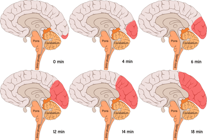
Cortical spreading depression (CSD) or spreading depolarization (SD) is a wave of electrophysiological hyperactivity followed by a wave of inhibition. [1] Spreading depolarization describes a phenomenon characterized by the appearance of depolarization waves of the neurons and neuroglia [2] that propagates across the cortex at a velocity of 1.5–9.5 mm/min. [3] [4] [5] [6]

Contents

CSD can be induced by hypoxic conditions and facilitates neuronal death in energy-compromised tissue. [7] CSD has also been implicated in migraine aura, where CSD is assumed to ascend in well-nourished tissue and is typically benign in most cases, although it may increase the probability in migraine patients to develop a stroke. [8]

Spreading depolarization within brainstem tissues regulating functions crucial for life has been implicated in sudden unexpected death in epilepsy, by way of ion channel mutations such as those seen in Dravet syndrome, a particularly severe form of childhood epilepsy that appears to carry an unusually high risk of SUDEP (sudden unexpected death in epilepsy).

Although the terms cortical spreading depression and spreading depolarization are often used as synonyms, a study found spreading depolarizations can produce variable effects on cortical activity in humans and rats, ranging from depressed to booming activity depending on SD depth. [9]

Uses of the term

Neuroscientists use the term cortical spreading depression to represent at least one of the following cortical processes:[ citation needed ]

The scintillating scotoma of migraine in humans may be related to the neurophysiologic phenomenon termed the spreading depression of Leão. [11]

Increased extracellular potassium ion concentration and excitatory glutamate contribute to the initiation and propagation of cortical spreading depression, which is the underlying cause of migraine aura. [12]

Chronic daily administration of migraine prophylactic drugs (topiramate, valproate, propranolol, amitriptyline, and methysergide) dose-dependently suppressed frequency of CSD induced by continuous cortical application of 1 M KCl solution. [13] However lamotrigine (a drug with specific anti-aura action, but no efficacy in migraine in general) has a marked suppressive effect which correlates with its rather selective action on the migraine aura. Valproate and riboflavin were shown to have no effect on the triggering of cortical spreading depression though they are effective in migraine without aura. [14] Taken together, these results are compatible with a causal role of cortical spreading depression in migraine with aura, but not in migraine without aura.[ citation needed ]

The folded structure of the cerebral cortex is capable of irregular and complex CSD propagation patterns. The irregularities of the folded cortex and the vasculature promote the presence of re-entrance waves, such as spirals and reverberating waves. [5] The expansion of the wave then is less predictable and it is affected by the concentration of different molecules and gradients on the cortex.[ citation needed ]

Its triggers and propagation mechanisms, as well as clinical manifestations of CSD, are a therapeutic target to reduce brain damage after a stroke or brain lesion. [15] [16] [17]

See also

Notes

  1. Dodick DW & Gargus JJ (August 2008). "Why migraines strike". Scientific American.
  2. Chuquet, Julien; Hollender, Liad; Nimchinsky, Esther A. (2007-04-11). "High-resolution in vivo imaging of the neurovascular unit during spreading depression". The Journal of Neuroscience. 27 (15): 4036–4044. doi:10.1523/JNEUROSCI.0721-07.2007. ISSN   1529-2401. PMC   6672520 . PMID   17428981.
  3. Ayata, Cenk; Lauritzen, Martin (2015-07-01). "Spreading Depression, Spreading Depolarizations, and the Cerebral Vasculature". Physiological Reviews. 95 (3): 953–993. doi:10.1152/physrev.00027.2014. ISSN   1522-1210. PMC   4491545 . PMID   26133935.
  4. Hartings, Dreier; Jed, Jens (2017-05-01). "Recording, analysis, and interpretation of spreading depolarizations in neurointensive care: Review and recommendations of the COSBID research group". Journal of Cerebral Blood Flow and Metabolism. 37 (5): 1595–1625. doi:10.1177/0271678X16654496. ISSN   1559-7016. PMC   5435289 . PMID   27317657.
  5. 1 2 Santos, Edgar; Schöll, Michael; Sánchez-Porras, Renán; Dahlem, Markus A.; Silos, Humberto; Unterberg, Andreas; Dickhaus, Hartmut; Sakowitz, Oliver W. (2014-10-01). "Radial, spiral and reverberating waves of spreading depolarization occur in the gyrencephalic brain". NeuroImage. 99: 244–255. doi:10.1016/j.neuroimage.2014.05.021. ISSN   1095-9572. PMID   24852458. S2CID   1347927.
  6. Porooshani H, Porooshani GH, Gannon L, Kyle GM (2004). "Speed of progression of migrainous visual aura measured by sequential field assessment". Neuro-Ophthalmology. 28 (2): 101–105. doi:10.1076/noph.28.2.101.23739. S2CID   72813214.
  7. Dreier, Jens P.; Reiffurth, Clemens (2015-05-20). "The stroke-migraine depolarization continuum". Neuron. 86 (4): 902–922. doi: 10.1016/j.neuron.2015.04.004 . ISSN   1097-4199. PMID   25996134.
  8. Santos, Edgar; Sánchez-Porras, Renán; Dohmen, Christian; Hertle, Daniel; Unterberg, Andreas W.; Sakowitz, Oliver W. (2012-04-01). "Spreading depolarizations in a case of migraine-related stroke". Cephalalgia: An International Journal of Headache. 32 (5): 433–436. doi:10.1177/0333102412441414. ISSN   1468-2982. PMID   22407661. S2CID   25190650.
  9. Nasretdinov, Azat; Vinokurova, Daria; Lemale, Coline L.; Burkhanova-Zakirova, Gulshat; Chernova, Ksenia; Makarova, Julia; Herreras, Oscar; Dreier, Jens P.; Khazipov, Roustem (25 November 2023). "Diversity of cortical activity changes beyond depression during Spreading Depolarizations". Nature Communications. 14 (1): 7729. doi:10.1038/s41467-023-43509-3. PMC   10676372 . PMID   38007508.
  10. Brennan KC, Beltrán-Parrazal L, López-Valdés HE, Theriot J, Toga AW, Charles AC (2007). "Distinct vascular conduction with cortical spreading depression". Journal of Neurophysiology . 97 (6): 4143–4151. doi:10.1152/jn.00028.2007. PMID   17329631. S2CID   20878873.
  11. Leão AAP (1944). "Spreading depression of activity in the cerebral cortex". J Neurophysiol. 7 (6): 359–390. doi:10.1152/jn.1944.7.6.359. S2CID   14270649.
  12. Richter and Lehmenkühler (2008)
  13. Ayata; et al. (Apr 2006). "Suppression of cortical spreading depression in migraine prophylaxis". Ann Neurol. 59 (4): 652–61. doi:10.1002/ana.20778. PMID   16450381. S2CID   31232538.
  14. Bogdanov; et al. (Feb 2011). "Migraine preventive drugs differentially affect cortical spreading depression in rat". Neurobiol. Dis. 41 (2): 430–5. doi:10.1016/j.nbd.2010.10.014. PMID   20977938. S2CID   207065722.
  15. Sánchez-Porras, Renán; Santos, Edgar; Schöll, Michael; Stock, Christian; Zheng, Zelong; Schiebel, Patrick; Orakcioglu, Berk; Unterberg, Andreas W.; Sakowitz, Oliver W. (2014-09-01). "The effect of ketamine on optical and electrical characteristics of spreading depolarizations in gyrencephalic swine cortex". Neuropharmacology. 84: 52–61. doi:10.1016/j.neuropharm.2014.04.018. ISSN   1873-7064. PMID   24796257. S2CID   21562145.
  16. Sánchez-Porras, R.; Zheng, Z.; Santos, E.; Schöll, M.; Unterberg, A. W.; Sakowitz, O. W. (2013-08-01). "The role of spreading depolarization in subarachnoid hemorrhage". European Journal of Neurology. 20 (8): 1121–1127. doi:10.1111/ene.12139. ISSN   1468-1331. PMID   23551588. S2CID   23615512.
  17. Kentar, Modar; Mann, Martina; Sahm, Felix; Olivares-Rivera, Arturo; Sanchez-Porras, Renan; Zerelles, Roland; Sakowitz, Oliver W.; Unterberg, Andreas W.; Santos, Edgar (2020-01-15). "Detection of spreading depolarizations in a middle cerebral artery occlusion model in swine". Acta Neurochirurgica. 162 (3): 581–592. doi:10.1007/s00701-019-04132-8. ISSN   0942-0940. PMID   31940093. S2CID   210196036.

Related Research Articles

<span class="mw-page-title-main">Migraine</span> Disorder resulting in recurrent moderate-severe headaches

Migraine is a genetically influenced complex neurological disorder characterized by episodes of moderate-to-severe headache, most often unilateral and generally associated with nausea and light and sound sensitivity. Other characterizing symptoms may include nausea, vomiting, cognitive dysfunction, allodynia, and dizziness. Exacerbation of headache symptoms during physical activity is another distinguishing feature. Up to one-third of migraine sufferers experience aura, a premonitory period of sensory disturbance widely accepted to be caused by cortical spreading depression at the onset of a migraine attack. Although primarily considered to be a headache disorder, migraine is highly heterogenous in its clinical presentation and is better thought of as a spectrum disease rather than a distinct clinical entity. Disease burden can range from episodic discrete attacks, consisting of as little as several lifetime attacks, to chronic disease.

<span class="mw-page-title-main">Cerebral cortex</span> Outer layer of the cerebrum of the mammalian brain

The cerebral cortex, also known as the cerebral mantle, is the outer layer of neural tissue of the cerebrum of the brain in humans and other mammals. It is the largest site of neural integration in the central nervous system. and plays a key role in attention, perception, awareness, thought, memory, language, and consciousness. The cerebral cortex is the part of the brain responsible for cognition.

<span class="mw-page-title-main">Transcranial magnetic stimulation</span> Brain stimulation using magnetic fields

Transcranial magnetic stimulation (TMS) is a noninvasive form of brain stimulation in which a changing magnetic field is used to induce an electric current at a specific area of the brain through electromagnetic induction. An electric pulse generator, or stimulator, is connected to a magnetic coil connected to the scalp. The stimulator generates a changing electric current within the coil which creates a varying magnetic field, inducing a current within a region in the brain itself.

<span class="mw-page-title-main">Alice in Wonderland syndrome</span> Neurological disorder that distorts perception of objects size and distance

Alice in Wonderland syndrome (AIWS), also known as Todd's syndrome or dysmetropsia, is a neurological disorder that distorts perception. People with this syndrome may experience distortions in their visual perception of objects, such as appearing smaller (micropsia) or larger (macropsia), or appearing to be closer (pelopsia) or farther (teleopsia) than they are. Distortion may also occur for senses other than vision.

<span class="mw-page-title-main">Neocortex</span> Mammalian structure involved in higher-order brain functions

The neocortex, also called the neopallium, isocortex, or the six-layered cortex, is a set of layers of the mammalian cerebral cortex involved in higher-order brain functions such as sensory perception, cognition, generation of motor commands, spatial reasoning and language. The neocortex is further subdivided into the true isocortex and the proisocortex.

<span class="mw-page-title-main">Visual snow syndrome</span> Visual impairment

Visual snow syndrome (VSS) is an uncommon neurological condition in which the primary symptom is that affected individuals see persistent flickering white, black, transparent, or coloured dots across the whole visual field.

<span class="mw-page-title-main">Cortical column</span> Group of neurons in the cortex of the brain

A cortical column is a group of neurons forming a cylindrical structure through the cerebral cortex of the brain perpendicular to the cortical surface. The structure was first identified by Mountcastle in 1957. He later identified minicolumns as the basic units of the neocortex which were arranged into columns. Each contains the same types of neurons, connectivity, and firing properties. Columns are also called hypercolumn, macrocolumn, functional column or sometimes cortical module. Neurons within a minicolumn (microcolumn) encode similar features, whereas a hypercolumn "denotes a unit containing a full set of values for any given set of receptive field parameters". A cortical module is defined as either synonymous with a hypercolumn (Mountcastle) or as a tissue block of multiple overlapping hypercolumns.

<span class="mw-page-title-main">Gyrus</span> Ridge on the cerebral cortex of the brain

In neuroanatomy, a gyrus is a ridge on the cerebral cortex. It is generally surrounded by one or more sulci. Gyri and sulci create the folded appearance of the brain in humans and other mammals.

<span class="mw-page-title-main">Aura (symptom)</span> Symptom of epilepsy and migraine

An aura is a perceptual disturbance experienced by some with epilepsy or migraine. An epileptic aura is a seizure.

Familial hemiplegic migraine (FHM) is an autosomal dominant type of hemiplegic migraine that typically includes weakness of half the body which can last for hours, days, or weeks. It can be accompanied by other symptoms, such as ataxia, coma, and paralysis. Migraine attacks may be provoked by minor head trauma. Some cases of minor head trauma in patients with hemiplegic migraine can develop into delayed cerebral edema, a life-threatening medical emergency. Clinical overlap occurs in some FHM patients with episodic ataxia type 2 and spinocerebellar ataxia type 6, benign familial infantile epilepsy, and alternating hemiplegia of childhood.

<span class="mw-page-title-main">Aristides Leão</span> Brazilian professor of neurophysiology (d. 1993)

Aristides Azevedo Pacheco Leão was a Brazilian neurophysiologist, researcher and university professor.
<span class="mw-page-title-main">Scintillating scotoma</span> Visual aura associated with migraine

Scintillating scotoma is a common visual aura that was first described by 19th-century physician Hubert Airy (1838–1903). Originating from the brain, it may precede a migraine headache, but can also occur acephalgically, also known as visual migraine or migraine aura. It is often confused with retinal migraine, which originates in the eyeball or socket.

Martinotti cells are small multipolar neurons with short branching dendrites. They are scattered throughout various layers of the cerebral cortex, sending their axons up to the cortical layer I where they form axonal arborization. The arbors transgress multiple columns in layer VI and make contacts with the distal tuft dendrites of pyramidal cells. Martinotti cells express somatostatin and sometimes calbindin, but not parvalbumin or vasoactive intestinal peptide. Furthermore, Martinotti cells in layer V have been shown to express the nicotinic acetylcholine receptor α2 subunit (Chrna2).

Recurrent thalamo-cortical resonance or Thalamocortical oscillation is an observed phenomenon of oscillatory neural activity between the thalamus and various cortical regions of the brain. It is proposed by Rodolfo Llinas and others as a theory for the integration of sensory information into the whole of perception in the brain. Thalamocortical oscillation is proposed to be a mechanism of synchronization between different cortical regions of the brain, a process known as temporal binding. This is possible through the existence of thalamocortical networks, groupings of thalamic and cortical cells that exhibit oscillatory properties.

<span class="mw-page-title-main">Spike-and-wave</span>

Spike-and-wave is a pattern of the electroencephalogram (EEG) typically observed during epileptic seizures. A spike-and-wave discharge is a regular, symmetrical, generalized EEG pattern seen particularly during absence epilepsy, also known as ‘petit mal’ epilepsy. The basic mechanisms underlying these patterns are complex and involve part of the cerebral cortex, the thalamocortical network, and intrinsic neuronal mechanisms.

<span class="mw-page-title-main">Retinal migraine</span> Medical condition

Retinal migraine is a retinal disease often accompanied by migraine headache and typically affects only one eye. It is caused by ischaemia or vascular spasm in or behind the affected eye.

The trigeminovascular system (TVS) refers to neurons and their axonal projections within the trigeminal nerve that project to the cranial meninges and meningeal blood vessels residing on the brain's surface. The term, introduced in 1983 denotes also the neuropeptides contained within axons that are released into the meninges to target vessels and surrounding cells.

<span class="mw-page-title-main">Illusory palinopsia</span> Subtype of palinopsia

Illusory palinopsia is a subtype of palinopsia, a visual disturbance defined as the persistence or recurrence of a visual image after the stimulus has been removed. Palinopsia is a broad term describing a heterogeneous group of symptoms, which is divided into hallucinatory palinopsia and illusory palinopsia. Illusory palinopsia is likely due to sustained awareness of a stimulus and is similar to a visual illusion: the distorted perception of a real external stimulus.

A migrainous infarction is a rare type of ischaemic stroke which occurs in correspondence with migraine aura symptoms. Symptoms include headaches, visual disturbances, strange sensations and dysphasia, all of which gradually worsen causing neurological changes which ultimately increase the risk of an ischaemic stroke. Typically, women under the age of 45 who experience migraine with aura (MA) are at the greatest risk for developing migrainous infarction, especially when combined with smoking and use of oral contraceptives.

<span class="mw-page-title-main">Martin Lauritzen</span> Danish neuroscientist and researcher

Martin Johannes Lauritzen is a Danish neuroscientist. He is a Professor of Translational Neurobiology at the Department of Neuroscience, University of Copenhagen, Denmark and also a Professor of Clinical Neurophysiology at the Department of Neurophysiology, Rigshospitalet.
References

Further reading