Medical ultrasound includes diagnostic techniques using ultrasound, as well as therapeutic applications of ultrasound. In diagnosis, it is used to create an image of internal body structures such as tendons, muscles, joints, blood vessels, and internal organs, to measure some characteristics or to generate an informative audible sound. The usage of ultrasound to produce visual images for medicine is called medical ultrasonography or simply sonography, or echography. The practice of examining pregnant women using ultrasound is called obstetric ultrasonography, and was an early development of clinical ultrasonography. The machine used is called an ultrasound machine, a sonograph or an echograph. The visual image formed using this technique is called an ultrasonogram, a sonogram or an echogram.

The skull is a bone protective cavity for the brain. The skull is composed of four types of bone i.e., cranial bones, facial bones, ear ossicles and hyoid bone, however two parts are more prominent: the cranium and the mandible. In humans, these two parts are the neurocranium (braincase) and the viscerocranium that includes the mandible as its largest bone. The skull forms the anterior-most portion of the skeleton and is a product of cephalisation—housing the brain, and several sensory structures such as the eyes, ears, nose, and mouth. In humans, these sensory structures are part of the facial skeleton.

A fontanelle is an anatomical feature of the infant human skull comprising soft membranous gaps (sutures) between the cranial bones that make up the calvaria of a fetus or an infant. Fontanelles allow for stretching and deformation of the neurocranium both during birth and later as the brain expands faster than the surrounding bone can grow. Premature complete ossification of the sutures is called craniosynostosis.

Hydrocephalus is a condition in which an accumulation of cerebrospinal fluid (CSF) occurs within the brain. This typically causes increased pressure inside the skull. Older people may have headaches, double vision, poor balance, urinary incontinence, personality changes, or mental impairment. In babies, it may be seen as a rapid increase in head size. Other symptoms may include vomiting, sleepiness, seizures, and downward pointing of the eyes.

Colpocephaly is a cephalic disorder involving the disproportionate enlargement of the occipital horns of the lateral ventricles and is usually diagnosed early after birth due to seizures. It is a nonspecific finding and is associated with multiple neurological syndromes, including agenesis of the corpus callosum, Chiari malformation, lissencephaly, and microcephaly. Although the exact cause of colpocephaly is not known yet, it is commonly believed to occur as a result of neuronal migration disorders during early brain development, intrauterine disturbances, perinatal injuries, and other central nervous system disorders. Individuals with colpocephaly have various degrees of motor disabilities, visual defects, spasticity, and moderate to severe intellectual disability. No specific treatment for colpocephaly exists, but patients may undergo certain treatments to improve their motor function or intellectual disability.

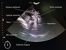
Obstetric ultrasonography, or prenatal ultrasound, is the use of medical ultrasonography in pregnancy, in which sound waves are used to create real-time visual images of the developing embryo or fetus in the uterus (womb). The procedure is a standard part of prenatal care in many countries, as it can provide a variety of information about the health of the mother, the timing and progress of the pregnancy, and the health and development of the embryo or fetus.
A molera is a "soft spot" on the top of a Chihuahua's skull; it is the equivalent to the bregmatic or anterior fontanelle in human babies, but unlike most mammals Chihuahua's fontanelle persist into maturity. Historically it has been very common amongst Chihuahuas and was regarded as a mark of purity for this miniature dog breed. It is still mentioned in many Chihuahua breed standards, however, it is considered a fault in European countries because of concern that this might reflect underlying malformations such as hydrocephalus and ventriculomegaly, Chiari-like malformation and syringomyelia. Fontanelles are fibrous, membrane-covered gaps that lie between the skull bones and at the intersection of the cranial sutures. The cranial sutures are the junctions between cranial bones. The fontanelles serve as the major sites of bone expansion during post-natal skull growth which accommodates the enlarging brain. The Chihuahua likely has a molera because of neuroparenchymal disproportion i.e. a proportionally big brain for the skull. This is likely because there is premature closure of the skull base cranial sutures. To accommodate the developing brain there is increased growth of the skull bone in a parallel plane giving the dog a characterised domed or "apple-headed" appearance.
Antenatal steroids, also known as antenatal corticosteroids, are medications administered to pregnant women expecting a preterm birth. When administered, these steroids accelerate the maturation of the fetus' lungs, which reduces the likelihood of infant respiratory distress syndrome and infant mortality. The effectiveness of this corticosteroid treatment on humans was first demonstrated in 1972 by Sir Graham Liggins and Ross Howie, during a randomized control trial using betamethasone.

Intraparenchymal hemorrhage (IPH) is one form of intracerebral bleeding in which there is bleeding within brain parenchyma. The other form is intraventricular hemorrhage (IVH).

Periventricular leukomalacia (PVL) is a form of white-matter brain injury, characterized by the necrosis of white matter near the lateral ventricles. It can affect newborns and fetuses; premature infants are at the greatest risk of neonatal encephalopathy which may lead to this condition. Affected individuals generally exhibit motor control problems or other developmental delays, and they often develop cerebral palsy or epilepsy later in life. The white matter in preterm born children is particularly vulnerable during the third trimester of pregnancy when white matter developing takes place and the myelination process starts around 30 weeks of gestational age.
The anterior fontanelle is the largest fontanelle, and is placed at the junction of the sagittal suture, coronal suture, and frontal suture; it is lozenge-shaped, and measures about 4 cm in its antero-posterior and 2.5 cm in its transverse diameter. The fontanelle allows the skull to deform during birth to ease its passage through the birth canal and for expansion of the brain after birth.
In anatomy, the germinal matrix is a highly cellular and highly vascularized region in the brain out from which cells migrate during brain development. The germinal matrix is the source of both neurons and glial cells and is most active between 8 and 28 weeks gestation. It is a fragile portion of the brain that may be damaged leading to a germinal matrix hemorrhage.
Germinal matrix hemorrhage is a bleeding into the subependymal germinal matrix with or without subsequent rupture into the lateral ventricle. Such intraventricular hemorrhage can occur due to perinatal asphyxia in preterm neonates.

Choroid plexus papilloma, also known as papilloma of the choroid plexus, is a rare benign neuroepithelial intraventricular WHO grade I lesion found in the choroid plexus. It leads to increased cerebrospinal fluid production, thus causing increased intracranial pressure and hydrocephalus.

Intraventricular hemorrhage (IVH), also known as intraventricular bleeding, is a bleeding into the brain's ventricular system, where the cerebrospinal fluid is produced and circulates through towards the subarachnoid space. It can result from physical trauma or from hemorrhagic stroke.
An asynclitic birth or asynclitism are terms used in obstetrics to refer to childbirth in which there is malposition of the head of the fetus in the uterus, relative to the birth canal. Asynclitic presentation is different from a shoulder presentation, in which the shoulder is presenting first. Many babies enter the pelvis in an asynclitic presentation, and most asynclitism corrects spontaneously as part of the normal birthing process.
Choroid plexus tumors are a rare type of cancer that occur from the brain tissue called choroid plexus of the brain. Choroid plexus tumors are uncommon tumors of the central nervous system that account for 0.5–0.6% of intracranial neoplasms in people of all ages. Choroid plexus papilloma, atypical choroid plexus papilloma, and choroid plexus carcinoma are the three World Health Organization types for these neoplasms. Children under the age of five account for 10% of cases of choroid plexus tumors. In children and adults, respectively, the lateral ventricle and the fourth ventricle are common locations, About 5% of all choroid plexus tumors are located in the third ventricle. Along with other unusual places such the cerebellopontine angle, the Luschka foramen, or brain parenchyma, the third ventricle is a rare location for choroid plexus tumors. Together, atypical choroid plexus papilloma, and choroid plexus carcinoma make up around 25% of all choroid plexus tumors. Although there have been reports of third ventricle choroid plexus papillomas in people in their fifth decade of life, only 14% of choroid plexus tumors are reported to arise in infants. Most findings indicate that choroid plexus tumors have no sex predilection.
Diffuse neonatal hemangiomatosis (DNH) is a potentially fatal disorder where multiple benign (non-cancerous) blood vessel tumors (hemangiomas) are present in the skin and other organs. The mortality rate of diffuse neonatal hemangiomatosis is 50-90%. This disease is normally found in female Caucasian infants. The most common site of internal organ damage, or lesions, is the liver, which can redirect blood away from the heart and cause arteriovenous shunting. This can cause high cardiac output, leading to further complications such as congestive heart failure. This condition affecting the liver is also known as infantile hepatic hemangioma (IHH). Other sites of internal organ damage can include the intestines, nervous system, lungs, and sometimes the skeletal system. Early detection and treatment with steroids results in most newborn babies with this disease remaining healthy, with serious problems developing for some individuals during the hemangioma's growth phase.
Joseph J. Volpe is an American physician, the Bronson Crothers Professor of Neurology, Emeritus at Harvard Medical School and Neurologist-in-Chief Emeritus at Boston Children's Hospital. He was an early contributor to the field of neonatal neurology and has authored several editions of an influential textbook, Neurology of the Newborn.

Functional ultrasound imaging (fUS) is a medical ultrasound imaging technique of detecting or measuring changes in neural activities or metabolism, for example, the loci of brain activity, typically through measuring blood flow or hemodynamic changes. The method can be seen as an extension of Doppler imaging.