The molars or molar teeth are large, flat teeth at the back of the mouth. They are more developed in mammals. They are used primarily to grind food during chewing. The name molar derives from Latin, molaris dens, meaning "millstone tooth", from mola, millstone and dens, tooth. Molars show a great deal of diversity in size and shape across mammal groups. The third molar of humans is sometimes vestigial.

The premolars, also called premolar teeth, or bicuspids, are transitional teeth located between the canine and molar teeth. In humans, there are two premolars per quadrant in the permanent set of teeth, making eight premolars total in the mouth. They have at least two cusps. Premolars can be considered transitional teeth during chewing, or mastication. They have properties of both the canines, that lie anterior and molars that lie posterior, and so food can be transferred from the canines to the premolars and finally to the molars for grinding, instead of directly from the canines to the molars.

In orthodontics, a malocclusion is a misalignment or incorrect relation between the teeth of the upper and lower dental arches when they approach each other as the jaws close. The English-language term dates from 1864; Edward Angle (1855-1930), the "father of modern orthodontics", popularised it. The word "malocclusion" derives from occlusion, and refers to the manner in which opposing teeth meet.

The maxillary first molar is the human tooth located laterally from both the maxillary second premolars of the mouth but mesial from both maxillary second molars.

The maxillary second molar is the tooth located distally from both the maxillary first molars of the mouth but mesial from both maxillary third molars. This is true only in permanent teeth. In deciduous (baby) teeth, the maxillary second molar is the last tooth in the mouth and does not have a third molar behind it. The function of this molar is similar to that of all molars in regard to grinding being the principal action during mastication, commonly known as chewing. There are usually four cusps on maxillary molars, two on the buccal and two palatal.

The mandibular first premolar is the tooth located laterally from both the mandibular canines of the mouth but mesial from both mandibular second premolars. The function of this premolar is similar to that of canines in regard to tearing being the principal action during mastication, commonly known as chewing. Mandibular first premolars have two cusps. The one large and sharp is located on the buccal side of the tooth. Since the lingual cusp is small and nonfunctional, the mandibular first premolar resembles a small canine. There are no deciduous (baby) mandibular premolars. Instead, the teeth that precede the permanent mandibular premolars are the deciduous mandibular molars.

The mandibular second premolar is the tooth located distally from both the mandibular first premolars of the mouth but mesial from both mandibular first molars. The function of this premolar is assist the mandibular first molar during mastication, commonly known as chewing. Mandibular second premolars have three cusps. There is one large cusp on the buccal side of the tooth. The lingual cusps are well developed and functional. Therefore, whereas the mandibular first premolar resembles a small canine, the mandibular second premolar is more alike to the first molar. There are no deciduous (baby) mandibular premolars. Instead, the teeth that precede the permanent mandibular premolars are the deciduous mandibular molars.
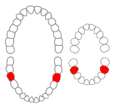
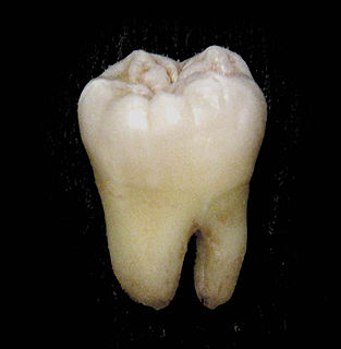
The mandibular first molar or six-year molar is the tooth located distally from both the mandibular second premolars of the mouth but mesial from both mandibular second molars. It is located on the mandibular (lower) arch of the mouth, and generally opposes the maxillary (upper) first molars and the maxillary 2nd premolar in normal class I occlusion. The function of this molar is similar to that of all molars in regard to grinding being the principal action during mastication, commonly known as chewing. There are usually five well-developed cusps on mandibular first molars: two on the buccal, two lingual, and one distal. The shape of the developmental and supplementary grooves, on the occlusal surface, are describes as being 'M' shaped. There are great differences between the deciduous (baby) mandibular molars and those of the permanent mandibular molars, even though their function are similar. The permanent mandibular molars are not considered to have any teeth that precede it. Despite being named molars, the deciduous molars are followed by permanent premolars.

The mandibular second molar is the tooth located distally from both the mandibular first molars of the mouth but mesial from both mandibular third molars. This is true only in permanent teeth. The function of this molar is similar to that of all molars in regard to grinding being the principal action during mastication, commonly known as chewing. Though there is more variation between individuals than that of the first mandibular molar, there are usually four cusps on mandibular second molars: two on the buccal and two lingual. There are great differences between the deciduous (baby) mandibular molars and those of the permanent mandibular molars, even though their function is similar. The permanent mandibular molars are not considered to have any teeth that precede them. Despite being named molars, the deciduous molars are followed by permanent premolars.

In anatomy, the Curve of Spee is defined as the curvature of the mandibular occlusal plane beginning at the canine and following the buccal cusps of the posterior teeth, continuing to the terminal molar. According to another definition the curve of Spee is an anatomic curvature of the occlusal alignment of the teeth, beginning at the tip of the lower incisor, following the buccal cusps of the natural premolars and molars and continuing to the anterior border of the ramus. It is named for the German embryologist Ferdinand Graf von Spee (1855–1937), who was first to describe the anatomic relations of human teeth in the sagittal plane.

The dental arches are the two arches of teeth, one on each jaw, that together constitute the dentition. In humans and many other species; the superior dental arch is a little larger than the inferior arch, so that in the normal condition the teeth in the maxilla slightly overlap those of the mandible both in front and at the sides. The way that the jaws, and thus the dental arches, approach each other when the mouth closes, which is called the occlusion, determines the occlusal relationship of opposing teeth, and it is subject to malocclusion if facial or dental development was imperfect.
Dental anatomy is a field of anatomy dedicated to the study of human tooth structures. The development, appearance, and classification of teeth fall within its purview. Tooth formation begins before birth, and the teeth's eventual morphology is dictated during this time. Dental anatomy is also a taxonomical science: it is concerned with the naming of teeth and the structures of which they are made, this information serving a practical purpose in dental treatment.
Occlusion, in a dental context, means simply the contact between teeth. More technically, it is the relationship between the maxillary (upper) and mandibular (lower) teeth when they approach each other, as occurs during chewing or at rest.
This is a list of definitions of commonly used terms of location and direction in dentistry. This set of terms provides orientation within the oral cavity, much as anatomical terms of location provide orientation throughout the body.

Post-canine megadontia is a relative enlargement of the molars and premolars compared to the size of the incisors and canines. This phenomenon is seen in some early hominid ancestors such as Paranthropus aethiopicus.

Crossbite is a form of malocclusion where a tooth has a more buccal or lingual position than its corresponding antagonist tooth in the upper or lower dental arch. In other words, crossbite is a lateral misalignment of the dental arches.
Serial extraction is the planned extraction of certain deciduous teeth and specific permanent teeth in an orderly sequence and predetermined pattern to guide the erupting permanent teeth into a more favorable position.
Tikitherium is an extinct genus of mammaliaforms from the Late Triassic. It is thought to be an insectivore and a close relative to Docodonta. Tikitherium refers to Tiki, the village located near the Tiki Formation where the specimen was found, and therium is Greek for “Beast”. The species was named copei in honor of Edward Drinker Cope for his pioneering discoveries towards understanding mammalian molars.
Orthodontic indices are one of the tools that are available for orthodontists to grade and assess malocclusion. Orthodontic indices can be useful for an epidemiologist to analyse prevalence and severity of malocclusion in any population.
Occlusion according to The Glossary of Prosthodontic Terms Ninth Edition is defined as 'the static relationship between the incising or masticating surfaces of the maxillary or mandibular teeth or tooth analogues'.


