Cystic artery

Last updated
Cystic artery
Gray532.png
The cystic artery branches from the right hepatic artery.
Bilebladder.png
Relationship to other vessels. (Note that relationships are highly variable -- see below.)
Details
SourceRight hepatic artery
BranchesSuperior branch
Deep branch
Vein Cystic vein
Supplies Gall bladder and cystic duct
Identifiers
Latin arteria cystica
TA98 A12.2.12.031
TA2 4230
FMA 14835
Anatomical terminology
3D medical animation still shot of cystic artery 3D Medical Animation still shot Cystic Artery.jpg
3D medical animation still shot of cystic artery

The cystic artery (also known as bachelor artery) is (usually) a branch of the right hepatic artery that provides arterial supply to the gallbladder and contributes arterial supply to the extrahepatic bile ducts.

Contents

Anatomy

The cystic artery usually has a diameter of less than 3mm. [1]

Origin

The cystic artery arises from the right hepatic artery in about 80% of cases. [1]

Course

It usually passes posterior to the common hepatic duct within the cystohepatic triangle. Within the triangle, it is usually superior to the cystic duct (if it does not pass superior to the cystic duct, it may be situated outside the triangle). [1]

Branches

Upon reaching the superior aspect of the neck of the gallbladder, it splits into superficial and deep branches. These branches then form an anastomotic network over the surface of the body and fundus of the gallbladder. [1]

It produces 2 to 4 minor branches (known as Calot’s arteries) that supply part of the cystic duct and cervix of the gallbladder before dividing into the major superficial and deep branches at the superior aspect of the gallbladder neck:

The cystic artery additionally emits fine branches that contribute to the arterial supply of the extrahepatic bile ducts. [1]

Variation

In the classic arrangement, occurring with a frequency of approximately 70%, a singular cystic artery originates from the geniculate flexure of the right hepatic artery in the upper portion of the hepatobiliary triangle. [2] A site of origin from a more proximal or distal portion of the right hepatic artery is also considered relatively normal.

Double cystic artery

When superficial and deep branches of the cystic artery do not share a common origin it is defined as a double cystic artery occurring with a frequency of 15%.

The deep branch consistently arises from the right hepatic artery which is generally also the source of origin of the superficial branch, however in some cases it has been found to initiate from the anterior segmental artery, middle hepatic artery, left hepatic artery, superior mesenteric artery, gastroduodenal artery or retroduodenal artery.

Approximately half of superficial cystic arteries have been shown to enter through the hepatobiliary triangle, while deep cystic arteries are often quite small in length and diameter. Tripling of the cystic artery is very rare, occurring in between 0-0.3% of cases.

Aberrant origin of the right hepatic artery

Unusual anatomy of the right hepatic can itself affect the path and form of the cystic artery, with the most frequent variation resulting from an aberrant origin of the right hepatic artery which is described in between 2-16% of cases.

Generally this anomalous source is the superior mesenteric artery or more rarely the abdominal aorta, producing what has been described as a "replacing right hepatic artery", passing through the hepatobiliary triangle and running posterior and parallel to the cystic duct. [3]

Because of the close proximity to the gallbladder to the (replacing) right hepatic artery a "caterpillar" or "Moynihan's" hump may form and this artery generally produces multiple short cystic branches rather than a single cystic artery. [4]

Left hepatic artery

The cystic artery can arise from the left hepatic artery, in which case it usually travels through a passage of liver parenchyma, approaching the neck of the gallbladder, before bifurcating into ascending (or superior) and descending (or inferior) branches. This arrangement is found in roughly 1% of cases.

This form of the cystic artery does not pass through the hepatobiliary triangle and has been found located both anteriorly and posteriorly to the common hepatic duct. [2]

Recurrent cystic artery

In less than 1% of patients a form known as a recurrent cystic artery is found – the cystic artery arises from the left hepatic artery and passes through either the cholecystoduodenal or cholecystocolic ligaments (connecting the gallbladder to the duodenum and transverse colon respectively), follows the right edge of the hepatoduodenal ligament, and connects to the fundus of the gallbladder before descending to supply the body and neck. [2]

Low-lying cystic artery

When a single cystic artery arises from gastroduodenal artery it forms a configuration known as low-lying cystic artery, as it passes inferior to the cystic duct through the cholecystoduodenal ligament, and therefore remains outside the hepatobiliary triangle. [2]

This condition has alternatively been described as an inferior cystic artery and transposition of the cystic artery and duct with a prevalence at approximately 5%.

In 25% of these patients with this course there will be a secondary arterial supply to the gallbladder, with the low-lying artery acting as the equivalent of the posterior branch. [4]

Clinical significance

It must be identified and ligated in operations that remove the gallbladder (cholecystectomies).

Related Research Articles

<span class="mw-page-title-main">Duodenum</span> First section of the small intestine

The duodenum is the first section of the small intestine in most higher vertebrates, including mammals, reptiles, and birds. In mammals it may be the principal site for iron absorption. The duodenum precedes the jejunum and ileum and is the shortest part of the small intestine.

Articles related to anatomy include:

<span class="mw-page-title-main">Portal vein</span> Short thick vein formed by the union of the superior mesenteric vein and the splenic vein

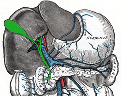
The portal vein or hepatic portal vein (HPV) is a blood vessel that carries blood from the gastrointestinal tract, gallbladder, pancreas and spleen to the liver. This blood contains nutrients and toxins extracted from digested contents. Approximately 75% of total liver blood flow is through the portal vein, with the remainder coming from the hepatic artery proper. The blood leaves the liver to the heart in the hepatic veins.

<span class="mw-page-title-main">Femoral artery</span> Large artery in the thigh

The femoral artery is a large artery in the thigh and the main arterial supply to the thigh and leg. The femoral artery gives off the deep femoral artery and descends along the anteromedial part of the thigh in the femoral triangle. It enters and passes through the adductor canal, and becomes the popliteal artery as it passes through the adductor hiatus in the adductor magnus near the junction of the middle and distal thirds of the thigh.

<span class="mw-page-title-main">Common bile duct</span> Gastrointestinal duct

The bile duct is a part of the biliary tract. It is formed by the union of the common hepatic duct and cystic duct. It ends by uniting with the pancreatic duct to form the hepatopancreatic ampulla. It possesses its own sphincter to enable regulation of bile flow.

<span class="mw-page-title-main">Cystic duct</span> Organ duct

The cystic duct is the duct that (typically) joins the gallbladder and the common hepatic duct; the union of the cystic duct and common hepatic duct forms the bile duct. Its length varies. It is often tortuous.

<span class="mw-page-title-main">Abdominal aorta</span> Largest artery in the abdomen

In human anatomy, the abdominal aorta is the largest artery in the abdominal cavity. As part of the aorta, it is a direct continuation of the descending aorta.

<span class="mw-page-title-main">Common hepatic duct</span> Exocrine duct

The common hepatic duct is the first part of the biliary tract. It joins the cystic duct coming from the gallbladder to form the common bile duct.

<span class="mw-page-title-main">Gastroduodenal artery</span>

In anatomy, the gastroduodenal artery is a small blood vessel in the abdomen. It supplies blood directly to the pylorus and proximal part of the duodenum. It also indirectly supplies the pancreatic head.

<span class="mw-page-title-main">Common carotid artery</span> One of the two arteries that supply the head and neck with blood

In anatomy, the left and right common carotid arteries (carotids) are arteries that supply the head and neck with oxygenated blood; they divide in the neck to form the external and internal carotid arteries.

<span class="mw-page-title-main">Hepatic artery proper</span>

The hepatic artery proper is the artery that supplies the liver and gallbladder. It raises from the common hepatic artery, a branch of the celiac artery.

<span class="mw-page-title-main">Occipital artery</span>

The occipital artery is a branch of the external carotid artery that provides arterial supply to the back of the scalp, sternocleidomastoid muscles, and deep muscles of the back and neck.

<span class="mw-page-title-main">Inferior phrenic arteries</span>

The inferior phrenic artery is a bilaterally paired artery of the abdominal cavity which represents the main source of arterial supply to the diaphragm. Each artery usually arises either from the coeliac trunk or the abdominal aorta, however, their origin is highly variable and the different sites of origin are different for the left artery and right artery. The superior suprarenal artery is a branch of the inferior phrenic artery.

<span class="mw-page-title-main">Posterior auricular artery</span> Small artery

The posterior auricular artery is a small artery that arises from the external carotid artery. It ascends along the side of the head. It supplies several muscles of the neck and several structures of the head.

<span class="mw-page-title-main">Superior pancreaticoduodenal artery</span> Artery

The superior pancreaticoduodenal artery is an artery that supplies blood to the duodenum and pancreas.

In anatomy, arterial tree is used to refer to all arteries and/or the branching pattern of the arteries. This article regards the human arterial tree. Starting from the aorta:

<span class="mw-page-title-main">Cystohepatic triangle</span> Space between the cystic duct, the common hepatic duct, and the liver

The cystohepatic triangle is an anatomic space bordered by the cystic duct inferiorly, the common hepatic duct medially, and the inferior surface of the liver superiorly.

<span class="mw-page-title-main">Hepatic plexus</span>

The hepatic plexus is a sympathetic and parasympathetic nerve plexus that provides innervation to the parenchyma of the liver as well as contributing innervation to some other abdominal structures.

<span class="mw-page-title-main">Cystic vein</span>

The cystic veins drain venous blood from the gallbladder and the cystic duct. The cystic veins either drain into various branches and tributaries of the hepatic portal vein.

<span class="mw-page-title-main">Outline of human anatomy</span> Overview of and topical guide to human anatomy

The following outline is provided as an overview of and topical guide to human anatomy:

References

  1. 1 2 3 4 5 Standring, Susan (2020). Gray's Anatomy: The Anatomical Basis of Clinical Practice (42th ed.). New York. pp. 1219–1220. ISBN   978-0-7020-7707-4. OCLC   1201341621.{{cite book}}: CS1 maint: location missing publisher (link)
  2. 1 2 3 4 Balija, M; Huis, M; Nikolic, V; Stulhofer, M (1999). "Laparoscopic visualization of the cystic artery anatomy". World Journal of Surgery. 23 (7): 703–7, discussion 707. doi:10.1007/pl00012372. PMID   10390590. S2CID   23174989.
  3. Mlakar, Boštjan; Gadžijev, Eldar M.; Ravnik, Dean; Hribernik, Marija (2003). "Anatomical Variations of the Cystic Artery". European Journal of Morphology. 41 (1): 31–4. doi:10.1076/ejom.41.1.31.28103. PMID   15121546.
  4. 1 2 Hugh, T. B.; Kelly, M. D.; Li, B (1992). "Laparoscopic anatomy of the cystic artery". American Journal of Surgery. 163 (6): 593–5. doi:10.1016/0002-9610(92)90564-8. PMID   1534464.