DNA unwinding element

Last updated
DNA unwinding at the DUE, allowing for formation of replication fork for DNA replication to occur. Unwound DNA Duplex.png
DNA unwinding at the DUE, allowing for formation of replication fork for DNA replication to occur.

A DNA unwinding element (DUE or DNAUE) is the initiation site for the opening of the double helix structure of the DNA at the origin of replication for DNA synthesis. [1] It is A-T rich and denatures easily due to its low helical stability, [2] which allows the single-strand region to be recognized by origin recognition complex.

Contents

DUEs are found in both prokaryotic and eukaryotic organisms, but were first discovered in yeast and bacteria origins, by Huang Kowalski. [3] [4] The DNA unwinding allows for access of replication machinery to the newly single strands. [1] In eukaryotes, DUEs are the binding site for DNA-unwinding element binding (DUE-B) proteins required for replication initiation. [3] In prokaryotes, DUEs are found in the form of tandem consensus sequences flanking the 5' end of DnaA binding domain. [2] The act of unwinding at these A-T rich elements occurs even in absence of any origin binding proteins due to negative supercoiling forces, making it an energetically favourable action. [2] DUEs are typically found spanning 30-100 bp of replication origins. [4] [5]

Function

The specific unwinding of the DUE allows for initiation complex assembly at the site of replication on single-stranded DNA, as discovered by Huang Kowalski. [4] The DNA helicase and associated enzymes are now able to bind to the unwound region, creating a replication fork start. The unwinding of this duplex strand region is associated with a low free energy requirement, due to helical instability caused by specific base-stacking interactions, in combination with counteracting supercoiling. [4] [6] Negative supercoiling allows the DNA to be stable upon melting, driven by reduction of torsional stress. [5] Found in the replication origins of both bacteria and yeast, as well as present in some mammalian ones. [5] Found to be between 30-100 bp long. [4] [5]

Prokaryotes

In prokaryotes, most of the time DNA replication is occurring from one single replication origin on one single strand of DNA sequence. Whether this genome is linear or circularized, bacteria have own machinery necessary for replication to occur. [7]

Process

In bacteria, the protein DnaA is the replication initiator. [8] It gets loaded onto oriC at a DnaA box sequence where it binds and assembles filaments to open duplex and recruit DnaB helicase with the help of DnaC. DnaA is highly conserved and has two DNA binding domains. Just upstream to this DnaA box, is three tandem 13-mer sequences. These tandem sequences, labelled L, M, R from 5' to 3' are the bacterial DUEs. Two out of three of these A-T rich regions (M and R) become unwound upon binding of DnaA to DnaA box, via close proximity to unwinding duplex. The final 13-mer sequence L, farthest from this DnaA box eventually gets unwound upon DnaB helicase encircling it. This forms a replication bubble for DNA replication to then proceed. [2]

Archaea use a simpler homolog of the eukaryotic origin recognition complex to find the origin of replication, at sequences termed the origin recognition box (ORB). [9]

Favourability

Unwinding of these three DUEs is a necessary step for DNA replication to initiate. The distant pull from duplex melting at the DnaA box sequence is what induces further melting at the M and R DUE sites. The more distant L site is then unwound by DnaB binding. Unwinding of these 13-mer sites is independent of oriC-binding proteins. It is the generation of negative supercoiling that causes the unwinding. [2]

The rates of DNA unwinding in the three E. coli DUEs were experimentally compared through nuclear resonance spectroscopy. In physiological conditions, the opening efficiency of each of the A-T rich sequences differed from one another. Largely due to the different distantly surrounding sequences. [2]

Additionally, melting of AT/TA base pairs were found to be much faster than that of GC/CG pairs (15-240s−1 vs. ~20s−1). This supports the idea that A-T sequences are evolutionarily favoured in DUE elements due to their ease of unwinding. [2]

Consensus Sequence

The three 13-mer sequences identified as DUEs in E. coli, are well-conserved at the origin of replication of all documented enteric bacteria. A general consensus sequence was made via comparison of conserved bacteria to form an 11 base sequence, GATCTnTTnTTTT. E. coli contains 9 bases of the 11 base consensus sequence in its oriC, within the 13-mer sequences. These sequences are found exclusively at the single origin of replication; not anywhere else within the genome sequence. [2]

Eukaryotes

Eukaryotic replication mechanisms work in relatively similar ways to that of prokaryotes, but is under more finely-tuned regulation. [10] There is a need to ensure that each DNA molecule is replicated only once and that this is occurring in the proper location at the proper time. [7] Operates in response to extracellular signals that coordinate initiation of division, differently from tissue to tissue. External signals trigger replication in S phase via production of cyclins which activate cyclin-dependent kinases (CDK) to form complexes. [10]

DNA replication in eukaryotes initiates upon origin recognition complex (ORC) binding to the origin. This occurs at G1 cell phase serving to drive the cell cycle forward into S phase. This binding allows for further factor binding to create a pre-replicative complex (pre-RC). Pre-RC triggered to initiate when cyclin-dependent kinase (CDK) and Dbf4-dependent kinase (DDK) bind to it. Initiation complexes then allow for recruitment of MCM helicase activator Cdc45 and subsequent unwinding of duplex at origin. [10] [11]

Replication in eukaryotes is initiated at multiple sites on the sequence, forming multiple replication forks simultaneously. This efficiency is required with the large genomes that they need to replicate. [10]

In eukaryotes, nucleosome structures can complicate replication initiation. [4] They can block access of DUE-B's to the DUE, thus suppressing transcription initiation. [4] Can impede on rate. The linear nature of eukaryotic DNA, vs prokaryotic circular DNA, though, is easier to unwind its duplex once has been properly unwound from nucleosome. [4] Activity of DUE can be modulated by transcription factors like ABF1. [4]

Yeast

A common yeast model system that well-represents eukaryotic replication is Saccharomyces cerevisiae . [12] It possesses autonomously replicating sequences (ARSs) that are transformed and maintained well in a plasmid. Some of these ARSs are seen to act as replication origins. These ARSs are composed of three domains A, B, and C. The A domain is where the ARS consensu [5] s sequence resides, coined an ACS. The B domain contains the DUE. Lastly, the C domain is necessary for facilitating protein-protein interactions. [12] ARSs are found distributed across 16 chromosomes, repeated every 30–40 kb. [12]

Between species, these ARS sequences are variable, but their A, B, and C domains are well conserved. [12] Any alterations in the DUE (domain B) causes lower overall function of the ARS as a whole in replication initiation. This was found via studies using imino exchange and NMR spectroscopy. [2]

Mammals

DUEs found in some mammalian replication origins to date. In general, very little mammalian origins of replication have been well-analyzed, so difficult to determine how prevalent the DUEs are, in their defined replication origins. [5]

Human cells still have very little detailing of their origins. [5] It is known that replication initiates in large initiation zone areas, associated with known proteins like the c-myc and β-globin gene. Ones with DUEs thought to act in nearly same way as yeast cells. [5]

DUE in origin of plasmids in mammalian cells, SV40, found to be associated with a T-ag hexamer, that introduces opposite supercoiling to increase favourability of strand unwinding. [4]

Mammals with DUEs have shown evidence of structure-forming abilities that provide single-stranded stability of unwound DNA. These include cruciforms, intramolecular triplexes, and more. [5]

DUE-binding proteins

DNA unwinding element proteins (DUE-Bs) are found in eukaryotes. [3]

They act to initiate strand separation by binding to DUE. [3] DUE-B sequence homologs found among a variety of animal species- fish, amphibians, and rodents. [3] DUE-B's have disordered C-terminal domains that bind to the DUE by recognition of this C-terminus. [3] No other sequence specificity involved in this interaction. [3] Confirmed by inducing mutations along length of DUE-B sequence, but in all cases dimerization abilities remaining intact. [3] Upon binding DNA, C-terminus becomes ordered, imparting a greater stability against protease degradation. [3] DUE-B's are 209 residues in total, 58 of which are disordered until bound to DUE. [3] DUE-B's hydrolyze ATP In order to function. [3] Also possess similar sequence to aminoacyl-tRNA synthetase, and were previously classified a such. [13] DUE-Bs form homodimers that create an extended beta-sheet secondary structure extending across it. [3] Two of these homodimers come together to form the overall asymmetric DUE-B structure. [3]

In formation of the pre-RC, Cdc45 is localized to the DUE for activity via interaction with a DUE-B. [11] Allowing for duplex unwinding and replication initiation. [11]

In humans, DUE-B's are 60 amino acids longer than its yeast ortholog counterparts. [13] Both localized mainly in the nucleus. [13]

DUE-B levels are in consistent quantity, regardless of cell cycle. [13] In S phase though, DUE-Bs can be temporarily phosphorylated to prevent premature replication. [13] DUE-B activity is covalently controlled. [13] The assembly of these DUE-Bs at the DUE regions is dependent on local kinase and phosphatase activity. [13] DUE-B's can also be down-regulated by siRNAs and have been implicated in extended G1 stages. [13]

Mutation Implications

Mutations that impair the unwinding at DUE sites directly impede DNA replication activity. [14] This can be a result of deletions/changes in the DUE region, the addition of reactive reagents, or the addition of specific nuclease. [4] DUE sites are relatively insensitive to point mutations though, maintaining their activity in when altering bases in protein binding sites. [4] In many cases, DUE activity can be partially regained by increasing temperature. [4] Can be regained by the re-addition of DUE site as well. [3]

If there is a severe enough mutation to DUE causing it to no longer be bound to DUE-B, Cdc45 cannot associate and will not bind to c-myc transcription factor. This can be recovered in disease-related (ATTCT)(n) length expansions of the DUE sequence. If DUE activity regained in excess, could cause dysregulated origin formation and cell cycle progression. [11]

In eukaryotes, when DUE-B's are knocked out, the cell will not go into S phase of its cycle, where DNA replication occurs. Increased apoptosis will result. [3] But, activity can be rescued by re-addition of the DUE-B's, even from a different species. This is because DUE-B's are homologous between species. [3] For example, if DUE-B in Xenopus egg are mutated, no DNA replication will occur, but can be saved by addition of HeLa DUE-B's to regain full functionality. [3]

Related Research Articles

<span class="mw-page-title-main">DNA replication</span> Biological process

In molecular biology, DNA replication is the biological process of producing two identical replicas of DNA from one original DNA molecule. DNA replication occurs in all living organisms acting as the most essential part of biological inheritance. This is essential for cell division during growth and repair of damaged tissues, while it also ensures that each of the new cells receives its own copy of the DNA. The cell possesses the distinctive property of division, which makes replication of DNA essential.

<span class="mw-page-title-main">RNA polymerase</span> Enzyme that synthesizes RNA from DNA

In molecular biology, RNA polymerase, or more specifically DNA-directed/dependent RNA polymerase (DdRP), is an enzyme that catalyzes the chemical reactions that synthesize RNA from a DNA template.

<span class="mw-page-title-main">Helicase</span> Class of enzymes to unpack an organisms genes

Helicases are a class of enzymes thought to be vital to all organisms. Their main function is to unpack an organism's genetic material. Helicases are motor proteins that move directionally along a nucleic acid phosphodiester backbone, separating two hybridized nucleic acid strands, using energy from ATP hydrolysis. There are many helicases, representing the great variety of processes in which strand separation must be catalyzed. Approximately 1% of eukaryotic genes code for helicases.

dnaB helicase

DnaB helicase is an enzyme in bacteria which opens the replication fork during DNA replication. Although the mechanism by which DnaB both couples ATP hydrolysis to translocation along DNA and denatures the duplex is unknown, a change in the quaternary structure of the protein involving dimerisation of the N-terminal domain has been observed and may occur during the enzymatic cycle. Initially when DnaB binds to dnaA, it is associated with dnaC, a negative regulator. After DnaC dissociates, DnaB binds dnaG.

<span class="mw-page-title-main">DnaA</span>

DnaA is a protein that activates initiation of DNA replication in bacteria. Based on the Replicon Model, a positively active initiator molecule contacts with a particular spot on a circular chromosome called the replicator to start DNA replication. It is a replication initiation factor which promotes the unwinding of DNA at oriC. The DnaA proteins found in all bacteria engage with the DnaA boxes to start chromosomal replication. In addition to the DnaA protein, its concentration, binding to DnaA-boxes, and binding of ATP or ADP, we will cover the regulation of the DnaA gene, the unique characteristics of the DnaA gene expression, promoter strength, and translation efficiency. The onset of the initiation phase of DNA replication is determined by the concentration of DnaA. DnaA accumulates during growth and then triggers the initiation of replication. Replication begins with active DnaA binding to 9-mer (9-bp) repeats upstream of oriC. Binding of DnaA leads to strand separation at the 13-mer repeats. This binding causes the DNA to loop in preparation for melting open by the helicase DnaB.

<span class="mw-page-title-main">Origin of replication</span> Sequence in a genome

The origin of replication is a particular sequence in a genome at which replication is initiated. Propagation of the genetic material between generations requires timely and accurate duplication of DNA by semiconservative replication prior to cell division to ensure each daughter cell receives the full complement of chromosomes. This can either involve the replication of DNA in living organisms such as prokaryotes and eukaryotes, or that of DNA or RNA in viruses, such as double-stranded RNA viruses. Synthesis of daughter strands starts at discrete sites, termed replication origins, and proceeds in a bidirectional manner until all genomic DNA is replicated. Despite the fundamental nature of these events, organisms have evolved surprisingly divergent strategies that control replication onset. Although the specific replication origin organization structure and recognition varies from species to species, some common characteristics are shared.

<span class="mw-page-title-main">Pre-replication complex</span>

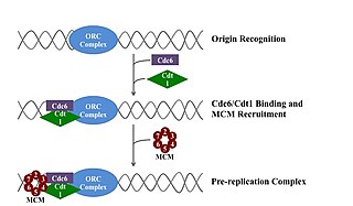
A pre-replication complex (pre-RC) is a protein complex that forms at the origin of replication during the initiation step of DNA replication. Formation of the pre-RC is required for DNA replication to occur. Complete and faithful replication of the genome ensures that each daughter cell will carry the same genetic information as the parent cell. Accordingly, formation of the pre-RC is a very important part of the cell cycle.

DNA gyrase, or simply gyrase, is an enzyme within the class of topoisomerase and is a subclass of Type II topoisomerases that reduces topological strain in an ATP dependent manner while double-stranded DNA is being unwound by elongating RNA-polymerase or by helicase in front of the progressing replication fork. It is the only known enzyme to actively contribute negative supercoiling to DNA, while it also is capable of relaxing positive supercoils. It does so by looping the template to form a crossing, then cutting one of the double helices and passing the other through it before releasing the break, changing the linking number by two in each enzymatic step. This process occurs in bacteria, whose single circular DNA is cut by DNA gyrase and the two ends are then twisted around each other to form supercoils. Gyrase is also found in eukaryotic plastids: it has been found in the apicoplast of the malarial parasite Plasmodium falciparum and in chloroplasts of several plants. Bacterial DNA gyrase is the target of many antibiotics, including nalidixic acid, novobiocin, albicidin, and ciprofloxacin.

<span class="mw-page-title-main">Replisome</span> Molecular complex

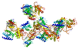
The replisome is a complex molecular machine that carries out replication of DNA. The replisome first unwinds double stranded DNA into two single strands. For each of the resulting single strands, a new complementary sequence of DNA is synthesized. The total result is formation of two new double stranded DNA sequences that are exact copies of the original double stranded DNA sequence.

A licensing factor is a protein or complex of proteins that allows an origin of replication to begin DNA replication at that site. Licensing factors primarily occur in eukaryotic cells, since bacteria use simpler systems to initiate replication. However, many archaea use homologues of eukaryotic licensing factors to initiate replication.

In molecular biology, origin recognition complex (ORC) is a multi-subunit DNA binding complex that binds in all eukaryotes and archaea in an ATP-dependent manner to origins of replication. The subunits of this complex are encoded by the ORC1, ORC2, ORC3, ORC4, ORC5 and ORC6 genes. ORC is a central component for eukaryotic DNA replication, and remains bound to chromatin at replication origins throughout the cell cycle.

<span class="mw-page-title-main">Prokaryotic DNA replication</span> DNA Replication in prokaryotes

Prokaryotic DNA Replication is the process by which a prokaryote duplicates its DNA into another copy that is passed on to daughter cells. Although it is often studied in the model organism E. coli, other bacteria show many similarities. Replication is bi-directional and originates at a single origin of replication (OriC). It consists of three steps: Initiation, elongation, and termination.

<span class="mw-page-title-main">Eukaryotic DNA replication</span> DNA replication in eukaryotic organisms

Eukaryotic DNA replication is a conserved mechanism that restricts DNA replication to once per cell cycle. Eukaryotic DNA replication of chromosomal DNA is central for the duplication of a cell and is necessary for the maintenance of the eukaryotic genome.

<span class="mw-page-title-main">Bacterial transcription</span>

Bacterial transcription is the process in which a segment of bacterial DNA is copied into a newly synthesized strand of messenger RNA (mRNA) with use of the enzyme RNA polymerase.

<span class="mw-page-title-main">Minichromosome maintenance</span>

The minichromosome maintenance protein complex (MCM) is a DNA helicase essential for genomic DNA replication. Eukaryotic MCM consists of six gene products, Mcm2–7, which form a heterohexamer. As a critical protein for cell division, MCM is also the target of various checkpoint pathways, such as the S-phase entry and S-phase arrest checkpoints. Both the loading and activation of MCM helicase are strictly regulated and are coupled to cell growth cycles. Deregulation of MCM function has been linked to genomic instability and a variety of carcinomas.

<span class="mw-page-title-main">Transcription factor II B</span> Mammalian protein found in Homo sapiens

Transcription factor II B (TFIIB) is a general transcription factor that is involved in the formation of the RNA polymerase II preinitiation complex (PIC) and aids in stimulating transcription initiation. TFIIB is localised to the nucleus and provides a platform for PIC formation by binding and stabilising the DNA-TBP complex and by recruiting RNA polymerase II and other transcription factors. It is encoded by the TFIIB gene, and is homologous to archaeal transcription factor B and analogous to bacterial sigma factors.

RNA polymerase II holoenzyme is a form of eukaryotic RNA polymerase II that is recruited to the promoters of protein-coding genes in living cells. It consists of RNA polymerase II, a subset of general transcription factors, and regulatory proteins known as SRB proteins.

<span class="mw-page-title-main">Circular chromosome</span> Type of chromosome

A circular chromosome is a chromosome in bacteria, archaea, mitochondria, and chloroplasts, in the form of a molecule of circular DNA, unlike the linear chromosome of most eukaryotes.

<span class="mw-page-title-main">Control of chromosome duplication</span>

In cell biology, eukaryotes possess a regulatory system that ensures that DNA replication occurs only once per cell cycle.

<span class="mw-page-title-main">Cruciform DNA</span>

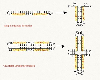
Cruciform DNA is a form of non-B DNA, or an alternative DNA structure. The formation of cruciform DNA requires the presence of palindromes called inverted repeat sequences. These inverted repeats contain a sequence of DNA in one strand that is repeated in the opposite direction on the other strand. As a result, inverted repeats are self-complementary and can give rise to structures such as hairpins and cruciforms. Cruciform DNA structures require at least a six nucleotide sequence of inverted repeats to form a structure consisting of a stem, branch point and loop in the shape of a cruciform, stabilized by negative DNA supercoiling.

References

  1. 1 2 Kowalski D, Eddy MJ (December 1989). "The DNA unwinding element: a novel, cis-acting component that facilitates opening of the Escherichia coli replication origin". The EMBO Journal. 8 (13): 4335–44. doi:10.1002/j.1460-2075.1989.tb08620.x. PMC   401646 . PMID   2556269.
  2. 1 2 3 4 5 6 7 8 9 Coman D, Russu IM (May 2005). "Base pair opening in three DNA-unwinding elements". The Journal of Biological Chemistry. 280 (21): 20216–21. doi: 10.1074/jbc.M502773200 . PMID   15784615.
  3. 1 2 3 4 5 6 7 8 9 10 11 12 13 14 15 16 17 Kemp M, Bae B, Yu JP, Ghosh M, Leffak M, Nair SK (April 2007). "Structure and function of the c-myc DNA-unwinding element-binding protein DUE-B". The Journal of Biological Chemistry. 282 (14): 10441–8. doi: 10.1074/jbc.M609632200 . PMID   17264083.
  4. 1 2 3 4 5 6 7 8 9 10 11 12 13 DePamphilis ML (1993). "Eukaryotic DNA replication: anatomy of an origin". Annual Review of Biochemistry. 62 (1): 29–63. doi:10.1146/annurev.bi.62.070193.000333. PMID   8352592.
  5. 1 2 3 4 5 6 7 8 9 Potaman VN, Pytlos MJ, Hashem VI, Bissler JJ, Leffak M, Sinden RR (2006). Wells RD, Ashizawa T (eds.). Genetic Instabilities and Neurological Diseases (Second ed.). Burlington: Academic Press. pp. 447–460. doi:10.1016/B978-012369462-1/50031-4. ISBN   9780123694621.
  6. Natale DA, Schubert AE, Kowalski D (April 1992). "DNA helical stability accounts for mutational defects in a yeast replication origin". Proceedings of the National Academy of Sciences of the United States of America. 89 (7): 2654–8. Bibcode:1992PNAS...89.2654N. doi: 10.1073/pnas.89.7.2654 . PMC   48720 . PMID   1557369.
  7. 1 2 Zyskind JW, Smith DW (2001). Brenner S, Miller JH (eds.). Encyclopedia of Genetics. New York: Academic Press. pp. 1381–1387. doi:10.1006/rwgn.2001.0938. ISBN   9780122270802.
  8. Chodavarapu S, Kaguni JM (2016-01-01). "Replication Initiation in Bacteria". In Kaguni LS, Oliveira MT (eds.). The Enzymes. DNA Replication Across Taxa. Vol. 39. Academic Press. pp. 1–30. doi:10.1016/bs.enz.2016.03.001. ISBN   9780128047354. PMC   5551690 . PMID   27241926.
  9. Bell SD (2012). "Archaeal Orc1/Cdc6 Proteins". The Eukaryotic Replisome: A Guide to Protein Structure and Function. Subcellular Biochemistry. Vol. 62. pp. 59–69. doi:10.1007/978-94-007-4572-8_4. ISBN   978-94-007-4571-1. PMID   22918580.
  10. 1 2 3 4 Bhagavan, N. V.; Ha, Chung-Eun (2015). Essentials of Medical Biochemistry (Second ed.). San Diego: Academic Press. pp. 401–417. ISBN   9780124166875.
  11. 1 2 3 4 Chowdhury A, Liu G, Kemp M, Chen X, Katrangi N, Myers S, Ghosh M, Yao J, Gao Y, Bubulya P, Leffak M (March 2010). "The DNA unwinding element binding protein DUE-B interacts with Cdc45 in preinitiation complex formation". Molecular and Cellular Biology. 30 (6): 1495–507. doi:10.1128/MCB.00710-09. PMC   2832489 . PMID   20065034.
  12. 1 2 3 4 Dhar MK, Sehgal S, Kaul S (May 2012). "Structure, replication efficiency and fragility of yeast ARS elements". Research in Microbiology. 163 (4): 243–53. doi:10.1016/j.resmic.2012.03.003. PMID   22504206.
  13. 1 2 3 4 5 6 7 8 Casper JM, Kemp MG, Ghosh M, Randall GM, Vaillant A, Leffak M (April 2005). "The c-myc DNA-unwinding element-binding protein modulates the assembly of DNA replication complexes in vitro". The Journal of Biological Chemistry. 280 (13): 13071–83. doi: 10.1074/jbc.M404754200 . PMID   15653697.
  14. Umek RM, Kowalski D (November 1990). "The DNA unwinding element in a yeast replication origin functions independently of easily unwound sequences present elsewhere on a plasmid". Nucleic Acids Research. 18 (22): 6601–5. doi:10.1093/nar/18.22.6601. PMC   332616 . PMID   2174542.