
Homininae, also called "African hominids" or "African apes", is a subfamily of Hominidae. It includes two tribes, with their extant as well as extinct species: 1) the tribe Hominini ―and 2) the tribe Gorillini (gorillas). Alternatively, the genus Pan is sometimes considered to belong to its own third tribe, Panini. Homininae comprises all hominids that arose after orangutans split from the line of great apes. The Homininae cladogram has three main branches, which lead to gorillas, and to humans and chimpanzees via the tribe Hominini and subtribes Hominina and Panina. There are two living species of Panina and two living species of gorillas, but only one extant human species. Traces of extinct Homo species, including Homo floresiensis have been found with dates as recent as 40,000 years ago. Organisms in this subfamily are described as hominine or hominines.

Homo rudolfensis is an extinct species of archaic human from the Early Pleistocene of East Africa about 2 million years ago (mya). Because H. rudolfensis coexisted with several other hominins, it is debated what specimens can be confidently assigned to this species beyond the lectotype skull KNM-ER 1470 and other partial skull aspects. No bodily remains are definitively assigned to H. rudolfensis. Consequently, both its generic classification and validity are debated without any wide consensus, with some recommending the species to actually belong to the genus Australopithecus as A. rudolfensis or Kenyanthropus as K. rudolfensis, or that it is synonymous with the contemporaneous and anatomically similar H. habilis.

Brodmann area 10 is the anterior-most portion of the prefrontal cortex in the human brain. BA10 was originally defined broadly in terms of its cytoarchitectonic traits as they were observed in the brains of cadavers, but because modern functional imaging cannot precisely identify these boundaries, the terms anterior prefrontal cortex, rostral prefrontal cortex and frontopolar prefrontal cortex are used to refer to the area in the most anterior part of the frontal cortex that approximately covers BA10—simply to emphasize the fact that BA10 does not include all parts of the prefrontal cortex.

The longitudinal fissure is the deep groove that separates the two cerebral hemispheres of the vertebrate brain. Lying within it is a continuation of the dura mater called the falx cerebri. The inner surfaces of the two hemispheres are convoluted by gyri and sulci just as is the outer surface of the brain.

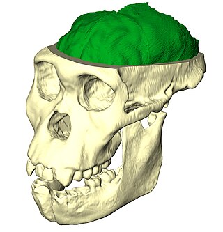
Homo floresiensis( also known as "Flores Man") is an extinct species of small archaic human that inhabited the island of Flores, Indonesia, until the arrival of modern humans about 50,000 years ago.

The Taung Child is the fossilised skull of a young Australopithecus africanus. It was discovered in 1924 by quarrymen working for the Northern Lime Company in Taung, South Africa. Raymond Dart described it as a new species in the journal Nature in 1925.

Liang Bua is a limestone cave on the island of Flores, Indonesia, slightly north of the town of Ruteng in Manggarai Regency, East Nusa Tenggara. The cave demonstrated archaeological and paleontological potential in the 1950s and 1960s as described by the Dutch missionary and archaeologist Theodor L. Verhoeven.
Pachygyria is a congenital malformation of the cerebral hemisphere. It results in unusually thick convolutions of the cerebral cortex. Typically, children have developmental delay and seizures, the onset and severity depending on the severity of the cortical malformation. Infantile spasms are common in affected children, as is intractable epilepsy.

The brain of Albert Einstein has been a subject of much research and speculation. Albert Einstein's brain was removed within seven and a half hours of his death. His apparent regularities or irregularities in the brain have been used to support various ideas about correlations in neuroanatomy with general or mathematical intelligence. Studies have suggested an increased number of glial cells in Einstein's brain.
The size of the brain is a frequent topic of study within the fields of anatomy, biological anthropology, animal science and evolution. Brain size is sometimes measured by weight and sometimes by volume. Neuroimaging intelligence testing can be used to study the volumetric measurements of the brain. Regarding "intelligence testing", a question that has been frequently investigated is the relation of brain size to intelligence. This question is controversial and will be addressed further in the section on intelligence. The measure of brain size and cranial capacity is not just important to humans, but to all mammals.

In human brain anatomy, an operculum, may refer to the frontal, temporal, or parietal operculum, which together cover the insula as the opercula of insula. It can also refer to the occipital operculum, part of the occipital lobe.

In neuroanatomy, a sulcus is a depression or groove in the cerebral cortex. It surrounds a gyrus, creating the characteristic folded appearance of the brain in humans and other mammals. The larger sulci are usually called fissures.
The empathising–systemising (E–S) theory is a theory on the psychological basis of autism and male–female neurological differences originally put forward by English clinical psychologist Simon Baron-Cohen. It classifies individuals based on abilities in empathic thinking (E) and systematic thinking (S). It measures skills using an Empathy Quotient (EQ) and Systemising Quotient (SQ) and attempts to explain the social and communication symptoms in autism spectrum disorders as deficits and delays in empathy combined with intact or superior systemising.

There is much to be discovered about the evolution of the brain and the principles that govern it. While much has been discovered, not everything currently known is well understood. The evolution of the brain has appeared to exhibit diverging adaptations within taxonomic classes such as Mammalia and more vastly diverse adaptations across other taxonomic classes. Brain to body size scales allometrically. This means as body size changes, so do other physiological, anatomical, and biochemical constructs connecting the brain to the body. Small bodied mammals have relatively large brains compared to their bodies whereas large mammals have a smaller brain to body ratios. If brain weight is plotted against body weight for primates, the regression line of the sample points can indicate the brain power of a primate species. Lemurs for example fall below this line which means that for a primate of equivalent size, we would expect a larger brain size. Humans lie well above the line indicating that humans are more encephalized than lemurs. In fact, humans are more encephalized compared to all other primates. This means that human brains have exhibited a larger evolutionary increase in its complexity relative to its size. Some of these evolutionary changes have been found to be linked to multiple genetic factors such as, proteins and other organelles.

In brain anatomy, the lunate sulcus or simian sulcus, also known as the sulcus lunatus, is a fissure in the occipital lobe variably found in humans and more often larger when present in apes and monkeys. The lunate sulcus marks the transition between V1 and V2.
Paleoneurobiology is the study of brain evolution by analysis of brain endocasts to determine endocranial traits and volumes. Considered a subdivision of neuroscience, paleoneurobiology combines techniques from other fields of study including paleontology and archaeology. It reveals specific insight concerning human evolution. The cranium is unique in that it grows in response to the growth of brain tissue rather than genetic guidance, as is the case with bones that support movement. Fossil skulls and their endocasts can be compared to each other, to the skulls and fossils of recently deceased individuals, and even compared to those of other species to make inferences about functional anatomy, physiology and phylogeny. Paleoneurobiology is in large part influenced by developments in neuroscience as a whole; without substantial knowledge about current functionality, it would be impossible to make inferences about the functionality of ancient brains.
The evolution of nervous systems dates back to the first development of nervous systems in animals. Neurons developed as specialized electrical signaling cells in multicellular animals, adapting the mechanism of action potentials present in motile single-celled and colonial eukaryotes. Primitive systems, like those found in protists, use chemical signalling for movement and sensitivity; data suggests these were precursors to modern neural cell types and their synapses. When some animals started living a mobile lifestyle and eating larger food particles externally, they developed ciliated epithelia, contractile muscles and coordinating & sensitive neurons for it in their outer layer.
Professor Michael John Morwood was a New Zealand archaeologist best known for discovering Homo floresiensis. In 2012, he received the Rhys Jones Medal by the Australian Archaeological Association.

Homo naledi is an extinct species of archaic human discovered in 2013 in the Rising Star Cave, Cradle of Humankind, South Africa dating to the Middle Pleistocene 335,000–236,000 years ago. The initial discovery comprises 1,550 specimens, representing 737 different elements, and at least 15 different individuals. Despite this exceptionally high number of specimens, their classification with other Homo species remains unclear.
Mata Menge is an early Middle Pleistocene paleoanthropological site located in the Ola Bula Formation in the So'a Basin on the island of Flores, Indonesia. Lithic artefacts and hominin remains have been discovered at the site. The level of sophistication of the Mata Menge lithic artefacts is described as being 'simple'.
