Decapentaplegic

Last updated
Decapentaplegic
Identifiers
Organism Drosophila melanogaster
SymbolDpp
UniProt P07713
Search for
Structures Swiss-model
Domains InterPro

Decapentaplegic (Dpp) is a key morphogen involved in the development of the fruit fly Drosophila melanogaster and is the first validated secreted morphogen. [1] It is known to be necessary for the correct patterning and development of the early Drosophila embryo and the fifteen imaginal discs, which are tissues that will become limbs and other organs and structures in the adult fly. It has also been suggested that Dpp plays a role in regulating the growth and size of tissues. Flies with mutations in decapentaplegic fail to form these structures correctly, hence the name (decapenta-, fifteen, -plegic, paralysis). Dpp is the Drosophila homolog of the vertebrate bone morphogenetic proteins (BMPs), which are members of the TGF-β superfamily, a class of proteins that are often associated with their own specific signaling pathway. Studies of Dpp in Drosophila have led to greater understanding of the function and importance of their homologs in vertebrates like humans.

Contents

Function in Drosophila

Morphogen concentration as a function of distance from the source (commonly referred to as the "French Flag Model"). The concentration of Dpp within certain boundaries and above certain thresholds lead to different cell fates. The exponential decay of the gradient allows the thresholds to not be affected by differences in tissue length. For example, with one set of parameters, an 80% drop in concentration will always occur 1/3 of the length of the tissue away, regardless of tissue size. FrenchFlag.png
Morphogen concentration as a function of distance from the source (commonly referred to as the "French Flag Model"). The concentration of Dpp within certain boundaries and above certain thresholds lead to different cell fates. The exponential decay of the gradient allows the thresholds to not be affected by differences in tissue length. For example, with one set of parameters, an 80% drop in concentration will always occur 1/3 of the length of the tissue away, regardless of tissue size.

Dpp is a classic morphogen, which means that it is present in a spatial concentration gradient in the tissues where it is found, and its presence as a gradient gives it functional meaning in how it affects development. The most studied tissues in which Dpp is found are the early embryo and the imaginal wing discs, which later form the wings of the fly. During embryonic development, Dpp is uniformly expressed at the dorsal side of the embryo, establishing a sharp concentration gradient. [2] In the imaginal discs, Dpp is strongly expressed in a narrow stripe of cells down the middle of the disc where the tissue marks the border between the anterior and posterior sides. Dpp diffuses from this stripe towards the edges of the tissue, forming a gradient as expected of a morphogen. However, although cells in the Dpp domain in the embryo do not proliferate, cells in the imaginal wing disc proliferate heavily, causing tissue growth. [1] Although gradient formation in the early embryo is well understood, how the Dpp morphogen gradient forms in the wing imaginal disc remains controversial.

Role and formation in embryonic development

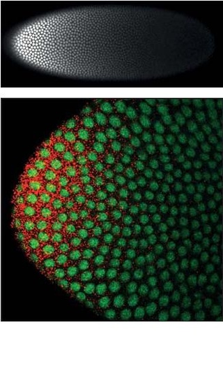
At the early blastoderm stage, Dpp signaling is uniform and low along the dorsal side. A sharp signaling profile emerges at the dorsal midline of the embryo during cellularization, with high levels of Dpp specifying the extraembryonic amnioserosa and low levels specifying the dorsal ectoderm. [3] Dpp signaling also incorporates a positive feedback mechanism that promotes future Dpp binding. [4] The morphogen gradient in embryos is established via a known active transport mechanism. [5] Gradient formation depends on the BMP inhibitors Short gastrulation (Sog) and Twisted gastrulation (Tsg), and other extracellular proteins such as Tolloid (Tld), and Screw (Scw). [6] [7] [8] Sog is produced in the ventral-lateral region of the embryo (perpendicular to the Dpp gradient) and forms a BMP-inhibiting gradient that prevents Dpp from binding to its receptor. [9] Sog and Tsg form a complex with Dpp and are actively transported toward the dorsal midline (middle of the embryo), following the Sog concentration gradient. Tld, a metalloprotease, releases Dpp from the complex by mediating Sog processing, activating Dpp signaling at the midline. [10] After gastrulation of the embryo, the Dpp gradient induces cardiac and visceral mesoderm formation. [11]

Signaling pathway

Dpp, like its vertebrate homologs, is a signaling molecule. In Drosophila, the receptor for Dpp is formed by two proteins, Thickveins (Tkv) and Punt. [12] Like Dpp itself, Tkv and Punt are highly similar to homologs in other species. When a cell receives a Dpp signal, the receptors are able to activate an intracellular protein called mothers against Dpp (mad) by phosphorylation. The initial discovery of mad in Drosophila paved the way for later experiments that identified the responder to TGF-β signaling in vertebrates, called SMADs. [13] Activated Mad is able to bind to DNA and act as a transcription factor to affect the expression of different genes in response to Dpp signaling. Genes activated by Dpp signaling include optomotor blind (omb) and spalt, and activity of these genes are often used as indicators of Dpp signaling in experiments. Another gene with a more complicated regulatory interaction with Dpp is brinker. Brinker is a transcription factor that represses the activation targets of Dpp, so in order to turn on these genes Dpp must repress brinker as well as activate the other targets. [14]

Role in imaginal wing disc

A picture illustrating the distribution of Dpp, shown in red, in the wing disc. Dpp is produced in a stripe just anterior of the anterior/posterior border and diffuses out to the edges of the tissue. Dpp wing expression.jpg
A picture illustrating the distribution of Dpp, shown in red, in the wing disc. Dpp is produced in a stripe just anterior of the anterior/posterior border and diffuses out to the edges of the tissue.

In the fly wing, the posterior and anterior halves of the tissue are populated by different kinds of cells that express different genes. Cells in the posterior but not the anterior express the transcription factor Engrailed (En). One of the genes activated by En is hedgehog (hh), a signaling factor. Hedgehog signaling instructs neighboring cells to express Dpp, but Dpp expression is also repressed by En. The result is that Dpp is only produced in a narrow stripe of cells immediately adjacent to but not within the posterior half of the tissue. [15] Dpp produced at this anterior/posterior border then diffuses out to the edges of the tissue, forming a spatial concentration gradient.

By reading their position along the gradient of Dpp, cells in the wing are able to determine their location relative to the anterior/posterior border, and they behave and develop accordingly.

It is possible that it is not actually the diffusion and gradient of Dpp that patterns tissues, but instead cells that receive Dpp signal instruct their neighbors on what to be, and those cells in turn signal their neighbors in a cascade through the tissue. Several experiments have been done to disprove this hypothesis and establish that it is actually the gradient of actual Dpp molecules that are responsible for patterning.

Mutant forms of the Dpp receptor Tkv exist that behave as if they are receiving high amounts of Dpp signal even in the absence of Dpp. Cells that contain this mutant receptor behave as if they are in an environment of high Dpp such as the area near the stripe of cells producing Dpp. By generating small patches of these cells in different parts of the wing tissue, investigators were able to distinguish how Dpp acts to pattern the tissue. If cells that receive a Dpp signal instruct their neighbors in a cascade, then additional tissue patterning centers should appear at the sites of the mutant cells that seem to receive high Dpp signaling but do not produce any Dpp themselves. However, if the physical presence of Dpp is necessary, then the cells near the mutants should not be affected at all. Experiments found the second case to be true, indicating that Dpp acts like a morphogen. [16]

The common way to assess differences in tissue patterning in the fly wing is to look at the pattern of veins in the wing. In flies where the ability of Dpp to diffuse through the tissue is impaired, the positioning of the veins is shifted from that in normal flies, and the wing is generally smaller. [17]

Dpp has also been proposed as a regulator of tissue growth and size, a classic problem in development. A problem common to organisms with multicellular organs that must grow from an initial size is how to know when to stop growing after the appropriate size is reached. Since Dpp is present in a gradient, it is conceivable that the slope of the gradient could be the measurement by which a tissue determines how large it is. If the amount of Dpp at the source is fixed and the amount at the edge of the tissue is zero, then the steepness of the gradient will decrease as the size of the tissue and the distance between the source and the edge increase. Experiments where an artificially steep gradient of Dpp is induced in wing tissue resulted in significantly increased amounts of cell proliferation, lending support to the steepness hypothesis. [18]

Formation of the Dpp gradient in the imaginal wing disc

The shape of the Dpp gradient is determined by four ligand kinetic parameters that are affected by biological parameters: [19] [20]

  1. The effective diffusion coefficient, which is dependent on extracellular diffusion, intracellular transport rates, and receptor binding/unbinding kinetics.
  2. The effective extracellular and intracellular degradation rates.
  3. The production rate, dependent on the Dpp production pathway.
  4. The immobile fraction (a parameter associated with the method used to measure Dpp kinetics, FRAP).

It is important to note that a single biological parameter can affect multiple kinetic parameters. For example, receptor levels will affect both the diffusion coefficient and the degradation rates. [21]

However, the mechanism by which the Dpp gradient is formed is still controversial, and no complete explanation has been proposed or proven. The four main categories of theories behind the formation of the gradient are free diffusion, restricted diffusion, transcytosis, and cytoneme-assisted transport.

An illustration of the assumptions made by each Dpp gradient formation model. A) Free Diffusion Model: Dpp freely diffuses through the ECM. B) Restricted Diffusion Model: Dpp diffuses through the ECM and interacts with proteoglycans and receptors. C) Transcytosis Model: Dpp passes through cells via endocytosis. D) Cytoneme-Mediated Transport Model: Dpp is directly routed to target cells via actin-based cytonemes. DppMorphModels.png
An illustration of the assumptions made by each Dpp gradient formation model. A) Free Diffusion Model: Dpp freely diffuses through the ECM. B) Restricted Diffusion Model: Dpp diffuses through the ECM and interacts with proteoglycans and receptors. C) Transcytosis Model: Dpp passes through cells via endocytosis. D) Cytoneme-Mediated Transport Model: Dpp is directly routed to target cells via actin-based cytonemes.

Free/restricted diffusion model

The free diffusion model assumes Dpp to diffuse freely through the extracellular matrix, degrading via receptor-mediated degradation events. FRAP assays have argued against this model by noting that diffusion of GFP-Dpp does not match that expected of a similarly sized molecule. [20] However, others have argued that a rate-limiting slow step further downstream of the process such as slow immobilization and/or slow degradation of Dpp itself could account for the observed differences in diffusion. [22] Single molecules of Dpp have been tracked using fluorescence correlation spectroscopy (FCS), showing that 65% of Dpp molecules diffuse rapidly (consistent with the free diffusion model) and 35% diffuse slowly (consistent with Dpp bound to receptors or glypicans).

The restricted diffusion model includes the effects of cell packing geometry and interactions with the extracellular matrix via binding events with receptors such as Tkv and the heparin sulfate proteoglycans dally and dally-like. [23] [24]

Transcytosis model

The transcytosis model assumes Dpp to be transported via repeated rounds of intracellular receptor-mediated endocytosis, with the gradient severity determined by endocytotic sorting of Dpp toward recycling through cells vs degradation. This model was initially based on an initial observation that Dpp could not accumulate across clones where a critical protein called dynamin necessary for endocytosis had been mutated into the shibire (shi) phenotype. [25] However, other experiments showed that Dpp was able to accumulate over shi clones, challenging the transcytosis model. [26] A revision of the theory behind the model proposes that endocytosis is not essential for Dpp movement but is involved in Dpp signaling. Dpp fails to move across cells with mutated dally and dally-like, two heparin sulfate proteoglycans (HSPGs) commonly found in the extracellular matrix. As a result, these results suggest that Dpp moves along the cell surface via restricted extracellular diffusion involving dally and dally-like, but the transport of Dpp itself does not rely on transcytosis. [27]

Cytoneme-mediated transport model

The cytoneme-mediated model suggests that Dpp is directly transported to target cells via actin-based filopodia called cytonemes that extend from the apical surface of Dpp-responding cells to the Dpp-producing source cells. [28] These cytonemes have been observed, but the dependence of the Dpp gradient on cytonemes has not been definitively proven in imaginal wing discs. However, Dpp is known to be required for and sufficient to extend and maintain cytonemes. Experiments analyzing the dynamics between Dpp and cytonemes have been conducted in the air sac primordium, where Dpp signaling was found to have a functional link with cytonemes. However, these experiments have not been replicated in imaginal wing discs.

Role in molluscs

Dpp is also found in molluscs, where it plays a key role in shell formation by controlling the shape of the conch. In bivalves, it is expressed until the protoconch has taken on the required shape, after which point its expression ceases. [29] It is also associated with shell formation in gastropods, [30] with an asymmetric distribution that may be associated with their coiling: shell growth appears to be inhibited where Dpp is expressed. [31]

Related Research Articles

Morphogenesis is the biological process that causes a cell, tissue or organism to develop its shape. It is one of three fundamental aspects of developmental biology along with the control of tissue growth and patterning of cellular differentiation.

<span class="mw-page-title-main">Sonic hedgehog protein</span> Signaling molecule in animals

Sonic hedgehog protein (SHH) is encoded for by the SHH gene. The protein is named after the video game character Sonic the Hedgehog.

<i>Drosophila</i> embryogenesis Embryogenesis of the fruit fly Drosophila, a popular model system

Drosophila embryogenesis, the process by which Drosophila embryos form, is a favorite model system for genetics and developmental biology. The study of its embryogenesis unlocked the century-long puzzle of how development was controlled, creating the field of evolutionary developmental biology. The small size, short generation time, and large brood size make it ideal for genetic studies. Transparent embryos facilitate developmental studies. Drosophila melanogaster was introduced into the field of genetic experiments by Thomas Hunt Morgan in 1909.

<span class="mw-page-title-main">Morphogen</span> Biological substance that guides development by non-uniform distribution

A morphogen is a substance whose non-uniform distribution governs the pattern of tissue development in the process of morphogenesis or pattern formation, one of the core processes of developmental biology, establishing positions of the various specialized cell types within a tissue. More specifically, a morphogen is a signaling molecule that acts directly on cells to produce specific cellular responses depending on its local concentration.

Axon guidance is a subfield of neural development concerning the process by which neurons send out axons to reach their correct targets. Axons often follow very precise paths in the nervous system, and how they manage to find their way so accurately is an area of ongoing research.

Compartments can be simply defined as separate, different, adjacent cell populations, which upon juxtaposition, create a lineage boundary. This boundary prevents cell movement from cells from different lineages across this barrier, restricting them to their compartment. Subdivisions are established by morphogen gradients and maintained by local cell-cell interactions, providing functional units with domains of different regulatory genes, which give rise to distinct fates. Compartment boundaries are found across species. In the hindbrain of vertebrate embryos, rhombomeres are compartments of common lineage outlined by expression of Hox genes. In invertebrates, the wing imaginal disc of Drosophila provides an excellent model for the study of compartments. Although other tissues, such as the abdomen, and even other imaginal discs are compartmentalized, much of our understanding of key concepts and molecular mechanisms involved in compartment boundaries has been derived from experimentation in the wing disc of the fruit fly.

<span class="mw-page-title-main">Bone morphogenetic protein 4</span> Human protein and coding gene

Bone morphogenetic protein 4 is a protein that in humans is encoded by BMP4 gene. BMP4 is found on chromosome 14q22-q23.

<span class="mw-page-title-main">Mothers against decapentaplegic homolog 1</span> Protein-coding gene in the species Homo sapiens

Mothers against decapentaplegic homolog 1 also known as SMAD family member 1 or SMAD1 is a protein that in humans is encoded by the SMAD1 gene.

Chordin is a protein with a prominent role in dorsal–ventral patterning during early embryonic development. In humans it is encoded for by the CHRD gene.

In the field of developmental biology, regional differentiation is the process by which different areas are identified in the development of the early embryo. The process by which the cells become specified differs between organisms.

<span class="mw-page-title-main">French flag model</span> Biological model

The French flag model is a conceptual definition of a morphogen, described by Lewis Wolpert in the 1960s. A morphogen is defined as a signaling molecule that acts directly on cells to produce specific cellular responses dependent on morphogen concentration. During early development, morphogen gradients generate different cell types in distinct spatial order. French flag patterning is often found in combination with others: vertebrate limb development is one of the many phenotypes exhibiting French flag patterning overlapped with a complementary pattern.

<span class="mw-page-title-main">Zone of polarizing activity</span>

The zone of polarizing activity (ZPA) is an area of mesenchyme that contains signals which instruct the developing limb bud to form along the anterior/posterior axis. Limb bud is undifferentiated mesenchyme enclosed by an ectoderm covering. Eventually, the limb bud develops into bones, tendons, muscles and joints. Limb bud development relies not only on the ZPA, but also many different genes, signals, and a unique region of ectoderm called the apical ectodermal ridge (AER). Research by Saunders and Gasseling in 1948 identified the AER and its subsequent involvement in proximal distal outgrowth. Twenty years later, the same group did transplantation studies in chick limb bud and identified the ZPA. It wasn't until 1993 that Todt and Fallon showed that the AER and ZPA are dependent on each other.

<span class="mw-page-title-main">Cytoneme</span>

Cytonemes are thin, cellular projections that are specialized for exchange of signaling proteins between cells. Cytonemes emanate from cells that make signaling proteins, extending directly to cells that receive signaling proteins. Cytonemes also extend directly from cells that receive signaling proteins to cells that make them.

The Nodal signaling pathway is a signal transduction pathway important in regional and cellular differentiation during embryonic development.

In evolutionary developmental biology, inversion refers to the hypothesis that during the course of animal evolution, the structures along the dorsoventral (DV) axis have taken on an orientation opposite that of the ancestral form.

Dally is the name of a gene that encodes a HS-modified-protein found in the fruit fly. The protein has to be processed after being codified, and in its mature form it is composed by 626 amino acids, forming a proteoglycan rich in heparin sulfate which is anchored to the cell surface via covalent linkage to glycophosphatidylinositol (GPI), so we can define it as a glypican. For its normal biosynthesis it requires sugarless (sgl), a gene that encodes an enzyme which plays a critical role in the process of modification of dally.

<i>Homeotic protein bicoid</i> Protein-coding gene in the species Drosophila melanogaster

Homeotic protein bicoid is encoded by the bcd maternal effect gene in Drosophilia. Homeotic protein bicoid concentration gradient patterns the anterior-posterior (A-P) axis during Drosophila embryogenesis. Bicoid was the first protein demonstrated to act as a morphogen. Although bicoid is important for the development of Drosophila and other higher dipterans, it is absent from most other insects, where its role is accomplished by other genes.

The Spemann-Mangold organizer is a group of cells that are responsible for the induction of the neural tissues during development in amphibian embryos. First described in 1924 by Hans Spemann and Hilde Mangold, the introduction of the organizer provided evidence that the fate of cells can be influenced by factors from other cell populations. This discovery significantly impacted the world of developmental biology and fundamentally changed the understanding of early development.

Thomas Lecuit, born 4 October 1971 in Saumur, is a French biologist specializing in the emergence of forms or morphogenesis. He is a professor at the Collège de France, holding the Dynamics of Life Chair. He leads a research team at the Institut de Biologie du Développement de Marseille (IBDM), and the Turing Centre for Living Systems, an interdisciplinary centre dedicated to the study of living organisms.

Gary Struhl is an American research scientist whose primary areas of research are developmental biology and genetics and genomics. He works as a professor at Columbia University Medical Center, teaching neuroscience within the Department of Genetics and Development.

References

  1. 1 2 Matsuda S, Harmansa S, Affolter M (February 2016). "BMP morphogen gradients in flies". Cytokine & Growth Factor Reviews. 27: 119–27. doi:10.1016/j.cytogfr.2015.11.003. PMID   26684043.
  2. O'Connor MB, Umulis D, Othmer HG, Blair SS (January 2006). "Shaping BMP morphogen gradients in the Drosophila embryo and pupal wing". Development. 133 (2): 183–93. doi:10.1242/dev.02214. PMC   6469686 . PMID   16368928.
  3. Wharton KA, Ray RP, Gelbart WM (February 1993). "An activity gradient of decapentaplegic is necessary for the specification of dorsal pattern elements in the Drosophila embryo". Development. 117 (2): 807–22. doi:10.1242/dev.117.2.807. PMID   8330541.
  4. Wang YC, Ferguson EL (March 2005). "Spatial bistability of Dpp-receptor interactions during Drosophila dorsal-ventral patterning". Nature. 434 (7030): 229–34. Bibcode:2005Natur.434..229W. doi:10.1038/nature03318. PMID   15759004. S2CID   4415152.
  5. Ferguson EL, Anderson KV (October 1992). "Decapentaplegic acts as a morphogen to organize dorsal-ventral pattern in the Drosophila embryo". Cell. 71 (3): 451–61. doi:10.1016/0092-8674(92)90514-D. PMID   1423606. S2CID   40423615.
  6. Arora K, Levine MS, O'Connor MB (November 1994). "The screw gene encodes a ubiquitously expressed member of the TGF-beta family required for specification of dorsal cell fates in the Drosophila embryo". Genes & Development. 8 (21): 2588–601. doi: 10.1101/gad.8.21.2588 . PMID   7958918.
  7. Francois V, Solloway M, O'Neill JW, Emery J, Bier E (November 1994). "Dorsal-ventral patterning of the Drosophila embryo depends on a putative negative growth factor encoded by the short gastrulation gene". Genes & Development. 8 (21): 2602–16. doi: 10.1101/gad.8.21.2602 . PMID   7958919.
  8. Ross JJ, Shimmi O, Vilmos P, Petryk A, Kim H, Gaudenz K, Hermanson S, Ekker SC, O'Connor MB, Marsh JL (March 2001). "Twisted gastrulation is a conserved extracellular BMP antagonist". Nature. 410 (6827): 479–83. Bibcode:2001Natur.410..479R. doi:10.1038/35068578. PMID   11260716. S2CID   24986331.
  9. Srinivasan S, Rashka KE, Bier E (January 2002). "Creation of a Sog morphogen gradient in the Drosophila embryo". Developmental Cell. 2 (1): 91–101. doi: 10.1016/S1534-5807(01)00097-1 . PMID   11782317.
  10. Marqués G, Musacchio M, Shimell MJ, Wünnenberg-Stapleton K, Cho KW, O'Connor MB (October 1997). "Production of a DPP activity gradient in the early Drosophila embryo through the opposing actions of the SOG and TLD proteins". Cell. 91 (3): 417–26. doi: 10.1016/S0092-8674(00)80425-0 . PMID   9363950. S2CID   16613162.
  11. Frasch M (March 1995). "Induction of visceral and cardiac mesoderm by ectodermal Dpp in the early Drosophila embryo". Nature. 374 (6521): 464–7. Bibcode:1995Natur.374..464F. doi:10.1038/374464a0. PMID   7700357. S2CID   4330159.
  12. Nellen D, Affolter M, Basler K (July 1994). "Receptor serine/threonine kinases implicated in the control of Drosophila body pattern by decapentaplegic" (PDF). Cell. 78 (2): 225–37. doi:10.1016/0092-8674(94)90293-3. PMID   8044837. S2CID   13467898.
  13. Sekelsky JJ, Newfeld SJ, Raftery LA, Chartoff EH, Gelbart WM (March 1995). "Genetic characterization and cloning of mothers against dpp, a gene required for decapentaplegic function in Drosophila melanogaster". Genetics. 139 (3): 1347–58. doi:10.1093/genetics/139.3.1347. PMC   1206461 . PMID   7768443.
  14. Campbell G, Tomlinson A (February 1999). "Transducing the Dpp morphogen gradient in the wing of Drosophila: regulation of Dpp targets by brinker". Cell. 96 (4): 553–62. doi: 10.1016/S0092-8674(00)80659-5 . PMID   10052457. S2CID   16296766.
  15. Zecca M, Basler K, Struhl G (August 1995). "Sequential organizing activities of engrailed, hedgehog and decapentaplegic in the Drosophila wing" (PDF). Development. 121 (8): 2265–78. doi:10.1242/dev.121.8.2265. PMID   7671794. Archived from the original (PDF) on 2021-10-28. Retrieved 2022-06-13.
  16. Affolter M, Basler K (September 2007). "The Decapentaplegic morphogen gradient: from pattern formation to growth regulation". Nature Reviews. Genetics. 8 (9): 663–74. doi:10.1038/nrg2166. PMID   17703237. S2CID   24005278.
  17. Crickmore MA, Mann RS (January 2007). "Hox control of morphogen mobility and organ development through regulation of glypican expression". Development. 134 (2): 327–34. doi:10.1242/dev.02737. PMID   17166918. S2CID   21491059.
  18. Rogulja D, Irvine KD (November 2005). "Regulation of cell proliferation by a morphogen gradient". Cell. 123 (3): 449–61. doi: 10.1016/j.cell.2005.08.030 . PMID   16269336. S2CID   18881009.
  19. Bollenbach T, Pantazis P, Kicheva A, Bökel C, González-Gaitán M, Jülicher F (March 2008). "Precision of the Dpp gradient". Development. 135 (6): 1137–46. doi: 10.1242/dev.012062 . PMID   18296653.
  20. 1 2 Kicheva A, Pantazis P, Bollenbach T, Kalaidzidis Y, Bittig T, Jülicher F, González-Gaitán M (January 2007). "Kinetics of morphogen gradient formation" (PDF). Science. 315 (5811): 521–5. Bibcode:2007Sci...315..521K. doi:10.1126/science.1135774. PMID   17255514. S2CID   2096679.
  21. Crickmore MA, Mann RS (July 2006). "Hox control of organ size by regulation of morphogen production and mobility". Science. 313 (5783): 63–8. Bibcode:2006Sci...313...63C. doi:10.1126/science.1128650. PMC   2628481 . PMID   16741075.
  22. Zhou S, Lo WC, Suhalim JL, Digman MA, Gratton E, Nie Q, Lander AD (April 2012). "Free extracellular diffusion creates the Dpp morphogen gradient of the Drosophila wing disc". Current Biology. 22 (8): 668–75. doi:10.1016/j.cub.2012.02.065. PMC   3338872 . PMID   22445299.
  23. Müller P, Rogers KW, Yu SR, Brand M, Schier AF (April 2013). "Morphogen transport". Development. 140 (8): 1621–38. doi:10.1242/dev.083519. PMC   3621481 . PMID   23533171.
  24. Lecuit T, Cohen SM (December 1998). "Dpp receptor levels contribute to shaping the Dpp morphogen gradient in the Drosophila wing imaginal disc". Development. 125 (24): 4901–7. doi:10.1242/dev.125.24.4901. PMID   9811574.
  25. Entchev EV, Schwabedissen A, González-Gaitán M (December 2000). "Gradient formation of the TGF-beta homolog Dpp". Cell. 103 (6): 981–91. doi: 10.1016/S0092-8674(00)00200-2 . PMID   11136982. S2CID   6100358.
  26. Schwank G, Dalessi S, Yang SF, Yagi R, de Lachapelle AM, Affolter M, Bergmann S, Basler K (July 2011). "Formation of the long range Dpp morphogen gradient". PLOS Biology. 9 (7): e1001111. doi: 10.1371/journal.pbio.1001111 . PMC   3144185 . PMID   21814489.
  27. Belenkaya TY, Han C, Yan D, Opoka RJ, Khodoun M, Liu H, Lin X (October 2004). "Drosophila Dpp morphogen movement is independent of dynamin-mediated endocytosis but regulated by the glypican members of heparan sulfate proteoglycans". Cell. 119 (2): 231–44. doi: 10.1016/j.cell.2004.09.031 . PMID   15479640. S2CID   10575655.
  28. Roy S, Huang H, Liu S, Kornberg TB (February 2014). "Cytoneme-mediated contact-dependent transport of the Drosophila decapentaplegic signaling protein". Science. 343 (6173): 1244624. doi:10.1126/science.1244624. PMC   4336149 . PMID   24385607.
  29. Kin K, Kakoi S, Wada H (May 2009). "A novel role for dpp in the shaping of bivalve shells revealed in a conserved molluscan developmental program". Developmental Biology. 329 (1): 152–66. doi:10.1016/j.ydbio.2009.01.021. hdl: 2241/102610 . PMID   19382296.
  30. Iijima M, Takeuchi T, Sarashina I, Endo K (May 2008). "Expression patterns of engrailed and dpp in the gastropod Lymnaea stagnalis". Development Genes and Evolution. 218 (5): 237–51. doi:10.1007/s00427-008-0217-0. PMID   18443822. S2CID   1045678.
  31. Kurita Y, Deguchi R, Wada H (December 2009). "Early development and cleavage pattern of the Japanese purple mussel, Septifer virgatus". Zoological Science. 26 (12): 814–20. doi:10.2108/zsj.26.814. hdl: 2241/113546 . PMID   19968468. S2CID   25868365.