Follistatin

Last updated
FST
PDB 2b0u EBI.jpg
Available structures
PDB Ortholog search: PDBe RCSB
Identifiers
Aliases FST , FS, follistatin
External IDs OMIM: 136470 MGI: 95586 HomoloGene: 7324 GeneCards: FST
Orthologs
SpeciesHumanMouse
Entrez
Ensembl
UniProt
RefSeq (mRNA)

NM_006350
NM_013409

NM_008046
NM_001301373
NM_001301375

RefSeq (protein)

NP_006341
NP_037541

NP_001288302
NP_001288304
NP_032072

Location (UCSC) Chr 5: 53.48 – 53.49 Mb Chr 13: 114.59 – 114.6 Mb
PubMed search [3] [4]
Wikidata
View/Edit Human View/Edit Mouse

Follistatin, also known as activin-bindings protein, is a protein that in humans is encoded by the FST gene. [5] [6] Follistatin is an autocrine glycoprotein that is expressed in nearly all tissues of higher animals. [6]

Contents

Its primary function is the binding and bioneutralization of members of the TGF-β superfamily, with a particular focus on activin, a paracrine hormone.

An earlier name for the same protein was FSH-suppressing protein (FSP). At the time of its initial isolation from follicular fluid, it was found to inhibit the anterior pituitary's secretion of follicle-stimulating hormone (FSH).

Biochemistry

Follistatin is part of the inhibin-activin-follistatin axis.

Three isoforms, FS-288, FS-300, and FS-315 have been reported. Two, FS-288 and FS-315, are created by alternative splicing of the primary mRNA transcript. FS-300 (porcine follistatin) is thought to be the product of posttranslational modification via truncation of the C-terminal domain from the primary amino-acid chain.

Although FS is ubiquitous, its highest concentration is in the female ovary, followed by the skin.

Follistatin is produced by folliculostellate (FS) cells of the anterior pituitary. FS cells make numerous contacts with the classical endocrine cells of the anterior pituitary including gonadotrophs.

Function

In the tissues activin has a strong role in cellular proliferation, thereby making follistatin the safeguard against uncontrolled cellular proliferation and also allowing it to function as an instrument of cellular differentiation. These roles are vital in tissue rebuilding and repair, and may account for follistatin's high presence in the skin.

In the blood, activin and follistatin are involved in the inflammatory response following tissue injury or pathogenic incursion. The source of follistatin in circulating blood plasma has yet to be determined, but due to its autocrine nature speculation suggests the endothelial cells lining all blood vessels, or the macrophages and monocytes circulating within the whole blood, may be sources.

Follistatin is involved in embryo development. It has inhibitory action on bone morphogenic proteins (BMPs); BMPs induce the ectoderm to become epidermal ectoderm. Inhibition of BMPs allows neuroectoderm to arise from ectoderm, a process which eventually forms the neural plate. Other inhibitors involved in this process are noggin and chordin.

Follistatin and BMPs are play a role in folliculogenesis within the ovary. The main role of follistatin in the oestrus/menstrus ovary appears to be progression of the follicle from early antral to antral/dominant. It is also involved in the promotion of cellular differentiation of the estrogen producing granulosa cells (GC) of the dominant follicle into the progesterone producing large lutein cells (LLC) of the corpus luteum.

Clinical significance

Follistatin is studied for its role in regulation of muscle growth in mice, as an antagonist to myostatin (also known as GDF-8, a TGF superfamily member) which inhibits excessive muscle growth. Lee and McPherron demonstrated that inhibition of GDF-8, either by genetic elimination (knockout mice) or by increasing the amount of follistatin, resulted in increased muscle mass. [7] [8] In 2009, research with macaque monkeys demonstrated that regulating follistatin via gene therapy also resulted in muscle growth and increases in strength. [9]

Increased levels of follistatin, by leading to increased muscle mass of certain core muscular groups, can increase life expectancy in cases of spinal muscular atrophy (SMA) in animal models. [10]

Elevated circulating follistatin levels are also associated with increased risk of type 2 diabetes , early death, heart failure, stroke and chronic kidney disease. It has been demonstrated that follistatin contributes to insulin resistance in type 2 diabetes development and nonalcoholic fatty liver disease (NAFLD). The genetic regulation of follistatin secretion from the liver is via Glucokinase regulatory protein (GCKR) identified by large GWAS studies. [11] [12]

It is also investigated for its involvement in polycystic ovary syndrome (PCOS), in part to resolve debate as to its direct role in this disease. [13]

Sporadic inclusion body myositis, a variant of inflammatory myopathy, involves muscle weakness. In one clinical trial, rAAV1.CMV.huFS344, 6 × 10¹¹ vg/kg, walk test results significantly improved versus untreated controls, along with decreased fibrosis and improved regeneration.

ACE-083, a follistatin-based fusion protein, was investigated for treatment focal or asymmetric myopathies. Intramuscular ACE-083 increased growth and force production in injected muscle in wild-type mice and mouse models of Charcot-Marie-Tooth disease (CMT) and Duchenne muscular dystrophy, without systemic effects or endocrine disruption. [14]

AAV-mediated FST reduced obesity-induced inflammatory adipokines and cytokines systemically and in synovial fluid. Mice receiving FST therapy were protected from post-traumatic osteoarthritis and bone remodeling from joint injury. [15]

In another mouse study, high dose animals showed significant quadricep growth.

Related Research Articles

<span class="mw-page-title-main">Myostatin</span> Mammalian and avian protein

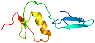
Myostatin is a protein that in humans is encoded by the MSTN gene. Myostatin is a myokine that is produced and released by myocytes and acts on muscle cells to inhibit muscle growth. Myostatin is a secreted growth differentiation factor that is a member of the TGF beta protein family.

<span class="mw-page-title-main">Hypothalamic–pituitary–gonadal axis</span> Concept of regarding the hypothalamus, pituitary gland and gonadal glands as a single entity

The hypothalamic–pituitary–gonadal axis refers to the hypothalamus, pituitary gland, and gonadal glands as if these individual endocrine glands were a single entity. Because these glands often act in concert, physiologists and endocrinologists find it convenient and descriptive to speak of them as a single system.

<span class="mw-page-title-main">Growth differentiation factor-9</span> Protein-coding gene in the species Homo sapiens

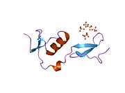
Growth/differentiation factor 9 is a protein that in humans is encoded by the GDF9 gene.

<span class="mw-page-title-main">Bone morphogenetic protein 4</span> Human protein and coding gene

Bone morphogenetic protein 4 is a protein that in humans is encoded by BMP4 gene. BMP4 is found on chromosome 14q22-q23.

<span class="mw-page-title-main">Bone morphogenetic protein 15</span> Protein-coding gene in humans

Bone morphogenetic protein 15 (BMP-15) is a protein that in humans is encoded by the BMP15 gene. It is involved in folliculogenesis, the process in which primordial follicles develop into pre-ovulatory follicles.

The transforming growth factor beta (TGFB) signaling pathway is involved in many cellular processes in both the adult organism and the developing embryo including cell growth, cell differentiation, cell migration, apoptosis, cellular homeostasis and other cellular functions. The TGFB signaling pathways are conserved. In spite of the wide range of cellular processes that the TGFβ signaling pathway regulates, the process is relatively simple. TGFβ superfamily ligands bind to a type II receptor, which recruits and phosphorylates a type I receptor. The type I receptor then phosphorylates receptor-regulated SMADs (R-SMADs) which can now bind the coSMAD SMAD4. R-SMAD/coSMAD complexes accumulate in the nucleus where they act as transcription factors and participate in the regulation of target gene expression.

<span class="mw-page-title-main">BMPR2</span> Protein-coding gene in the species Homo sapiens

Bone morphogenetic protein receptor type II or BMPR2 is a serine/threonine receptor kinase encoded by the BMPR2 gene. It binds bone morphogenetic proteins, members of the TGF beta superfamily of ligands, which are involved in paracrine signaling. BMPs are involved in a host of cellular functions including osteogenesis, cell growth and cell differentiation. Signaling in the BMP pathway begins with the binding of a BMP to the type II receptor. This causes the recruitment of a BMP type I receptor, which the type II receptor phosphorylates. The type I receptor phosphorylates an R-SMAD, a transcriptional regulator.

<span class="mw-page-title-main">ACVR1</span> Protein-coding gene

Activin A receptor, type I (ACVR1) is a protein which in humans is encoded by the ACVR1 gene; also known as ALK-2. ACVR1 has been linked to the 2q23-24 region of the genome. This protein is important in the bone morphogenic protein (BMP) pathway which is responsible for the development and repair of the skeletal system. While knock-out models with this gene are in progress, the ACVR1 gene has been connected to fibrodysplasia ossificans progressiva, an extremely rare progressive genetic disease characterized by heterotopic ossification of muscles, tendons and ligaments. It is a bone morphogenetic protein receptor, type 1.

<span class="mw-page-title-main">ACVR2A</span> Protein-coding gene in the species Homo sapiens

Activin receptor type-2A is a protein that in humans is encoded by the ACVR2A gene. ACVR2A is an activin type 2 receptor.

<span class="mw-page-title-main">ACVR2B</span> Protein-coding gene in the species Homo sapiens

Activin receptor type-2B is a protein that in humans is encoded by the ACVR2B gene. ACVR2B is an activin type 2 receptor.

<span class="mw-page-title-main">GDF11</span> Protein-coding gene in humans

Growth differentiation factor 11 (GDF11), also known as bone morphogenetic protein 11 (BMP-11), is a protein that in humans is encoded by the growth differentiation factor 11 gene. GDF11 is a member of the Transforming growth factor beta family.

<span class="mw-page-title-main">INHBA</span> Protein-coding gene in the species Homo sapiens

Inhibin, beta A, also known as INHBA, is a protein which in humans is encoded by the INHBA gene. INHBA is a subunit of both activin and inhibin, two closely related glycoproteins with opposing biological effects.

<span class="mw-page-title-main">INHA</span> Protein and coding gene in humans

Inhibin, alpha, also known as INHA, is a protein which in humans is encoded by the INHA gene.

<span class="mw-page-title-main">FSTL3</span> Protein-coding gene in the species Homo sapiens

Follistatin-related protein 3 is a protein that in humans is encoded by the FSTL3 gene.

<span class="mw-page-title-main">STRAP</span> Protein-coding gene in the species Homo sapiens

Serine-threonine kinase receptor-associated protein is an enzyme that in humans is encoded by the STRAP gene.

<span class="mw-page-title-main">FSTL1</span> Protein-coding gene in the species Homo sapiens

Follistatin-related protein 1 is a protein that in humans is encoded by the FSTL1 gene.

<span class="mw-page-title-main">Activin and inhibin</span> Regulators of feedback on FSH-production

Activin and inhibin are two closely related protein complexes that have almost directly opposite biological effects. Identified in 1986, activin enhances FSH biosynthesis and secretion, and participates in the regulation of the menstrual cycle. Many other functions have been found to be exerted by activin, including roles in cell proliferation, differentiation, apoptosis, metabolism, homeostasis, immune response, wound repair, and endocrine function. Conversely, inhibin downregulates FSH synthesis and inhibits FSH secretion. The existence of inhibin was hypothesized as early as 1916; however, it was not demonstrated to exist until Neena Schwartz and Cornelia Channing's work in the mid-1970s, after which both proteins were molecularly characterized ten years later.

A myokine is one of several hundred cytokines or other small proteins and proteoglycan peptides that are produced and released by skeletal muscle cells in response to muscular contractions. They have autocrine, paracrine and/or endocrine effects; their systemic effects occur at picomolar concentrations.

Ovarian follicle activation can be defined as primordial follicles in the ovary moving from a quiescent (inactive) to a growing phase. The primordial follicle in the ovary is what makes up the “pool” of follicles that will be induced to enter growth and developmental changes that change them into pre-ovulatory follicles, ready to be released during ovulation. The process of development from a primordial follicle to a pre-ovulatory follicle is called folliculogenesis.

Myostatin inhibitors are a class of drugs that work by blocking the effects of myostatin, which inhibits muscle growth. In animal models and limited human studies, myostatin inhibitors have increased muscle size. They are being developed to treat obesity, sarcopenia, muscular dystrophy, and other illnesses.

References

  1. 1 2 3 GRCh38: Ensembl release 89: ENSG00000134363 Ensembl, May 2017
  2. 1 2 3 GRCm38: Ensembl release 89: ENSMUSG00000021765 Ensembl, May 2017
  3. "Human PubMed Reference:". National Center for Biotechnology Information, U.S. National Library of Medicine.
  4. "Mouse PubMed Reference:". National Center for Biotechnology Information, U.S. National Library of Medicine.
  5. Ueno N, Ling N, Ying SY, Esch F, Shimasaki S, Guillemin R (December 1987). "Isolation and partial characterization of follistatin: a single-chain Mr 35,000 monomeric protein that inhibits the release of follicle-stimulating hormone". Proceedings of the National Academy of Sciences of the United States of America. 84 (23): 8282–8286. Bibcode:1987PNAS...84.8282U. doi: 10.1073/pnas.84.23.8282 . PMC   299526 . PMID   3120188.
  6. 1 2 Tortoriello DV, Sidis Y, Holtzman DA, Holmes WE, Schneyer AL (August 2001). "Human follistatin-related protein: a structural homologue of follistatin with nuclear localization". Endocrinology. 142 (8): 3426–3434. doi: 10.1210/endo.142.8.8319 . PMID   11459787.
  7. Lee SJ, McPherron AC (July 2001). "Regulation of myostatin activity and muscle growth". Proceedings of the National Academy of Sciences of the United States of America. 98 (16): 9306–9311. Bibcode:2001PNAS...98.9306L. doi: 10.1073/pnas.151270098 . PMC   55416 . PMID   11459935.
  8. "'Mighty mice' made mightier" . Retrieved 2008-02-26.
  9. "Success Boosting Monkey Muscle Could Help Humans". NPR. 11 Nov 2009. Retrieved 2009-11-12.
  10. Rose FF, Mattis VB, Rindt H, Lorson CL (March 2009). "Delivery of recombinant follistatin lessens disease severity in a mouse model of spinal muscular atrophy". Human Molecular Genetics. 18 (6): 997–1005. doi:10.1093/hmg/ddn426. PMC   2649020 . PMID   19074460.
  11. Wu C, Borné Y, Gao R, López Rodriguez M, Roell WC, Wilson JM, et al. (November 2021). "Elevated circulating follistatin associates with an increased risk of type 2 diabetes". Nature Communications. 12 (1): 6486. Bibcode:2021NatCo..12.6486W. doi:10.1038/s41467-021-26536-w. PMC   8580990 . PMID   34759311.
  12. Pan J, Nilsson J, Engström G, De Marinis Y (2024). "Elevated circulating follistatin associates with increased risk of mortality and cardiometabolic disorders". Nutr Metab Cardiovasc Dis. 34 (2): 418–425. doi: 10.1016/j.numecd.2023.09.012 . PMID   38000997.
  13. Asteria C (October 2000). "Identification of follistatin as a possible trait-causing gene in polycystic ovary syndrome". European Journal of Endocrinology. 143 (4): 467–9. doi: 10.1530/eje.0.1430467 . PMID   11022191.
  14. Pearsall RS, Davies MV, Cannell M, Li J, Widrick J, Mulivor AW, et al. (August 2019). "Follistatin-based ligand trap ACE-083 induces localized hypertrophy of skeletal muscle with functional improvement in models of neuromuscular disease". Scientific Reports. 9 (1): 11392. Bibcode:2019NatSR...911392P. doi:10.1038/s41598-019-47818-w. PMC   6684588 . PMID   31388039.
  15. Tang R, Harasymowicz NS, Wu CL, Collins KH, Choi YR, Oswald SJ, et al. (May 2020). "Gene therapy for follistatin mitigates systemic metabolic inflammation and post-traumatic arthritis in high-fat diet-induced obesity". Science Advances. 6 (19): eaaz7492. Bibcode:2020SciA....6.7492T. doi:10.1126/sciadv.aaz7492. PMC   7209997 . PMID   32426485.

Further reading