Transferrin receptor

Last updated
Transferrin receptor 1
1cx8.jpg
Transferrin receptor 1, dimer, Human
Identifiers
Symbol TFRC
Alt. symbolsCD71, TFR1
NCBI gene 7037
HGNC 11763
OMIM 190010
RefSeq NM_003234
UniProt P02786
Other data
Locus Chr. 3 q29
Search for
Structures Swiss-model
Domains InterPro
Transferrin receptor 2
Identifiers
Symbol TFR2
Alt. symbolsHFE3, TFRC2
NCBI gene 7036
HGNC 11762
OMIM 604720
RefSeq NM_003227
UniProt Q9UP52
Other data
Locus Chr. 7 q22
Search for
Structures Swiss-model
Domains InterPro

Transferrin receptor (TfR) is a carrier protein for transferrin. It is needed for the import of iron into the cell and is regulated in response to intracellular iron concentration. It imports iron by internalizing the transferrin-iron complex through receptor-mediated endocytosis. [1] The existence of a receptor for transferrin iron uptake has been recognized since the late 1950s. [2] Earlier two transferrin receptors in humans, transferrin receptor 1 and transferrin receptor 2 had been characterized and until recently cellular iron uptake was believed to occur chiefly via these two well documented transferrin receptors. Both these receptors are transmembrane glycoproteins. TfR1 is a high affinity ubiquitously expressed receptor while expression of TfR2 is restricted to certain cell types and is unaffected by intracellular iron concentrations. TfR2 binds to transferrin with a 25-30 fold lower affinity than TfR1. [3] [4] Although TfR1 mediated iron uptake is the major pathway for iron acquisition by most cells and especially developing erythrocytes, several studies have indicated that the uptake mechanism varies depending upon the cell type. It is also reported that Tf uptake exists independent of these TfRs although the mechanisms are not well characterized. [5] [6] [7] [8] The multifunctional glycolytic enzyme glyceraldehyde 3-phosphate dehydrogenase (GAPDH, EC 1.2.1.12) has been shown to utilize post translational modifications to exhibit higher order moonlighting behavior wherein it switches its function as a holo or apo transferrin receptor leading to either iron delivery or iron export respectively. [9] [10] [11]

Contents

Post-transcriptional regulation

Low iron concentrations promote increased levels of transferrin receptor, to increase iron intake into the cell. Thus, transferrin receptor maintains cellular iron homeostasis.

TfR production in the cell is regulated according to iron levels by iron-responsive element-binding proteins, IRP1 and IRP2. In the absence of iron, one of these proteins (generally IRP2) binds to the hairpin like structure (IRE) that is in the 3' UTR of the TfR mRNA. Once binding occurs, the mRNA is stabilized and degradation is inhibited.

See also

Related Research Articles

<span class="mw-page-title-main">Endocytosis</span> Cellular process

Endocytosis is a cellular process in which substances are brought into the cell. The material to be internalized is surrounded by an area of cell membrane, which then buds off inside the cell to form a vesicle containing the ingested material. Endocytosis includes pinocytosis and phagocytosis. It is a form of active transport.

<span class="mw-page-title-main">Steroid hormone</span> Substance with biological function

A steroid hormone is a steroid that acts as a hormone. Steroid hormones can be grouped into two classes: corticosteroids and sex steroids. Within those two classes are five types according to the receptors to which they bind: glucocorticoids and mineralocorticoids and androgens, estrogens, and progestogens. Vitamin D derivatives are a sixth closely related hormone system with homologous receptors. They have some of the characteristics of true steroids as receptor ligands.

<span class="mw-page-title-main">Transferrin</span> Mammalian protein found in Homo sapiens

Transferrins are glycoproteins found in vertebrates which bind to and consequently mediate the transport of iron (Fe) through blood plasma. They are produced in the liver and contain binding sites for two Fe3+ ions. Human transferrin is encoded by the TF gene and produced as a 76 kDa glycoprotein.

<span class="mw-page-title-main">Endosome</span> Vacuole to which materials ingested by endocytosis are delivered

Endosomes are a collection of intracellular sorting organelles in eukaryotic cells. They are parts of endocytic membrane transport pathway originating from the trans Golgi network. Molecules or ligands internalized from the plasma membrane can follow this pathway all the way to lysosomes for degradation or can be recycled back to the cell membrane in the endocytic cycle. Molecules are also transported to endosomes from the trans Golgi network and either continue to lysosomes or recycle back to the Golgi apparatus.

<span class="mw-page-title-main">Receptor-mediated endocytosis</span>

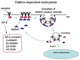
Receptor-mediated endocytosis (RME), also called clathrin-mediated endocytosis, is a process by which cells absorb metabolites, hormones, proteins – and in some cases viruses – by the inward budding of the plasma membrane (invagination). This process forms vesicles containing the absorbed substances and is strictly mediated by receptors on the surface of the cell. Only the receptor-specific substances can enter the cell through this process.

<span class="mw-page-title-main">Omigapil</span> Drug developed by Novartis

Omigapil is a drug that was developed by Novartis and tested in clinical trials for its ability to help treat Parkinson's disease (PD) and amyotrophic lateral sclerosis (ALS). The development for PD and ALS have been terminated due to lack of benefit, but Santhera Pharmaceuticals bought the compound for development for the treatment of congenital muscular dystrophy (CMD).

<span class="mw-page-title-main">Human iron metabolism</span> Iron metabolism in the body

Human iron metabolism is the set of chemical reactions that maintain human homeostasis of iron at the systemic and cellular level. Iron is both necessary to the body and potentially toxic. Controlling iron levels in the body is a critically important part of many aspects of human health and disease. Hematologists have been especially interested in systemic iron metabolism, because iron is essential for red blood cells, where most of the human body's iron is contained. Understanding iron metabolism is also important for understanding diseases of iron overload, such as hereditary hemochromatosis, and iron deficiency, such as iron-deficiency anemia.

<span class="mw-page-title-main">Glyceraldehyde 3-phosphate dehydrogenase</span> Enzyme of the glycolysis metabolic pathway

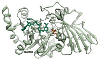
Glyceraldehyde 3-phosphate dehydrogenase is an enzyme of about 37kDa that catalyzes the sixth step of glycolysis and thus serves to break down glucose for energy and carbon molecules. In addition to this long established metabolic function, GAPDH has recently been implicated in several non-metabolic processes, including transcription activation, initiation of apoptosis, ER to Golgi vesicle shuttling, and fast axonal, or axoplasmic transport. In sperm, a testis-specific isoenzyme GAPDHS is expressed.

Transcytosis is a type of transcellular transport in which various macromolecules are transported across the interior of a cell. Macromolecules are captured in vesicles on one side of the cell, drawn across the cell, and ejected on the other side. Examples of macromolecules transported include IgA, transferrin, and insulin. While transcytosis is most commonly observed in epithelial cells, the process is also present elsewhere. Blood capillaries are a well-known site for transcytosis, though it occurs in other cells, including neurons, osteoclasts and M cells of the intestine.

<span class="mw-page-title-main">Fructose-bisphosphate aldolase</span>

Fructose-bisphosphate aldolase, often just aldolase, is an enzyme catalyzing a reversible reaction that splits the aldol, fructose 1,6-bisphosphate, into the triose phosphates dihydroxyacetone phosphate (DHAP) and glyceraldehyde 3-phosphate (G3P). Aldolase can also produce DHAP from other (3S,4R)-ketose 1-phosphates such as fructose 1-phosphate and sedoheptulose 1,7-bisphosphate. Gluconeogenesis and the Calvin cycle, which are anabolic pathways, use the reverse reaction. Glycolysis, a catabolic pathway, uses the forward reaction. Aldolase is divided into two classes by mechanism.

<span class="mw-page-title-main">Iron-responsive element-binding protein</span> Protein family

The iron-responsive element-binding proteins, also known as IRE-BP, IRBP, IRP and IFR , bind to iron-responsive elements (IREs) in the regulation of human iron metabolism.

<span class="mw-page-title-main">Iron in biology</span> Use of Iron by organisms

Iron is an important biological element. It is used in both the ubiquitous Iron-sulfur proteins and in Vertebrates it is used in Hemoglobin which is essential for Blood and oxygen transport.

<span class="mw-page-title-main">Transferrin receptor 2</span> Mammalian protein found in Homo sapiens

Transferrin receptor 2 (TfR2) is a protein that in humans is encoded by the TFR2 gene. This protein is involved in the uptake of transferrin-bound iron into cells by endocytosis, although its role is minor compared to transferrin receptor 1.

<span class="mw-page-title-main">Transferrin receptor 1</span> Protein-coding gene in the species Homo sapiens

Transferrin receptor protein 1 (TfR1), also known as Cluster of Differentiation 71 (CD71), is a protein that in humans is encoded by the TFRC gene. TfR1 is required for iron import from transferrin into cells by endocytosis.

<span class="mw-page-title-main">PRKCI</span> Protein-coding gene in the species Homo sapiens

Protein kinase C iota type is an enzyme that in humans is encoded by the PRKCI gene.

<span class="mw-page-title-main">GAPDHS</span> Protein-coding gene in the species Homo sapiens

Glyceraldehyde-3-phosphate dehydrogenase, spermatogenic or glyceraldehyde-3-phosphate dehydrogenase, testis-specific is an enzyme that in humans is encoded by the GAPDHS gene.

Soluble transferrin receptor conventionally refers to the cleaved extracellular portion of transferrin receptor 1 that is released into serum. This receptor is a protein dimer of two identical subunits, linked together by two pairs of disulfide bonds. Its molecular mass 190,000 Dalton.

<span class="mw-page-title-main">Protein moonlighting</span> Proteins performing more than one function

Protein moonlighting is a phenomenon by which a protein can perform more than one function. It is an excellent example of gene sharing.

<span class="mw-page-title-main">Tyrosine phosphorylation</span> Phosphorylation of peptidyl-tyrosine

Tyrosine phosphorylation is the addition of a phosphate (PO43−) group to the amino acid tyrosine on a protein. It is one of the main types of protein phosphorylation. This transfer is made possible through enzymes called tyrosine kinases. Tyrosine phosphorylation is a key step in signal transduction and the regulation of enzymatic activity.

<span class="mw-page-title-main">ACO1</span> Protein-coding gene in the species Homo sapiens

Aconitase 1, soluble is a protein that in humans is encoded by the ACO1 gene.

References

  1. Qian ZM, Li H, Sun H, Ho K (December 2002). "Targeted drug delivery via the transferrin receptor-mediated endocytosis pathway". Pharmacological Reviews. 54 (4): 561–87. doi:10.1124/pr.54.4.561. PMID   12429868. S2CID   12453356.; Figure 3: The cycle of transferrin and transferrin receptor 1-mediated cellular iron uptake.
  2. Jandl JH, Inman JK, Simmons RL, Allen DW (January 1959). "Transfer of iron from serum iron-binding protein to human reticulocytes". The Journal of Clinical Investigation. 38 (1, Part 1): 161–85. doi:10.1172/JCI103786. PMC   444123 . PMID   13620780.
  3. Kawabata H, Germain RS, Vuong PT, Nakamaki T, Said JW, Koeffler HP (June 2000). "Transferrin receptor 2-alpha supports cell growth both in iron-chelated cultured cells and in vivo". The Journal of Biological Chemistry. 275 (22): 16618–25. doi: 10.1074/jbc.M908846199 . PMID   10748106.
  4. West AP, Bennett MJ, Sellers VM, Andrews NC, Enns CA, Bjorkman PJ (December 2000). "Comparison of the interactions of transferrin receptor and transferrin receptor 2 with transferrin and the hereditary hemochromatosis protein HFE". The Journal of Biological Chemistry. 275 (49): 38135–8. doi: 10.1074/jbc.C000664200 . PMID   11027676.
  5. Gkouvatsos K, Papanikolaou G, Pantopoulos K (March 2012). "Regulation of iron transport and the role of transferrin". Biochimica et Biophysica Acta (BBA) - General Subjects. 1820 (3): 188–202. doi:10.1016/j.bbagen.2011.10.013. PMID   22085723.
  6. Trinder D, Zak O, Aisen P (June 1996). "Transferrin receptor-independent uptake of differic transferrin by human hepatoma cells with antisense inhibition of receptor expression". Hepatology. 23 (6): 1512–20. doi: 10.1053/jhep.1996.v23.pm0008675172 . PMID   8675172.
  7. Kozyraki R, Fyfe J, Verroust PJ, Jacobsen C, Dautry-Varsat A, Gburek J, Willnow TE, Christensen EI, Moestrup SK (October 2001). "Megalin-dependent cubilin-mediated endocytosis is a major pathway for the apical uptake of transferrin in polarized epithelia". Proceedings of the National Academy of Sciences of the United States of America. 98 (22): 12491–6. Bibcode:2001PNAS...9812491K. doi: 10.1073/pnas.211291398 . PMC   60081 . PMID   11606717.
  8. Yang J, Goetz D, Li JY, Wang W, Mori K, Setlik D, Du T, Erdjument-Bromage H, Tempst P, Strong R, Barasch J (November 2002). "An iron delivery pathway mediated by a lipocalin". Molecular Cell. 10 (5): 1045–56. doi: 10.1016/s1097-2765(02)00710-4 . PMID   12453413.
  9. Sirover MA (December 2014). "Structural analysis of glyceraldehyde-3-phosphate dehydrogenase functional diversity". The International Journal of Biochemistry & Cell Biology. 57: 20–6. doi:10.1016/j.biocel.2014.09.026. PMC   4268148 . PMID   25286305.
  10. Boradia VM, Raje M, Raje CI (December 2014). "Protein moonlighting in iron metabolism: glyceraldehyde-3-phosphate dehydrogenase (GAPDH)". Biochemical Society Transactions. 42 (6): 1796–801. doi:10.1042/BST20140220. PMID   25399609.
  11. Sheokand N, Malhotra H, Kumar S, Tillu VA, Chauhan AS, Raje CI, Raje M (October 2014). "Moonlighting cell-surface GAPDH recruits apotransferrin to effect iron egress from mammalian cells" (PDF). Journal of Cell Science. 127 (Pt 19): 4279–91. doi: 10.1242/jcs.154005 . PMID   25074810. S2CID   9917899.

Further reading