Estrogen

Last updated

Estrogen
Drug class
Estradiol.svg
Estradiol, the major estrogen sex hormone in humans and a widely used medication
Class identifiers
Use Contraception, menopause, hypogonadism, prostate cancer, breast cancer, others
ATC code G03C
Biological target Estrogen receptors (ERα, ERβ, mERs (e.g., GPER, others))
External links
MeSH D004967
Legal status
In Wikidata

Estrogen (British English : oestrogen; see spelling differences) is a category of sex hormone responsible for the development and regulation of the female reproductive system and secondary sex characteristics. [1] [2] There are three major endogenous estrogens that have estrogenic hormonal activity: estrone (E1), estradiol (E2), and estriol (E3). [1] [3] Estradiol, an estrane, is the most potent and prevalent. [1] Another estrogen called estetrol (E4) is produced only during pregnancy.

Contents

Estrogens are synthesized in all vertebrates [4] and some insects. [5] Quantitatively, estrogens circulate at lower levels than androgens in both men and women. [6] While estrogen levels are significantly lower in males than in females, estrogens nevertheless have important physiological roles in males. [7]

Like all steroid hormones, estrogens readily diffuse across the cell membrane. Once inside the cell, they bind to and activate estrogen receptors (ERs) which in turn modulate the expression of many genes. [8] Additionally, estrogens bind to and activate rapid-signaling membrane estrogen receptors (mERs), [9] [10] such as GPER (GPR30). [11]

In addition to their role as natural hormones, estrogens are used as medications, for instance in menopausal hormone therapy and hormonal birth control.

Synthetic and natural estrogens have been found in the environment and are referred to as xenoestrogens. Estrogens are among the wide range of endocrine-disrupting compounds (EDCs) and can cause health issues and reproductive disfunction in both wildlife and humans. [12] [13]

Types and examples

The four major naturally occurring estrogens are estrone (E1), estradiol (E2), estriol (E3), and estetrol (E4). Estradiol (E2) is the predominant estrogen during reproductive years both in terms of absolute serum levels as well as in terms of estrogenic activity. During menopause, estrone is the predominant circulating estrogen and during pregnancy estriol is the predominant circulating estrogen in terms of serum levels. Given by subcutaneous injection in mice, estradiol is about 10-fold more potent than estrone and about 100-fold more potent than estriol. [14] Thus, estradiol is the most important estrogen in non-pregnant females who are between the menarche and menopause stages of life. However, during pregnancy this role shifts to estriol, and postmenopause, estrone becomes the primary form of estrogen in the body. Another type of estrogen called estetrol (E4) is produced only during pregnancy. All of the different forms of estrogen are synthesized from androgens, specifically testosterone and androstenedione, by the enzyme aromatase.

Minor endogenous estrogens, the biosyntheses of which do not involve aromatase, include 27-hydroxycholesterol, dehydroepiandrosterone (DHEA), 7-oxo-DHEA, 7α-hydroxy-DHEA, 16α-hydroxy-DHEA, 7β-hydroxyepiandrosterone, androstenedione (A4), androstenediol (A5), 3α-androstanediol, and 3β-androstanediol. [15] [16] Some estrogen metabolites, such as the catechol estrogens 2-hydroxyestradiol, 2-hydroxyestrone, 4-hydroxyestradiol, and 4-hydroxyestrone, as well as 16α-hydroxyestrone, are also estrogens with varying degrees of activity. [17] The biological importance of these minor estrogens is not entirely clear.

Biological function

Reference ranges for the blood content of estradiol, the primary type of estrogen, during the menstrual cycle Estradiol during menstrual cycle.png
Reference ranges for the blood content of estradiol, the primary type of estrogen, during the menstrual cycle

The actions of estrogen are mediated by the estrogen receptor (ER), a dimeric nuclear protein that binds to DNA and controls gene expression. Like other steroid hormones, estrogen enters passively into the cell where it binds to and activates the estrogen receptor. The estrogen:ER complex binds to specific DNA sequences called a hormone response element to activate the transcription of target genes (in a study using an estrogen-dependent breast cancer cell line as model, 89 such genes were identified). [19] Since estrogen enters all cells, its actions are dependent on the presence of the ER in the cell. The ER is expressed in specific tissues including the ovary, uterus and breast. The metabolic effects of estrogen in postmenopausal women have been linked to the genetic polymorphism of the ER. [20]

While estrogens are present in both men and women, they are usually present at significantly higher levels in women of reproductive age. They promote the development of female secondary sexual characteristics, such as breasts, darkening and enlargement of nipples, [21] and thickening of the endometrium and other aspects of regulating the menstrual cycle. In males, estrogen regulates certain functions of the reproductive system important to the maturation of sperm [22] [23] [24] and may be necessary for a healthy libido. [25]

Affinities of estrogen receptor ligands for the ERα and ERβ
Ligand Other names Relative binding affinities (RBA, %)a Absolute binding affinities (Ki, nM)aAction
ERα ERβ ERα ERβ
Estradiol E2; 17β-Estradiol1001000.115 (0.04–0.24)0.15 (0.10–2.08)Estrogen
Estrone E1; 17-Ketoestradiol16.39 (0.7–60)6.5 (1.36–52)0.445 (0.3–1.01)1.75 (0.35–9.24)Estrogen
Estriol E3; 16α-OH-17β-E212.65 (4.03–56)26 (14.0–44.6)0.45 (0.35–1.4)0.7 (0.63–0.7)Estrogen
Estetrol E4; 15α,16α-Di-OH-17β-E24.03.04.919Estrogen
Alfatradiol 17α-Estradiol20.5 (7–80.1)8.195 (2–42)0.2–0.520.43–1.2Metabolite
16-Epiestriol 16β-Hydroxy-17β-estradiol7.795 (4.94–63)50 ? ?Metabolite
17-Epiestriol 16α-Hydroxy-17α-estradiol55.45 (29–103)79–80 ? ?Metabolite
16,17-Epiestriol 16β-Hydroxy-17α-estradiol1.013 ? ?Metabolite
2-Hydroxyestradiol 2-OH-E222 (7–81)11–352.51.3Metabolite
2-Methoxyestradiol 2-MeO-E20.0027–2.01.0 ? ?Metabolite
4-Hydroxyestradiol 4-OH-E213 (8–70)7–561.01.9Metabolite
4-Methoxyestradiol 4-MeO-E22.01.0 ? ?Metabolite
2-Hydroxyestrone 2-OH-E12.0–4.00.2–0.4 ? ?Metabolite
2-Methoxyestrone 2-MeO-E1<0.001–<1<1 ? ?Metabolite
4-Hydroxyestrone 4-OH-E11.0–2.01.0 ? ?Metabolite
4-Methoxyestrone 4-MeO-E1<1<1 ? ?Metabolite
16α-Hydroxyestrone 16α-OH-E1; 17-Ketoestriol2.0–6.535 ? ?Metabolite
2-Hydroxyestriol 2-OH-E32.01.0 ? ?Metabolite
4-Methoxyestriol 4-MeO-E31.01.0 ? ?Metabolite
Estradiol sulfate E2S; Estradiol 3-sulfate<1<1 ? ?Metabolite
Estradiol disulfate Estradiol 3,17β-disulfate0.0004 ? ? ?Metabolite
Estradiol 3-glucuronide E2-3G0.0079 ? ? ?Metabolite
Estradiol 17β-glucuronide E2-17G0.0015 ? ? ?Metabolite
Estradiol 3-gluc. 17β-sulfate E2-3G-17S0.0001 ? ? ?Metabolite
Estrone sulfate E1S; Estrone 3-sulfate<1<1>10>10Metabolite
Estradiol benzoate EB; Estradiol 3-benzoate10 ? ? ?Estrogen
Estradiol 17β-benzoate E2-17B11.332.6 ? ?Estrogen
Estrone methyl ether Estrone 3-methyl ether0.145 ? ? ?Estrogen
ent-Estradiol 1-Estradiol1.31–12.349.44–80.07 ? ?Estrogen
Equilin 7-Dehydroestrone13 (4.0–28.9)13.0–490.790.36Estrogen
Equilenin 6,8-Didehydroestrone2.0–157.0–200.640.62Estrogen
17β-Dihydroequilin 7-Dehydro-17β-estradiol7.9–1137.9–1080.090.17Estrogen
17α-Dihydroequilin 7-Dehydro-17α-estradiol18.6 (18–41)14–320.240.57Estrogen
17β-Dihydroequilenin 6,8-Didehydro-17β-estradiol35–6890–1000.150.20Estrogen
17α-Dihydroequilenin 6,8-Didehydro-17α-estradiol20490.500.37Estrogen
Δ8-Estradiol 8,9-Dehydro-17β-estradiol68720.150.25Estrogen
Δ8-Estrone 8,9-Dehydroestrone19320.520.57Estrogen
Ethinylestradiol EE; 17α-Ethynyl-17β-E2120.9 (68.8–480)44.4 (2.0–144)0.02–0.050.29–0.81Estrogen
Mestranol EE 3-methyl ether ?2.5 ? ?Estrogen
Moxestrol RU-2858; 11β-Methoxy-EE35–435–200.52.6Estrogen
Methylestradiol 17α-Methyl-17β-estradiol7044 ? ?Estrogen
Diethylstilbestrol DES; Stilbestrol129.5 (89.1–468)219.63 (61.2–295)0.040.05Estrogen
Hexestrol Dihydrodiethylstilbestrol153.6 (31–302)60–2340.060.06Estrogen
Dienestrol Dehydrostilbestrol37 (20.4–223)56–4040.050.03Estrogen
Benzestrol (B2) 114 ? ? ?Estrogen
Chlorotrianisene TACE1.74 ?15.30 ?Estrogen
Triphenylethylene TPE0.074 ? ? ?Estrogen
Triphenylbromoethylene TPBE2.69 ? ? ?Estrogen
Tamoxifen ICI-46,4743 (0.1–47)3.33 (0.28–6)3.4–9.692.5SERM
Afimoxifene 4-Hydroxytamoxifen; 4-OHT100.1 (1.7–257)10 (0.98–339)2.3 (0.1–3.61)0.04–4.8SERM
Toremifene 4-Chlorotamoxifen; 4-CT ? ?7.14–20.315.4SERM
Clomifene MRL-4125 (19.2–37.2)120.91.2SERM
Cyclofenil F-6066; Sexovid151–152243 ? ?SERM
Nafoxidine U-11,000A30.9–44160.30.8SERM
Raloxifene 41.2 (7.8–69)5.34 (0.54–16)0.188–0.5220.2SERM
Arzoxifene LY-353,381 ? ?0.179 ?SERM
Lasofoxifene CP-336,15610.2–16619.00.229 ?SERM
Ormeloxifene Centchroman ? ?0.313 ?SERM
Levormeloxifene 6720-CDRI; NNC-460,0201.551.88 ? ?SERM
Ospemifene Deaminohydroxytoremifene0.82–2.630.59–1.22 ? ?SERM
Bazedoxifene  ? ?0.053 ?SERM
Etacstil GW-56384.3011.5 ? ?SERM
ICI-164,384 63.5 (3.70–97.7)1660.20.08Antiestrogen
Fulvestrant ICI-182,78043.5 (9.4–325)21.65 (2.05–40.5)0.421.3Antiestrogen
Propylpyrazoletriol PPT49 (10.0–89.1)0.120.4092.8ERα agonist
16α-LE2 16α-Lactone-17β-estradiol14.6–570.0890.27131ERα agonist
16α-Iodo-E2 16α-Iodo-17β-estradiol30.22.30 ? ?ERα agonist
Methylpiperidinopyrazole MPP110.05 ? ?ERα antagonist
Diarylpropionitrile DPN0.12–0.256.6–1832.41.7ERβ agonist
8β-VE2 8β-Vinyl-17β-estradiol0.3522.0–8312.90.50ERβ agonist
Prinaberel ERB-041; WAY-202,0410.2767–72 ? ?ERβ agonist
ERB-196 WAY-202,196 ?180 ? ?ERβ agonist
Erteberel SERBA-1; LY-500,307 ? ?2.680.19ERβ agonist
SERBA-2  ? ?14.51.54ERβ agonist
Coumestrol 9.225 (0.0117–94)64.125 (0.41–185)0.14–80.00.07–27.0Xenoestrogen
Genistein 0.445 (0.0012–16)33.42 (0.86–87)2.6–1260.3–12.8Xenoestrogen
Equol 0.2–0.2870.85 (0.10–2.85) ? ?Xenoestrogen
Daidzein 0.07 (0.0018–9.3)0.7865 (0.04–17.1)2.085.3Xenoestrogen
Biochanin A 0.04 (0.022–0.15)0.6225 (0.010–1.2)1748.9Xenoestrogen
Kaempferol 0.07 (0.029–0.10)2.2 (0.002–3.00) ? ?Xenoestrogen
Naringenin 0.0054 (<0.001–0.01)0.15 (0.11–0.33) ? ?Xenoestrogen
8-Prenylnaringenin 8-PN4.4 ? ? ?Xenoestrogen
Quercetin <0.001–0.010.002–0.040 ? ?Xenoestrogen
Ipriflavone <0.01<0.01 ? ?Xenoestrogen
Miroestrol 0.39 ? ? ?Xenoestrogen
Deoxymiroestrol 2.0 ? ? ?Xenoestrogen
β-Sitosterol <0.001–0.0875<0.001–0.016 ? ?Xenoestrogen
Resveratrol <0.001–0.0032 ? ? ?Xenoestrogen
α-Zearalenol 48 (13–52.5) ? ? ?Xenoestrogen
β-Zearalenol 0.6 (0.032–13) ? ? ?Xenoestrogen
Zeranol α-Zearalanol48–111 ? ? ?Xenoestrogen
Taleranol β-Zearalanol16 (13–17.8)140.80.9Xenoestrogen
Zearalenone ZEN7.68 (2.04–28)9.45 (2.43–31.5) ? ?Xenoestrogen
Zearalanone ZAN0.51 ? ? ?Xenoestrogen
Bisphenol A BPA0.0315 (0.008–1.0)0.135 (0.002–4.23)19535Xenoestrogen
Endosulfan EDS<0.001–<0.01<0.01 ? ?Xenoestrogen
Kepone Chlordecone0.0069–0.2 ? ? ?Xenoestrogen
o,p'-DDT 0.0073–0.4 ? ? ?Xenoestrogen
p,p'-DDT 0.03 ? ? ?Xenoestrogen
Methoxychlor p,p'-Dimethoxy-DDT0.01 (<0.001–0.02)0.01–0.13 ? ?Xenoestrogen
HPTE Hydroxychlor; p,p'-OH-DDT1.2–1.7 ? ? ?Xenoestrogen
Testosterone T; 4-Androstenolone<0.0001–<0.01<0.002–0.040>5000>5000Androgen
Dihydrotestosterone DHT; 5α-Androstanolone0.01 (<0.001–0.05)0.0059–0.17221–>500073–1688Androgen
Nandrolone 19-Nortestosterone; 19-NT0.010.2376553Androgen
Dehydroepiandrosterone DHEA; Prasterone0.038 (<0.001–0.04)0.019–0.07245–1053163–515Androgen
5-Androstenediol A5; Androstenediol6173.60.9Androgen
4-Androstenediol 0.50.62319Androgen
4-Androstenedione A4; Androstenedione<0.01<0.01>10000>10000Androgen
3α-Androstanediol 3α-Adiol0.070.326048Androgen
3β-Androstanediol 3β-Adiol3762Androgen
Androstanedione 5α-Androstanedione<0.01<0.01>10000>10000Androgen
Etiocholanedione 5β-Androstanedione<0.01<0.01>10000>10000Androgen
Methyltestosterone 17α-Methyltestosterone<0.0001 ? ? ?Androgen
Ethinyl-3α-androstanediol 17α-Ethynyl-3α-adiol4.0<0.07 ? ?Estrogen
Ethinyl-3β-androstanediol 17α-Ethynyl-3β-adiol505.6 ? ?Estrogen
Progesterone P4; 4-Pregnenedione<0.001–0.6<0.001–0.010 ? ?Progestogen
Norethisterone NET; 17α-Ethynyl-19-NT0.085 (0.0015–<0.1)0.1 (0.01–0.3)1521084Progestogen
Norethynodrel 5(10)-Norethisterone0.5 (0.3–0.7)<0.1–0.221453Progestogen
Tibolone 7α-Methylnorethynodrel0.5 (0.45–2.0)0.2–0.076 ? ?Progestogen
Δ4-Tibolone 7α-Methylnorethisterone0.069–<0.10.027–<0.1 ? ?Progestogen
3α-Hydroxytibolone 2.5 (1.06–5.0)0.6–0.8 ? ?Progestogen
3β-Hydroxytibolone 1.6 (0.75–1.9)0.070–0.1 ? ?Progestogen
Footnotes:a = (1) Binding affinity values are of the format "median (range)" (# (#–#)), "range" (#–#), or "value" (#) depending on the values available. The full sets of values within the ranges can be found in the Wiki code. (2) Binding affinities were determined via displacement studies in a variety of in-vitro systems with labeled estradiol and human ERα and ERβ proteins (except the ERβ values from Kuiper et al. (1997), which are rat ERβ). Sources: See template page.
Relative affinities of estrogens for steroid hormone receptors and blood proteins
Estrogen Relative binding affinities (%)
ER Tooltip Estrogen receptor AR Tooltip Androgen receptor PR Tooltip Progesterone receptor GR Tooltip Glucocorticoid receptor MR Tooltip Mineralocorticoid receptor SHBG Tooltip Sex hormone-binding globulin CBG Tooltip Corticosteroid binding globulin
Estradiol 1007.92.60.60.138.7–12<0.1
Estradiol benzoate  ? ? ? ? ?<0.1–0.16<0.1
Estradiol valerate 2 ? ? ? ? ? ?
Estrone 11–35<1<1<1<12.7<0.1
Estrone sulfate 22 ? ? ? ? ?
Estriol 10–15<1<1<1<1<0.1<0.1
Equilin 40 ? ? ? ? ?0
Alfatradiol 15<1<1<1<1 ? ?
Epiestriol 20<1<1<1<1 ? ?
Ethinylestradiol 100–1121–315–251–3<10.18<0.1
Mestranol 1 ? ? ? ?<0.1<0.1
Methylestradiol 671–33–251–3<1 ? ?
Moxestrol 12<0.10.83.2<0.1<0.2<0.1
Diethylstilbestrol  ? ? ? ? ?<0.1<0.1
Notes: Reference ligands (100%) were progesterone for the PR Tooltip progesterone receptor, testosterone for the AR Tooltip androgen receptor, estradiol for the ER Tooltip estrogen receptor, dexamethasone for the GR Tooltip glucocorticoid receptor, aldosterone for the MR Tooltip mineralocorticoid receptor, dihydrotestosterone for SHBG Tooltip sex hormone-binding globulin, and cortisol for CBG Tooltip Corticosteroid-binding globulin. Sources: See template.
Affinities and estrogenic potencies of estrogen esters and ethers at the estrogen receptors
Estrogen Other names RBA Tooltip Relative binding affinity (%)a REP (%)b
ER ERα ERβ
Estradiol E2100100100
Estradiol 3-sulfate E2S; E2-3S ?0.020.04
Estradiol 3-glucuronide E2-3G ?0.020.09
Estradiol 17β-glucuronide E2-17G ?0.0020.0002
Estradiol benzoate EB; Estradiol 3-benzoate101.10.52
Estradiol 17β-acetate E2-17A31–4524 ?
Estradiol diacetate EDA; Estradiol 3,17β-diacetate ?0.79 ?
Estradiol propionate EP; Estradiol 17β-propionate19–262.6 ?
Estradiol valerate EV; Estradiol 17β-valerate2–110.04–21 ?
Estradiol cypionate EC; Estradiol 17β-cypionate ?c4.0 ?
Estradiol palmitate Estradiol 17β-palmitate0 ? ?
Estradiol stearate Estradiol 17β-stearate0 ? ?
Estrone E1; 17-Ketoestradiol115.3–3814
Estrone sulfate E1S; Estrone 3-sulfate20.0040.002
Estrone glucuronide E1G; Estrone 3-glucuronide ?<0.0010.0006
Ethinylestradiol EE; 17α-Ethynylestradiol10017–150129
Mestranol EE 3-methyl ether11.3–8.20.16
Quinestrol EE 3-cyclopentyl ether ?0.37 ?
Footnotes:a = Relative binding affinities (RBAs) were determined via in-vitro displacement of labeled estradiol from estrogen receptors (ERs) generally of rodent uterine cytosol. Estrogen esters are variably hydrolyzed into estrogens in these systems (shorter ester chain length -> greater rate of hydrolysis) and the ER RBAs of the esters decrease strongly when hydrolysis is prevented. b = Relative estrogenic potencies (REPs) were calculated from half-maximal effective concentrations (EC50) that were determined via in-vitro β‐galactosidase (β-gal) and green fluorescent protein (GFP) production assays in yeast expressing human ERα and human ERβ. Both mammalian cells and yeast have the capacity to hydrolyze estrogen esters. c = The affinities of estradiol cypionate for the ERs are similar to those of estradiol valerate and estradiol benzoate (figure). Sources: See template page.
Selected biological properties of endogenous estrogens in rats
Estrogen ER Tooltip Estrogen receptor RBA Tooltip relative binding affinity (%) Uterine weight (%) Uterotrophy LH Tooltip Luteinizing hormone levels (%) SHBG Tooltip Sex hormone-binding globulin RBA Tooltip relative binding affinity (%)
Control100100
Estradiol (E2) 100506 ± 20+++12–19100
Estrone (E1) 11 ± 8490 ± 22+++ ?20
Estriol (E3) 10 ± 4468 ± 30+++8–183
Estetrol (E4) 0.5 ± 0.2 ?Inactive ?1
17α-Estradiol 4.2 ± 0.8 ? ? ? ?
2-Hydroxyestradiol 24 ± 7285 ± 8+b31–6128
2-Methoxyestradiol 0.05 ± 0.04101Inactive ?130
4-Hydroxyestradiol 45 ± 12 ? ? ? ?
4-Methoxyestradiol 1.3 ± 0.2260++ ?9
4-Fluoroestradiol a180 ± 43 ?+++ ? ?
2-Hydroxyestrone 1.9 ± 0.8130 ± 9Inactive110–1428
2-Methoxyestrone 0.01 ± 0.00103 ± 7Inactive95–100120
4-Hydroxyestrone 11 ± 4351++21–5035
4-Methoxyestrone 0.13 ± 0.04338++65–9212
16α-Hydroxyestrone 2.8 ± 1.0552 ± 42+++7–24<0.5
2-Hydroxyestriol 0.9 ± 0.3302+b ? ?
2-Methoxyestriol 0.01 ± 0.00 ?Inactive ?4
Notes: Values are mean ± SD or range. ERRBA = Relative binding affinity to estrogen receptors of rat uterine cytosol. Uterine weight = Percentage change in uterine wet weight of ovariectomized rats after 72 hours with continuous administration of 1 μg/hour via subcutaneously implanted osmotic pumps. LH levels = Luteinizing hormone levels relative to baseline of ovariectomized rats after 24 to 72 hours of continuous administration via subcutaneous implant. Footnotes:a = Synthetic (i.e., not endogenous). b = Atypical uterotrophic effect which plateaus within 48 hours (estradiol's uterotrophy continues linearly up to 72 hours). Sources: See template.

Overview of actions

Female pubertal development

Estrogens are responsible for the development of female secondary sexual characteristics during puberty, including breast development, widening of the hips, and female fat distribution. Conversely, androgens are responsible for pubic and body hair growth, as well as acne and axillary odor.

Breast development

Estrogen, in conjunction with growth hormone (GH) and its secretory product insulin-like growth factor 1 (IGF-1), is critical in mediating breast development during puberty, as well as breast maturation during pregnancy in preparation of lactation and breastfeeding. [44] [45] Estrogen is primarily and directly responsible for inducing the ductal component of breast development, [46] [47] [48] as well as for causing fat deposition and connective tissue growth. [46] [47] It is also indirectly involved in the lobuloalveolar component, by increasing progesterone receptor expression in the breasts [46] [48] [49] and by inducing the secretion of prolactin. [50] [51] Allowed for by estrogen, progesterone and prolactin work together to complete lobuloalveolar development during pregnancy. [47] [52]

Androgens such as testosterone powerfully oppose estrogen action in the breasts, such as by reducing estrogen receptor expression in them. [53] [54]

Female reproductive system

Estrogens are responsible for maturation and maintenance of the vagina and uterus, and are also involved in ovarian function, such as maturation of ovarian follicles. In addition, estrogens play an important role in regulation of gonadotropin secretion. For these reasons, estrogens are required for female fertility.

Neuroprotection and DNA repair

Estrogen regulated DNA repair mechanisms in the brain have neuroprotective effects. [55] Estrogen regulates the transcription of DNA base excision repair genes as well as the translocation of the base excision repair enzymes between different subcellular compartments.

Brain and behavior

Sex drive

Estrogens are involved in libido (sex drive) in both women and men.

Cognition

Verbal memory scores are frequently used as one measure of higher level cognition. These scores vary in direct proportion to estrogen levels throughout the menstrual cycle, pregnancy, and menopause. Furthermore, estrogens when administered shortly after natural or surgical menopause prevents decreases in verbal memory. In contrast, estrogens have little effect on verbal memory if first administered years after menopause. [56] Estrogens also have positive influences on other measures of cognitive function. [57] However the effect of estrogens on cognition is not uniformly favorable and is dependent on the timing of the dose and the type of cognitive skill being measured. [58]

The protective effects of estrogens on cognition may be mediated by estrogen's anti-inflammatory effects in the brain. [59] Studies have also shown that the Met allele gene and level of estrogen mediates the efficiency of prefrontal cortex dependent working memory tasks. [60] [61] Researchers have urged for further research to illuminate the role of estrogen and its potential for improvement on cognitive function. [62]

Mental health

Estrogen is considered to play a significant role in women's mental health. Sudden estrogen withdrawal, fluctuating estrogen, and periods of sustained low estrogen levels correlate with a significant lowering of mood. Clinical recovery from postpartum, perimenopause, and postmenopause depression has been shown to be effective after levels of estrogen were stabilized and/or restored. [63] [64] [65] Menstrual exacerbation (including menstrual psychosis) is typically triggered by low estrogen levels, [66] and is often mistaken for premenstrual dysphoric disorder. [67]

Compulsions in male lab mice, such as those in obsessive-compulsive disorder (OCD), may be caused by low estrogen levels. When estrogen levels were raised through the increased activity of the enzyme aromatase in male lab mice, OCD rituals were dramatically decreased. Hypothalamic protein levels in the gene COMT are enhanced by increasing estrogen levels which are believed to return mice that displayed OCD rituals to normal activity. Aromatase deficiency is ultimately suspected which is involved in the synthesis of estrogen in humans and has therapeutic implications in humans having obsessive-compulsive disorder. [68]

Local application of estrogen in the rat hippocampus has been shown to inhibit the re-uptake of serotonin. Contrarily, local application of estrogen has been shown to block the ability of fluvoxamine to slow serotonin clearance, suggesting that the same pathways which are involved in SSRI efficacy may also be affected by components of local estrogen signaling pathways. [69]

Parenthood

Studies have also found that fathers had lower levels of cortisol and testosterone but higher levels of estrogen (estradiol) than did non-fathers. [70]

Binge eating

Estrogen may play a role in suppressing binge eating. Hormone replacement therapy using estrogen may be a possible treatment for binge eating behaviors in females. Estrogen replacement has been shown to suppress binge eating behaviors in female mice. [71] The mechanism by which estrogen replacement inhibits binge-like eating involves the replacement of serotonin (5-HT) neurons. Women exhibiting binge eating behaviors are found to have increased brain uptake of neuron 5-HT, and therefore less of the neurotransmitter serotonin in the cerebrospinal fluid. [72] Estrogen works to activate 5-HT neurons, leading to suppression of binge like eating behaviors. [71]

It is also suggested that there is an interaction between hormone levels and eating at different points in the female menstrual cycle. Research has predicted increased emotional eating during hormonal flux, which is characterized by high progesterone and estradiol levels that occur during the mid-luteal phase. It is hypothesized that these changes occur due to brain changes across the menstrual cycle that are likely a genomic effect of hormones. These effects produce menstrual cycle changes, which result in hormone release leading to behavioral changes, notably binge and emotional eating. These occur especially prominently among women who are genetically vulnerable to binge eating phenotypes. [73]

Binge eating is associated with decreased estradiol and increased progesterone. [74] Klump et al. [75] Progesterone may moderate the effects of low estradiol (such as during dysregulated eating behavior), but that this may only be true in women who have had clinically diagnosed binge episodes (BEs). Dysregulated eating is more strongly associated with such ovarian hormones in women with BEs than in women without BEs. [75]

The implantation of 17β-estradiol pellets in ovariectomized mice significantly reduced binge eating behaviors and injections of GLP-1 in ovariectomized mice decreased binge-eating behaviors. [71]

The associations between binge eating, menstrual-cycle phase and ovarian hormones correlated. [74] [76] [77]

Masculinization in rodents

In rodents, estrogens (which are locally aromatized from androgens in the brain) play an important role in psychosexual differentiation, for example, by masculinizing territorial behavior; [78] the same is not true in humans. [79] In humans, the masculinizing effects of prenatal androgens on behavior (and other tissues, with the possible exception of effects on bone) appear to act exclusively through the androgen receptor. [80] Consequently, the utility of rodent models for studying human psychosexual differentiation has been questioned. [81]

Skeletal system

Estrogens are responsible for both the pubertal growth spurt, which causes an acceleration in linear growth, and epiphyseal closure, which limits height and limb length, in both females and males. In addition, estrogens are responsible for bone maturation and maintenance of bone mineral density throughout life. Due to hypoestrogenism, the risk of osteoporosis increases during menopause.

Cardiovascular system

Women are less impacted by heart disease due to vasculo-protective action of estrogen which helps in preventing atherosclerosis. [82] It also helps in maintaining the delicate balance between fighting infections and protecting arteries from damage thus lowering the risk of cardiovascular disease. [83] During pregnancy, high levels of estrogens increase coagulation and the risk of venous thromboembolism. Estrogen has been shown to upregulate the peptide hormone adropin. [35]

Absolute and relative incidence of venous thromboembolism (VTE) during pregnancy and the postpartum period
Absolute incidence of first VTE per 10,000 person–years during pregnancy and the postpartum period
Swedish data ASwedish data BEnglish dataDanish data
Time periodNRate (95% CI)NRate (95% CI)NRate (95% CI)NRate (95% CI)
Outside pregnancy11054.2 (4.0–4.4)10153.8 (?)14803.2 (3.0–3.3)28953.6 (3.4–3.7)
Antepartum99520.5 (19.2–21.8)69014.2 (13.2–15.3)1569.9 (8.5–11.6)49110.7 (9.7–11.6)
  Trimester 120713.6 (11.8–15.5)17211.3 (9.7–13.1)234.6 (3.1–7.0)614.1 (3.2–5.2)
  Trimester 227517.4 (15.4–19.6)17811.2 (9.7–13.0)305.8 (4.1–8.3)755.7 (4.6–7.2)
  Trimester 351329.2 (26.8–31.9)34019.4 (17.4–21.6)10318.2 (15.0–22.1)35519.7 (17.7–21.9)
Around delivery115154.6 (128.8–185.6)79106.1 (85.1–132.3)34142.8 (102.0–199.8)
Postpartum64942.3 (39.2–45.7)50933.1 (30.4–36.1)13527.4 (23.1–32.4)21817.5 (15.3–20.0)
  Early postpartum58475.4 (69.6–81.8)46059.3 (54.1–65.0)17746.8 (39.1–56.1)19930.4 (26.4–35.0)
  Late postpartum658.5 (7.0–10.9)496.4 (4.9–8.5)187.3 (4.6–11.6)3193.2 (1.9–5.0)
Incidence rate ratios (IRRs) of first VTE during pregnancy and the postpartum period
Swedish data ASwedish data BEnglish dataDanish data
Time periodIRR* (95% CI)IRR* (95% CI)IRR (95% CI)†IRR (95% CI)†
Outside pregnancy
Reference (i.e., 1.00)
Antepartum5.08 (4.66–5.54)3.80 (3.44–4.19)3.10 (2.63–3.66)2.95 (2.68–3.25)
  Trimester 13.42 (2.95–3.98)3.04 (2.58–3.56)1.46 (0.96–2.20)1.12 (0.86–1.45)
  Trimester 24.31 (3.78–4.93)3.01 (2.56–3.53)1.82 (1.27–2.62)1.58 (1.24–1.99)
  Trimester 37.14 (6.43–7.94)5.12 (4.53–5.80)5.69 (4.66–6.95)5.48 (4.89–6.12)
Around delivery37.5 (30.9–44.45)27.97 (22.24–35.17)44.5 (31.68–62.54)
Postpartum10.21 (9.27–11.25)8.72 (7.83–9.70)8.54 (7.16–10.19)4.85 (4.21–5.57)
  Early postpartum19.27 (16.53–20.21)15.62 (14.00–17.45)14.61 (12.10–17.67)8.44 (7.27–9.75)
  Late postpartum2.06 (1.60–2.64)1.69 (1.26–2.25)2.29 (1.44–3.65)0.89 (0.53–1.39)
Notes: Swedish data A = Using any code for VTE regardless of confirmation. Swedish data B = Using only algorithm-confirmed VTE. Early postpartum = First 6 weeks after delivery. Late postpartum = More than 6 weeks after delivery. * = Adjusted for age and calendar year. † = Unadjusted ratio calculated based on the data provided. Source: [84]

Immune system

The effect of estrogen on the immune system is in general described as Th2 favoring, rather than suppressive, as is the case of the effect of male sex hormone - testosterone. [85] Indeed, women respond better to vaccines, infections and are generally less likely to develop cancer, the tradeoff of this is that they are more likely to develop an autoimmune disease. [86] The Th2 shift manifests itself in a decrease of cellular immunity and increase in humoral immunity (antibody production) shifts it from cellular to humoral by downregulating cell-mediated immunity and enhancing Th2 immune response by stimulating IL-4 production and Th2 differentiation. [85] [87] Type 1 and type 17 immune responses are downregulated, likely to be at least partially due to IL-4, which inhibits Th1. Effect of estrogen on different immune cells' cell types is in line with its Th2 bias. Activity of basophils, eosinophils, M2 macrophages and is enhanced, whereas activity of NK cells is downregulated. Conventional dendritic cells are biased towards Th2 under the influence of estrogen, whereas plasmacytoid dendritic cells, key players in antiviral defence, have increased IFN-g secretion. [87] Estrogen also influences B cells by increasing their survival, proliferation, differentiation and function, which corresponds with higher antibody and B cell count generally detected in women. [88]

On a molecular level estrogen induces the above-mentioned effects on cell via acting on intracellular receptors termed ER α and ER β, which upon ligation form either homo or heterodimers. The genetic and nongenetic targets of the receptors differ between homo and heterodimers. [89] Ligation of these receptors allows them to translocate to the nucleus and act as transcription factors either by binding estrogen response elements (ERE) on DNA or binding DNA together with other transcriptional factors e.g. Nf-kB or AP-1, both of which result in RNA polymerase recruitment and further chromatin remodelation. [89] A non-transcriptional response to oestrogen stimulation was also documented (termed membrane-initiated steroid signalling, MISS). This pathway stimulates the ERK and PI3K/AKT pathways, which are known to increase cellular proliferation and affect chromatin remodelation. [89]

Associated conditions

Researchers have implicated estrogens in various estrogen-dependent conditions, such as ER-positive breast cancer, as well as a number of genetic conditions involving estrogen signaling or metabolism, such as estrogen insensitivity syndrome, aromatase deficiency, and aromatase excess syndrome.

High estrogen can amplify stress-hormone responses in stressful situations. [90]

Biochemistry

Biosynthesis

Steroidogenesis, showing estrogens at bottom right as in pink triangle Steroidogenesis.svg
Steroidogenesis, showing estrogens at bottom right as in pink triangle

Estrogens, in females, are produced primarily by the ovaries, and during pregnancy, the placenta. [92] Follicle-stimulating hormone (FSH) stimulates the ovarian production of estrogens by the granulosa cells of the ovarian follicles and corpora lutea. Some estrogens are also produced in smaller amounts by other tissues such as the liver, pancreas, bone, adrenal glands, skin, brain, adipose tissue, [93] and the breasts. [94] These secondary sources of estrogens are especially important in postmenopausal women. [95] The pathway of estrogen biosynthesis in extragonadal tissues is different. These tissues are not able to synthesize C19 steroids, and therefore depend on C19 supplies from other tissues [95] and the level of aromatase. [96]

In females, synthesis of estrogens starts in theca interna cells in the ovary, by the synthesis of androstenedione from cholesterol. Androstenedione is a substance of weak androgenic activity which serves predominantly as a precursor for more potent androgens such as testosterone as well as estrogen. This compound crosses the basal membrane into the surrounding granulosa cells, where it is converted either immediately into estrone, or into testosterone and then estradiol in an additional step. The conversion of androstenedione to testosterone is catalyzed by 17β-hydroxysteroid dehydrogenase (17β-HSD), whereas the conversion of androstenedione and testosterone into estrone and estradiol, respectively is catalyzed by aromatase, enzymes which are both expressed in granulosa cells. In contrast, granulosa cells lack 17α-hydroxylase and 17,20-lyase, whereas theca cells express these enzymes and 17β-HSD but lack aromatase. Hence, both granulosa and theca cells are essential for the production of estrogen in the ovaries.

Estrogen levels vary through the menstrual cycle, with levels highest near the end of the follicular phase just before ovulation.

Note that in males, estrogen is also produced by the Sertoli cells when FSH binds to their FSH receptors.

Production rates, secretion rates, clearance rates, and blood levels of major sex hormones
SexSex hormoneReproductive
phase
Blood
production rate
Gonadal
secretion rate
Metabolic
clearance rate
Reference range (serum levels)
SI unitsNon-SI units
Men Androstenedione
2.8 mg/day1.6 mg/day2200 L/day2.8–7.3 nmol/L80–210 ng/dL
Testosterone
6.5 mg/day6.2 mg/day950 L/day6.9–34.7 nmol/L200–1000 ng/dL
Estrone
150 μg/day110 μg/day2050 L/day37–250 pmol/L10–70 pg/mL
Estradiol
60 μg/day50 μg/day1600 L/day<37–210 pmol/L10–57 pg/mL
Estrone sulfate
80 μg/dayInsignificant167 L/day600–2500 pmol/L200–900 pg/mL
Women Androstenedione
3.2 mg/day2.8 mg/day2000 L/day3.1–12.2 nmol/L89–350 ng/dL
Testosterone
190 μg/day60 μg/day500 L/day0.7–2.8 nmol/L20–81 ng/dL
Estrone Follicular phase110 μg/day80 μg/day2200 L/day110–400 pmol/L30–110 pg/mL
Luteal phase260 μg/day150 μg/day2200 L/day310–660 pmol/L80–180 pg/mL
Postmenopause40 μg/dayInsignificant1610 L/day22–230 pmol/L6–60 pg/mL
Estradiol Follicular phase90 μg/day80 μg/day1200 L/day<37–360 pmol/L10–98 pg/mL
Luteal phase250 μg/day240 μg/day1200 L/day699–1250 pmol/L190–341 pg/mL
Postmenopause6 μg/dayInsignificant910 L/day<37–140 pmol/L10–38 pg/mL
Estrone sulfate Follicular phase100 μg/dayInsignificant146 L/day700–3600 pmol/L250–1300 pg/mL
Luteal phase180 μg/dayInsignificant146 L/day1100–7300 pmol/L400–2600 pg/mL
Progesterone Follicular phase2 mg/day1.7 mg/day2100 L/day0.3–3 nmol/L0.1–0.9 ng/mL
Luteal phase25 mg/day24 mg/day2100 L/day19–45 nmol/L6–14 ng/mL
Notes and sources
Notes: "The concentration of a steroid in the circulation is determined by the rate at which it is secreted from glands, the rate of metabolism of precursor or prehormones into the steroid, and the rate at which it is extracted by tissues and metabolized. The secretion rate of a steroid refers to the total secretion of the compound from a gland per unit time. Secretion rates have been assessed by sampling the venous effluent from a gland over time and subtracting out the arterial and peripheral venous hormone concentration. The metabolic clearance rate of a steroid is defined as the volume of blood that has been completely cleared of the hormone per unit time. The production rate of a steroid hormone refers to entry into the blood of the compound from all possible sources, including secretion from glands and conversion of prohormones into the steroid of interest. At steady state, the amount of hormone entering the blood from all sources will be equal to the rate at which it is being cleared (metabolic clearance rate) multiplied by blood concentration (production rate = metabolic clearance rate × concentration). If there is little contribution of prohormone metabolism to the circulating pool of steroid, then the production rate will approximate the secretion rate." Sources: See template.

Distribution

Estrogens are plasma protein bound to albumin and/or sex hormone-binding globulin in the circulation.

Metabolism

Estrogens are metabolized via hydroxylation by cytochrome P450 enzymes such as CYP1A1 and CYP3A4 and via conjugation by estrogen sulfotransferases (sulfation) and UDP-glucuronyltransferases (glucuronidation). In addition, estradiol is dehydrogenated by 17β-hydroxysteroid dehydrogenase into the much less potent estrogen estrone. These reactions occur primarily in the liver, but also in other tissues.

Excretion

Estrogens are inactivated primarily by the kidneys and liver and excreted via the gastrointestinal tract [97] in the form of conjugates, found in feces, bile, and urine. [98]

Medical use

Estrogens are used as medications, mainly in hormonal contraception, hormone replacement therapy [99]

Chemistry

The estrogen steroid hormones are estrane steroids.

History

In 1929, Adolf Butenandt and Edward Adelbert Doisy independently isolated and purified estrone, the first estrogen to be discovered. [100] Then, estriol and estradiol were discovered in 1930 and 1933, respectively. Shortly following their discovery, estrogens, both natural and synthetic, were introduced for medical use. Examples include estriol glucuronide (Emmenin, Progynon), estradiol benzoate, conjugated estrogens (Premarin), diethylstilbestrol, and ethinylestradiol.

The word estrogen derives from Ancient Greek. It is derived from "oestros" [101] (a periodic state of sexual activity in female mammals), and genos (generating). [101] It was first published in the early 1920s and referenced as "oestrin". [102] With the years, American English adapted the spelling of estrogen to fit with its phonetic pronunciation.

Society and culture

Etymology

The name estrogen is derived from the Greek οἶστρος (oîstros), literally meaning "verve" or "inspiration" but figuratively sexual passion or desire, [103] and the suffix -gen , meaning "producer of".

Environment

A range of synthetic and natural substances that possess estrogenic activity have been identified in the environment and are referred to xenoestrogens. [104]

Estrogens are among the wide range of endocrine-disrupting compounds (EDCs) because they have high estrogenic potency. When an EDC makes its way into the environment, it may cause male reproductive dysfunction to wildlife and humans. [12] [13] The estrogen excreted from farm animals makes its way into fresh water systems. [105] [106] During the germination period of reproduction the fish are exposed to low levels of estrogen which may cause reproductive dysfunction to male fish. [107] [108]

Cosmetics

Some hair shampoos on the market include estrogens and placental extracts; others contain phytoestrogens. In 1998, there were case reports of four prepubescent African-American girls developing breasts after exposure to these shampoos. [109] In 1993, the FDA determined that not all over-the-counter topically applied hormone-containing drug products for human use are generally recognized as safe and effective and are misbranded. An accompanying proposed rule deals with cosmetics, concluding that any use of natural estrogens in a cosmetic product makes the product an unapproved new drug and that any cosmetic using the term "hormone" in the text of its labeling or in its ingredient statement makes an implied drug claim, subjecting such a product to regulatory action. [110]

In addition to being considered misbranded drugs, products claiming to contain placental extract may also be deemed to be misbranded cosmetics if the extract has been prepared from placentas from which the hormones and other biologically active substances have been removed and the extracted substance consists principally of protein. The FDA recommends that this substance be identified by a name other than "placental extract" and describing its composition more accurately because consumers associate the name "placental extract" with a therapeutic use of some biological activity. [110]

See also

Related Research Articles

<span class="mw-page-title-main">Testosterone</span> Primary male sex hormone

Testosterone is the primary male sex hormone and androgen in males. In humans, testosterone plays a key role in the development of male reproductive tissues such as testicles and prostate, as well as promoting secondary sexual characteristics such as increased muscle and bone mass, and the growth of body hair. It is associated with increased aggression, sex drive, dominance, courtship display, and a wide range of behavioral characteristics. In addition, testosterone in both sexes is involved in health and well-being, where it has a significant effect on overall mood, cognition, social and sexual behavior, metabolism and energy output, the cardiovascular system, and in the prevention of osteoporosis. Insufficient levels of testosterone in men may lead to abnormalities including frailty, accumulation of adipose fat tissue within the body, anxiety and depression, sexual performance issues, and bone loss.

<span class="mw-page-title-main">Estradiol</span> Chemical compound

Estradiol (E2), also spelled oestradiol, is an estrogen steroid hormone and the major female sex hormone. It is involved in the regulation of female reproductive cycles such as estrous and menstrual cycles. Estradiol is responsible for the development of female secondary sexual characteristics such as the breasts, widening of the hips and a female pattern of fat distribution. It is also important in the development and maintenance of female reproductive tissues such as the mammary glands, uterus and vagina during puberty, adulthood and pregnancy. It also has important effects in many other tissues including bone, fat, skin, liver, and the brain.

<span class="mw-page-title-main">Androgen</span> Any steroid hormone that promotes male characteristics

An androgen is any natural or synthetic steroid hormone that regulates the development and maintenance of male characteristics in vertebrates by binding to androgen receptors. This includes the embryological development of the primary male sex organs, and the development of male secondary sex characteristics at puberty. Androgens are synthesized in the testes, the ovaries, and the adrenal glands.

<span class="mw-page-title-main">Androstenedione</span> Endogenous weak androgen

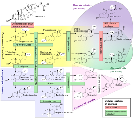
Androstenedione, or 4-androstenedione, also known as androst-4-ene-3,17-dione, is an endogenous weak androgen steroid hormone and intermediate in the biosynthesis of estrone and of testosterone from dehydroepiandrosterone (DHEA). It is closely related to androstenediol (androst-5-ene-3β,17β-diol).

<span class="mw-page-title-main">Estrone</span> Chemical compound

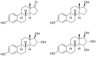
Estrone (E1), also spelled oestrone, is a steroid, a weak estrogen, and a minor female sex hormone. It is one of three major endogenous estrogens, the others being estradiol and estriol. Estrone, as well as the other estrogens, are synthesized from cholesterol and secreted mainly from the gonads, though they can also be formed from adrenal androgens in adipose tissue. Relative to estradiol, both estrone and estriol have far weaker activity as estrogens. Estrone can be converted into estradiol, and serves mainly as a precursor or metabolic intermediate of estradiol. It is both a precursor and metabolite of estradiol.

<span class="mw-page-title-main">Estriol</span> Chemical compound

Estriol (E3), also spelled oestriol, is a steroid, a weak estrogen, and a minor female sex hormone. It is one of three major endogenous estrogens, the others being estradiol and estrone. Levels of estriol in women who are not pregnant are almost undetectable. However, during pregnancy, estriol is synthesized in very high quantities by the placenta and is the most produced estrogen in the body by far, although circulating levels of estriol are similar to those of other estrogens due to a relatively high rate of metabolism and excretion. Relative to estradiol, both estriol and estrone have far weaker activity as estrogens.

<span class="mw-page-title-main">Aromatase inhibitor</span> Class of drugs

Aromatase inhibitors (AIs) are a class of drugs used in the treatment of breast cancer in postmenopausal women and in men, and gynecomastia in men. They may also be used off-label to reduce estrogen conversion when supplementing testosterone exogenously. They may also be used for chemoprevention in women at high risk for breast cancer.

<span class="mw-page-title-main">Estrogen receptor</span> Proteins activated by the hormone estrogen

Estrogen receptors (ERs) are a group of proteins found inside cells. They are receptors that are activated by the hormone estrogen (17β-estradiol). Two classes of ER exist: nuclear estrogen receptors, which are members of the nuclear receptor family of intracellular receptors, and membrane estrogen receptors (mERs), which are mostly G protein-coupled receptors. This article refers to the former (ER).

<span class="mw-page-title-main">Estrogen insensitivity syndrome</span> Medical condition

Estrogen insensitivity syndrome (EIS), or estrogen resistance, is a form of congenital estrogen deficiency or hypoestrogenism which is caused by a defective estrogen receptor (ER) – specifically, the estrogen receptor alpha (ERα) – that results in an inability of estrogen to mediate its biological effects in the body. Congenital estrogen deficiency can alternatively be caused by a defect in aromatase, the enzyme responsible for the biosynthesis of estrogens, a condition which is referred to as aromatase deficiency and is similar in symptomatology to EIS.

17β-Hydroxysteroid dehydrogenases, also 17-ketosteroid reductases (17-KSR), are a group of alcohol oxidoreductases which catalyze the reduction of 17-ketosteroids and the dehydrogenation of 17β-hydroxysteroids in steroidogenesis and steroid metabolism. This includes interconversion of DHEA and androstenediol, androstenedione and testosterone, and estrone and estradiol.

<span class="mw-page-title-main">Estrone sulfate</span> Chemical compound

Estrone sulfate, also known as E1S, E1SO4 and estrone 3-sulfate, is a natural, endogenous steroid and an estrogen ester and conjugate.

<span class="mw-page-title-main">Aromatase deficiency</span> Medical condition

Aromatase deficiency is a rare condition characterized by extremely low levels or complete absence of the enzyme aromatase activity in the body. It is an autosomal recessive disease resulting from various mutations of gene CYP19 (P450arom) which can lead to ambiguous genitalia and delayed puberty in females, continued linear growth into adulthood and osteoporosis in males and virilization in pregnant mothers. As of 2020, fewer than 15 cases have been identified in genetically male individuals and at least 30 cases in genetically female individuals.

Breast development, also known as mammogenesis, is a complex biological process in primates that takes place throughout a female's life.

<span class="mw-page-title-main">Catamenial epilepsy</span> Epilepsy exacerbated during certain phases of the menstrual cycle

Catamenial epilepsy is a form of epilepsy in women where seizures are exacerbated during certain phases of the menstrual cycle. In rare cases, seizures occur only during certain parts of the cycle; in most cases, seizures occur more frequently during certain parts of the cycle. Catamenial epilepsy is underlain by hormonal fluctuations of the menstrual cycle where estrogens promote seizures and progesterone counteracts seizure activity.

Sexual motivation is influenced by hormones such as testosterone, estrogen, progesterone, oxytocin, and vasopressin. In most mammalian species, sex hormones control the ability and motivation to engage in sexual behaviours.

<span class="mw-page-title-main">Aromatase excess syndrome</span> Medical condition

Aromatase excess syndrome is a rarely diagnosed genetic and endocrine syndrome which is characterized by an overexpression of aromatase, the enzyme responsible for the biosynthesis of the estrogen sex hormones from the androgens, in turn resulting in excessive levels of circulating estrogens and, accordingly, symptoms of hyperestrogenism. It affects both sexes, manifesting itself in males as marked or complete phenotypical feminization and in females as hyperfeminization.

<span class="mw-page-title-main">Gynecomastia</span> Endocrine system disorder of human male breast

Gynecomastia is the abnormal non-cancerous enlargement of one or both breasts in males due to the growth of breast tissue as a result of a hormone imbalance between estrogens and androgens. Gynecomastia can cause significant psychological distress or unease.

<span class="mw-page-title-main">16α-Hydroxyestrone</span> Chemical compound

16α-Hydroxyestrone (16α-OH-E1), or hydroxyestrone, also known as estra-1,3,5(10)-triene-3,16α-diol-17-one, is an endogenous steroidal estrogen and a major metabolite of estrone, as well as an intermediate in the biosynthesis of estriol. It is a potent estrogen similarly to estrone, and it has been suggested that the ratio of 16α-hydroxyestrone to 2-hydroxyestrone, the latter being much less estrogenic in comparison and even antiestrogenic in the presence of more potent estrogens like estradiol, may be involved in the pathophysiology of breast cancer. Conversely, 16α-hydroxyestrone may help to protect against osteoporosis.

<span class="mw-page-title-main">Estrogen (medication)</span> Type of medication

An estrogen (E) is a type of medication which is used most commonly in hormonal birth control and menopausal hormone therapy, and as part of feminizing hormone therapy for transgender women. They can also be used in the treatment of hormone-sensitive cancers like breast cancer and prostate cancer and for various other indications. Estrogens are used alone or in combination with progestogens. They are available in a wide variety of formulations and for use by many different routes of administration. Examples of estrogens include bioidentical estradiol, natural conjugated estrogens, synthetic steroidal estrogens like ethinylestradiol, and synthetic nonsteroidal estrogens like diethylstilbestrol. Estrogens are one of three types of sex hormone agonists, the others being androgens/anabolic steroids like testosterone and progestogens like progesterone.

The pharmacology of estradiol, an estrogen medication and naturally occurring steroid hormone, concerns its pharmacodynamics, pharmacokinetics, and various routes of administration.

References

  1. 1 2 3 Huether SE, McCance KL (2019). Understanding Pathophysiology. Elsevier Health Sciences. p. 767. ISBN   978-0-32-367281-8. Estrogen is a generic term for any of three similar hormones derived from cholesterol: estradiol, estrone, and estriol.
  2. Satoskar RS, Rege N, Bhandarkar SD (2017). Pharmacology and Pharmacotherapeutics. Elsevier Health Sciences. p. 943. ISBN   978-8-13-124941-3. The natural estrogens are steroids. However, typical estrogenic activity is also shown by chemicals which are not steroids. Hence, the term 'estrogen' is used as a generic term to describe all the compounds having estrogenic activity.
  3. Delgado BJ, Lopez-Ojeda W (20 December 2021). "Estrogen". StatPearls [Internet]. StatPearls Publishing. PMID   30855848. Estrogen is a steroid hormone associated with the female reproductive organs and is responsible for the development of female sexual characteristics. Estrogen is often referred to as estrone, estradiol, and estriol. ... Synthetic estrogen is also available for clinical use, designed to increase absorption and effectiveness by altering the estrogen chemical structure for topical or oral administration. Synthetic steroid estrogens include ethinyl estradiol, estradiol valerate, estropipate, conjugate esterified estrogen, and quinestrol.
  4. Ryan KJ (August 1982). "Biochemistry of aromatase: significance to female reproductive physiology". Cancer Research. 42 (8 Suppl): 3342s–3344s. PMID   7083198.
  5. Mechoulam R, Brueggemeier RW, Denlinger DL (September 2005). "Estrogens in insects". Cellular and Molecular Life Sciences. 40 (9): 942–944. doi:10.1007/BF01946450. S2CID   31950471.
  6. Burger HG (April 2002). "Androgen production in women". Fertility and Sterility. 77 (Suppl 4): S3–S5. doi: 10.1016/S0015-0282(02)02985-0 . PMID   12007895.
  7. Lombardi G, Zarrilli S, Colao A, Paesano L, Di Somma C, Rossi F, De Rosa M (June 2001). "Estrogens and health in males". Molecular and Cellular Endocrinology. 178 (1–2): 51–55. doi:10.1016/S0303-7207(01)00420-8. PMID   11403894. S2CID   36834775.
  8. Whitehead SA, Nussey S (2001). Endocrinology: an integrated approach. Oxford: BIOS: Taylor & Francis. ISBN   978-1-85996-252-7. PMID   20821847.
  9. Soltysik K, Czekaj P (April 2013). "Membrane estrogen receptors - is it an alternative way of estrogen action?". Journal of Physiology and Pharmacology. 64 (2): 129–142. PMID   23756388.
  10. Micevych PE, Kelly MJ (2012). "Membrane estrogen receptor regulation of hypothalamic function". Neuroendocrinology. 96 (2): 103–110. doi:10.1159/000338400. PMC   3496782 . PMID   22538318.
  11. Prossnitz ER, Arterburn JB, Sklar LA (February 2007). "GPR30: A G protein-coupled receptor for estrogen". Molecular and Cellular Endocrinology. 265–266: 138–142. doi:10.1016/j.mce.2006.12.010. PMC   1847610 . PMID   17222505.
  12. 1 2 Wang S, Huang W, Fang G, Zhang Y, Qiao H (2008). "Analysis of steroidal estrogen residues in food and environmental samples". International Journal of Environmental Analytical Chemistry. 88 (1): 1–25. Bibcode:2008IJEAC..88....1W. doi:10.1080/03067310701597293. S2CID   93975613.
  13. 1 2 Korach KD (1998). Reproductive and developmental toxicology. New York: Marcel Dekker. ISBN   0-585-15807-X. OCLC   44957536.
  14. A. Labhart (6 December 2012). Clinical Endocrinology: Theory and Practice. Springer Science & Business Media. pp. 548–. ISBN   978-3-642-96158-8.
  15. Baker ME (March 2013). "What are the physiological estrogens?". Steroids. 78 (3): 337–340. doi:10.1016/j.steroids.2012.12.011. PMID   23313336. S2CID   11803629.
  16. Miller KK, Al-Rayyan N, Ivanova MM, Mattingly KA, Ripp SL, Klinge CM, Prough RA (January 2013). "DHEA metabolites activate estrogen receptors alpha and beta". Steroids. 78 (1): 15–25. doi:10.1016/j.steroids.2012.10.002. PMC   3529809 . PMID   23123738.
  17. Bhavnani BR, Nisker JA, Martin J, Aletebi F, Watson L, Milne JK (2000). "Comparison of pharmacokinetics of a conjugated equine estrogen preparation (premarin) and a synthetic mixture of estrogens (C.E.S.) in postmenopausal women". Journal of the Society for Gynecologic Investigation. 7 (3): 175–183. doi:10.1016/s1071-5576(00)00049-6. PMID   10865186.
  18. Häggström M (2014). "Reference ranges for estradiol, progesterone, luteinizing hormone and follicle-stimulating hormone during the menstrual cycle". WikiJournal of Medicine. 1 (1). doi: 10.15347/wjm/2014.001 . ISSN   2002-4436.
  19. Lin CY, Ström A, Vega VB, Kong SL, Yeo AL, Thomsen JS, et al. (2004). "Discovery of estrogen receptor alpha target genes and response elements in breast tumor cells". Genome Biology. 5 (9): R66. doi: 10.1186/gb-2004-5-9-r66 . PMC   522873 . PMID   15345050.
  20. Darabi M, Ani M, Panjehpour M, Rabbani M, Movahedian A, Zarean E (2011). "Effect of estrogen receptor β A1730G polymorphism on ABCA1 gene expression response to postmenopausal hormone replacement therapy". Genetic Testing and Molecular Biomarkers. 15 (1–2): 11–15. doi:10.1089/gtmb.2010.0106. PMID   21117950.
  21. Lauwers, J.; Shinskie, D. (2004). Counseling the Nursing Mother: A Lactation Consultant's Guide. Jones & Bartlett Learning, LLC. p. 93. ISBN   978-0-7637-2765-9 . Retrieved 12 October 2023.
  22. Raloff J (6 December 1997). "Science News Online (12/6/97): Estrogen's Emerging Manly Alter Ego". Science News. Retrieved 4 March 2008.
  23. Hess RA, Bunick D, Lee KH, Bahr J, Taylor JA, Korach KS, Lubahn DB (December 1997). "A role for oestrogens in the male reproductive system". Nature. 390 (6659): 509–512. Bibcode:1997Natur.390..509H. doi:10.1038/37352. PMC   5719867 . PMID   9393999.
  24. "Estrogen Linked To Sperm Count, Male Fertility". Science Blog. Archived from the original on 7 May 2007. Retrieved 4 March 2008.
  25. Hill RA, Pompolo S, Jones ME, Simpson ER, Boon WC (December 2004). "Estrogen deficiency leads to apoptosis in dopaminergic neurons in the medial preoptic area and arcuate nucleus of male mice". Molecular and Cellular Neurosciences. 27 (4): 466–476. doi:10.1016/j.mcn.2004.04.012. PMID   15555924. S2CID   25280077.
  26. Chidi-Ogbolu N, Baar K (2018). "Effect of Estrogen on Musculoskeletal Performance and Injury Risk". Frontiers in Physiology. 9: 1834. doi: 10.3389/fphys.2018.01834 . PMC   6341375 . PMID   30697162.
  27. Lowe DA, Baltgalvis KA, Greising SM (April 2010). "Mechanisms behind estrogen's beneficial effect on muscle strength in females". Exercise and Sport Sciences Reviews. 38 (2): 61–67. doi:10.1097/JES.0b013e3181d496bc. PMC   2873087 . PMID   20335737.
  28. Max SR (December 1984). "Androgen-estrogen synergy in rat levator ani muscle: glucose-6-phosphate dehydrogenase". Molecular and Cellular Endocrinology. 38 (2–3): 103–107. doi:10.1016/0303-7207(84)90108-4. PMID   6510548. S2CID   24198956.
  29. Koot RW, Amelink GJ, Blankenstein MA, Bär PR (1991). "Tamoxifen and oestrogen both protect the rat muscle against physiological damage". The Journal of Steroid Biochemistry and Molecular Biology. 40 (4–6): 689–695. doi:10.1016/0960-0760(91)90292-d. PMID   1958566. S2CID   44446541.
  30. Haizlip, KM; Harrison, BC; Leinwand, LA (January 2015). "Sex-based differences in skeletal muscle kinetics and fiber-type composition". Physiology. 30 (1): 30–9. doi:10.1152/physiol.00024.2014. PMC   4285578 . PMID   25559153. "Supplementation with estrogen increases the type-IIX percentage composition in the plantaris back to 42%. (70)"
  31. Frank AP, de Souza Santos R, Palmer BF, Clegg DJ (October 2019). "Determinants of body fat distribution in humans may provide insight about obesity-related health risks". Journal of Lipid Research. 60 (10): 1710–1719. doi: 10.1194/jlr.R086975 . PMC   6795075 . PMID   30097511.
  32. Brown LM, Gent L, Davis K, Clegg DJ (September 2010). "Metabolic impact of sex hormones on obesity". Brain Research. 1350: 77–85. doi:10.1016/j.brainres.2010.04.056. PMC   2924463 . PMID   20441773.
  33. Janssen I, Powell LH, Kazlauskaite R, Dugan SA (March 2010). "Testosterone and visceral fat in midlife women: the Study of Women's Health Across the Nation (SWAN) fat patterning study". Obesity. 18 (3): 604–610. doi:10.1038/oby.2009.251. PMC   2866448 . PMID   19696765.
  34. Rubinow KB (2017). "Estrogens and Body Weight Regulation in Men". Sex and Gender Factors Affecting Metabolic Homeostasis, Diabetes and Obesity. Advances in Experimental Medicine and Biology. Vol. 1043. Springer. pp. 285–313. doi:10.1007/978-3-319-70178-3_14. ISBN   978-3-319-70177-6. PMC   5835337 . PMID   29224100.
  35. 1 2 Stokar J, Gurt I, Cohen-Kfir E, Yakubovsky O, Hallak N, Benyamini H, et al. (June 2022). "Hepatic adropin is regulated by estrogen and contributes to adverse metabolic phenotypes in ovariectomized mice". Molecular Metabolism. 60: 101482. doi:10.1016/j.molmet.2022.101482. PMC   9044006 . PMID   35364299.
  36. Pawlina, W. (2023). Histology: A Text and Atlas: With Correlated Cell and Molecular Biology. Wolters Kluwer Health. p. 1481. ISBN   978-1-9751-8152-9 . Retrieved 12 October 2023.
  37. Greenberg, J.S.; Bruess, C.E.; Oswalt, S.B. (2014). Exploring the Dimensions of Human Sexuality. Jones & Bartlett Learning. p. 248. ISBN   978-1-4496-4851-0 . Retrieved 12 October 2023.
  38. Massaro D, Massaro GD (December 2004). "Estrogen regulates pulmonary alveolar formation, loss, and regeneration in mice" (PDF). American Journal of Physiology. Lung Cellular and Molecular Physiology. 287 (6): L1154–L1159. doi:10.1152/ajplung.00228.2004. PMID   15298854. S2CID   24642944. Archived from the original (PDF) on 25 February 2019.
  39. Christensen A, Bentley GE, Cabrera R, Ortega HH, Perfito N, Wu TJ, Micevych P (July 2012). "Hormonal regulation of female reproduction". Hormone and Metabolic Research. 44 (8): 587–591. doi:10.1055/s-0032-1306301. PMC   3647363 . PMID   22438212.
  40. Handa RJ, Ogawa S, Wang JM, Herbison AE (January 2012). "Roles for oestrogen receptor β in adult brain function". Journal of Neuroendocrinology. 24 (1): 160–173. doi:10.1111/j.1365-2826.2011.02206.x. PMC   3348521 . PMID   21851428.
  41. Kow LM, Pfaff DW (May 1998). "Mapping of neural and signal transduction pathways for lordosis in the search for estrogen actions on the central nervous system". Behavioural Brain Research. 92 (2): 169–180. doi:10.1016/S0166-4328(97)00189-7. PMID   9638959. S2CID   28276218.
  42. Warnock JK, Swanson SG, Borel RW, Zipfel LM, Brennan JJ (2005). "Combined esterified estrogens and methyltestosterone versus esterified estrogens alone in the treatment of loss of sexual interest in surgically menopausal women". Menopause. 12 (4): 374–384. doi:10.1097/01.GME.0000153933.50860.FD. PMID   16037752. S2CID   24557071.
  43. Heiman JR, Rupp H, Janssen E, Newhouse SK, Brauer M, Laan E (May 2011). "Sexual desire, sexual arousal and hormonal differences in premenopausal US and Dutch women with and without low sexual desire". Hormones and Behavior. 59 (5): 772–779. doi:10.1016/j.yhbeh.2011.03.013. PMID   21514299. S2CID   20807391.
  44. Brisken C, O'Malley B (December 2010). "Hormone action in the mammary gland". Cold Spring Harbor Perspectives in Biology. 2 (12): a003178. doi:10.1101/cshperspect.a003178. PMC   2982168 . PMID   20739412.
  45. Kleinberg DL (February 1998). "Role of IGF-I in normal mammary development". Breast Cancer Research and Treatment. 47 (3): 201–208. doi:10.1023/a:1005998832636. PMID   9516076. S2CID   30440069.
  46. 1 2 3 Johnson LR (2003). Essential Medical Physiology. Academic Press. p. 770. ISBN   978-0-12-387584-6.
  47. 1 2 3 Norman AW, Henry HL (30 July 2014). Hormones. Academic Press. p. 311. ISBN   978-0-08-091906-5.
  48. 1 2 Coad J, Dunstall M (2011). Anatomy and Physiology for Midwives, with Pageburst online access,3: Anatomy and Physiology for Midwives. Elsevier Health Sciences. p. 413. ISBN   978-0-7020-3489-3.
  49. Haslam SZ, Osuch JR (1 January 2006). Hormones and Breast Cancer in Post-Menopausal Women. IOS Press. p. 69. ISBN   978-1-58603-653-9.
  50. Silbernagl S, Despopoulos A (1 January 2011). Color Atlas of Physiology. Thieme. pp. 305–. ISBN   978-3-13-149521-1.
  51. Fadem B (2007). High-yield Comprehensive USMLE Step 1 Review. Lippincott Williams & Wilkins. pp. 445–. ISBN   978-0-7817-7427-7.
  52. Blackburn S (14 April 2014). Maternal, Fetal, & Neonatal Physiology. Elsevier Health Sciences. pp. 146–. ISBN   978-0-323-29296-2.
  53. Strauss JF, Barbieri RL (13 September 2013). Yen and Jaffe's Reproductive Endocrinology. Elsevier Health Sciences. pp. 236–. ISBN   978-1-4557-2758-2.
  54. Wilson CB, Nizet V, Maldonado Y, Remington JS, Klein JO (24 February 2015). Remington and Klein's Infectious Diseases of the Fetus and Newborn Infant. Elsevier Health Sciences. pp. 190–. ISBN   978-0-323-24147-2.
  55. Zárate S, Stevnsner T, Gredilla R (2017). "Role of Estrogen and Other Sex Hormones in Brain Aging. Neuroprotection and DNA Repair". Frontiers in Aging Neuroscience. 9: 430. doi: 10.3389/fnagi.2017.00430 . PMC   5743731 . PMID   29311911.
  56. Sherwin BB (February 2012). "Estrogen and cognitive functioning in women: lessons we have learned". Behavioral Neuroscience. 126 (1): 123–127. doi:10.1037/a0025539. PMC   4838456 . PMID   22004260.
  57. Hara Y, Waters EM, McEwen BS, Morrison JH (July 2015). "Estrogen Effects on Cognitive and Synaptic Health Over the Lifecourse". Physiological Reviews. 95 (3): 785–807. doi:10.1152/physrev.00036.2014. PMC   4491541 . PMID   26109339.
  58. Korol DL, Pisani SL (August 2015). "Estrogens and cognition: Friends or foes?: An evaluation of the opposing effects of estrogens on learning and memory". Hormones and Behavior. 74: 105–115. doi:10.1016/j.yhbeh.2015.06.017. PMC   4573330 . PMID   26149525.
  59. Au A, Feher A, McPhee L, Jessa A, Oh S, Einstein G (January 2016). "Estrogens, inflammation and cognition". Frontiers in Neuroendocrinology. 40: 87–100. doi: 10.1016/j.yfrne.2016.01.002 . PMID   26774208.
  60. Jacobs E, D'Esposito M (April 2011). "Estrogen shapes dopamine-dependent cognitive processes: implications for women's health". The Journal of Neuroscience. 31 (14): 5286–5293. doi:10.1523/JNEUROSCI.6394-10.2011. PMC   3089976 . PMID   21471363.
  61. Colzato LS, Hommel B (1 January 2014). "Effects of estrogen on higher-order cognitive functions in unstressed human females may depend on individual variation in dopamine baseline levels". Frontiers in Neuroscience. 8: 65. doi: 10.3389/fnins.2014.00065 . PMC   3985021 . PMID   24778605.
  62. Hogervorst E (March 2013). "Estrogen and the brain: does estrogen treatment improve cognitive function?". Menopause International. 19 (1): 6–19. doi:10.1177/1754045312473873. PMID   27951525. S2CID   10122688.
  63. Douma SL, Husband C, O'Donnell ME, Barwin BN, Woodend AK (2005). "Estrogen-related mood disorders: reproductive life cycle factors". ANS. Advances in Nursing Science. 28 (4): 364–375. doi:10.1097/00012272-200510000-00008. PMID   16292022. S2CID   9172877.
  64. Osterlund MK, Witt MR, Gustafsson JA (December 2005). "Estrogen action in mood and neurodegenerative disorders: estrogenic compounds with selective properties-the next generation of therapeutics". Endocrine. 28 (3): 235–242. doi:10.1385/ENDO:28:3:235. PMID   16388113. S2CID   8205014.
  65. Lasiuk GC, Hegadoren KM (October 2007). "The effects of estradiol on central serotonergic systems and its relationship to mood in women". Biological Research for Nursing. 9 (2): 147–160. doi:10.1177/1099800407305600. PMID   17909167. S2CID   37965502.
  66. Grigoriadis S, Seeman MV (June 2002). "The role of estrogen in schizophrenia: implications for schizophrenia practice guidelines for women". Canadian Journal of Psychiatry. 47 (5): 437–442. doi: 10.1177/070674370204700504 . PMID   12085678.
  67. "PMDD/PMS". The Massachusetts General Hospital Center for Women's Mental Health. Retrieved 12 January 2019.
  68. Hill RA, McInnes KJ, Gong EC, Jones ME, Simpson ER, Boon WC (February 2007). "Estrogen deficient male mice develop compulsive behavior". Biological Psychiatry. 61 (3): 359–366. doi:10.1016/j.biopsych.2006.01.012. PMID   16566897. S2CID   22669945.
  69. Benmansour S, Weaver RS, Barton AK, Adeniji OS, Frazer A (April 2012). "Comparison of the effects of estradiol and progesterone on serotonergic function". Biological Psychiatry. 71 (7): 633–641. doi:10.1016/j.biopsych.2011.11.023. PMC   3307822 . PMID   22225849.
  70. Berg SJ, Wynne-Edwards KE (June 2001). "Changes in testosterone, cortisol, and estradiol levels in men becoming fathers". Mayo Clinic Proceedings. 76 (6): 582–592. doi:10.4065/76.6.582. PMID   11393496.
  71. 1 2 3 Cao X, Xu P, Oyola MG, Xia Y, Yan X, Saito K, et al. (October 2014). "Estrogens stimulate serotonin neurons to inhibit binge-like eating in mice". The Journal of Clinical Investigation. 124 (10): 4351–4362. doi:10.1172/JCI74726. PMC   4191033 . PMID   25157819.
  72. Jimerson DC, Lesem MD, Kaye WH, Hegg AP, Brewerton TD (September 1990). "Eating disorders and depression: is there a serotonin connection?". Biological Psychiatry. 28 (5): 443–454. doi:10.1016/0006-3223(90)90412-u. PMID   2207221. S2CID   31058047.
  73. Klump KL, Keel PK, Racine SE, Burt SA, Burt AS, Neale M, et al. (February 2013). "The interactive effects of estrogen and progesterone on changes in emotional eating across the menstrual cycle". Journal of Abnormal Psychology. 122 (1): 131–137. doi:10.1037/a0029524. PMC   3570621 . PMID   22889242.
  74. 1 2 Edler C, Lipson SF, Keel PK (January 2007). "Ovarian hormones and binge eating in bulimia nervosa". Psychological Medicine. 37 (1): 131–141. doi:10.1017/S0033291706008956. PMID   17038206. S2CID   36609028.
  75. 1 2 Klump KL, Racine SE, Hildebrandt B, Burt SA, Neale M, Sisk CL, et al. (September 2014). "Ovarian Hormone Influences on Dysregulated Eating: A Comparison of Associations in Women with versus without Binge Episodes". Clinical Psychological Science. 2 (4): 545–559. doi:10.1177/2167702614521794. PMC   4203460 . PMID   25343062.
  76. Klump KL, Keel PK, Culbert KM, Edler C (December 2008). "Ovarian hormones and binge eating: exploring associations in community samples". Psychological Medicine. 38 (12): 1749–1757. doi:10.1017/S0033291708002997. PMC   2885896 . PMID   18307829.
  77. Lester NA, Keel PK, Lipson SF (January 2003). "Symptom fluctuation in bulimia nervosa: relation to menstrual-cycle phase and cortisol levels". Psychological Medicine. 33 (1): 51–60. doi:10.1017/s0033291702006815. PMID   12537036. S2CID   21497515.
  78. Wu MV, Manoli DS, Fraser EJ, Coats JK, Tollkuhn J, Honda S, et al. (October 2009). "Estrogen masculinizes neural pathways and sex-specific behaviors". Cell. 139 (1): 61–72. doi:10.1016/j.cell.2009.07.036. PMC   2851224 . PMID   19804754.
  79. Rochira V, Carani C (October 2009). "Aromatase deficiency in men: a clinical perspective". Nature Reviews. Endocrinology. 5 (10): 559–568. doi:10.1038/nrendo.2009.176. PMID   19707181. S2CID   22116130.
  80. Wilson JD (September 2001). "Androgens, androgen receptors, and male gender role behavior" (PDF). Hormones and Behavior. 40 (2): 358–366. doi:10.1006/hbeh.2001.1684. PMID   11534997. S2CID   20480423. Archived from the original (PDF) on 26 February 2019.
  81. Baum MJ (November 2006). "Mammalian animal models of psychosexual differentiation: when is 'translation' to the human situation possible?". Hormones and Behavior. 50 (4): 579–588. doi:10.1016/j.yhbeh.2006.06.003. PMID   16876166. S2CID   7465192.
  82. Rosano GM, Panina G (1999). "Oestrogens and the heart". Therapie. 54 (3): 381–385. PMID   10500455.
  83. Nadkarni S, Cooper D, Brancaleone V, Bena S, Perretti M (November 2011). "Activation of the annexin A1 pathway underlies the protective effects exerted by estrogen in polymorphonuclear leukocytes". Arteriosclerosis, Thrombosis, and Vascular Biology. 31 (11): 2749–2759. doi:10.1161/ATVBAHA.111.235176. PMC   3357483 . PMID   21836070.
  84. Abdul Sultan A, West J, Stephansson O, Grainge MJ, Tata LJ, Fleming KM, Humes D, Ludvigsson JF (November 2015). "Defining venous thromboembolism and measuring its incidence using Swedish health registries: a nationwide pregnancy cohort study". BMJ Open. 5 (11): e008864. doi:10.1136/bmjopen-2015-008864. PMC   4654387 . PMID   26560059.
  85. 1 2 Foo YZ, Nakagawa S, Rhodes G, Simmons LW (February 2017). "The effects of sex hormones on immune function: a meta-analysis" (PDF). Biological Reviews of the Cambridge Philosophical Society. 92 (1): 551–571. doi:10.1111/brv.12243. PMID   26800512. S2CID   37931012.
  86. Taneja V (27 August 2018). "Sex Hormones Determine Immune Response". Frontiers in Immunology. 9: 1931. doi: 10.3389/fimmu.2018.01931 . PMC   6119719 . PMID   30210492.
  87. 1 2 Roved J, Westerdahl H, Hasselquist D (February 2017). "Sex differences in immune responses: Hormonal effects, antagonistic selection, and evolutionary consequences". Hormones and Behavior. 88: 95–105. doi:10.1016/j.yhbeh.2016.11.017. PMID   27956226. S2CID   9137227.
  88. Khan D, Ansar Ahmed S (6 January 2016). "The Immune System Is a Natural Target for Estrogen Action: Opposing Effects of Estrogen in Two Prototypical Autoimmune Diseases". Frontiers in Immunology. 6: 635. doi: 10.3389/fimmu.2015.00635 . PMC   4701921 . PMID   26779182.
  89. 1 2 3 Kovats S (April 2015). "Estrogen receptors regulate innate immune cells and signaling pathways". Cellular Immunology. 294 (2): 63–69. doi:10.1016/j.cellimm.2015.01.018. PMC   4380804 . PMID   25682174.
  90. Prior JC (2018). Estrogen's Storm Season: stories of perimenopause. Vancouver, British Columbia: CeMCOR (Centre for Menstrual Cycle and Ovulation Research). ISBN   9780973827521 . Retrieved 24 July 2021. [...] high estrogen amplifies your stress hormone responses to stressful things [...]
  91. Häggström M, Richfield D (2014). "Diagram of the pathways of human steroidogenesis". WikiJournal of Medicine. 1 (1). doi: 10.15347/wjm/2014.005 . ISSN   2002-4436.
  92. Marieb E (2013). Anatomy & physiology. Benjamin-Cummings. p. 903. ISBN   978-0-321-88760-3.
  93. Hemsell DL, Grodin JM, Brenner PF, Siiteri PK, MacDonald PC (March 1974). "Plasma precursors of estrogen. II. Correlation of the extent of conversion of plasma androstenedione to estrone with age". The Journal of Clinical Endocrinology and Metabolism. 38 (3): 476–479. doi: 10.1210/jcem-38-3-476 . PMID   4815174.
  94. Barakat R, Oakley O, Kim H, Jin J, Ko CJ (September 2016). "Extra-gonadal sites of estrogen biosynthesis and function". BMB Reports. 49 (9): 488–496. doi:10.5483/BMBRep.2016.49.9.141. PMC   5227141 . PMID   27530684.
  95. 1 2 Nelson LR, Bulun SE (September 2001). "Estrogen production and action". Journal of the American Academy of Dermatology. 45 (3 Suppl): S116–S124. doi:10.1067/mjd.2001.117432. PMID   11511861.
  96. Labrie F, Bélanger A, Luu-The V, Labrie C, Simard J, Cusan L, et al. (1998). "DHEA and the intracrine formation of androgens and estrogens in peripheral target tissues: its role during aging". Steroids. 63 (5–6): 322–328. doi:10.1016/S0039-128X(98)00007-5. PMID   9618795. S2CID   37344052.
  97. Trachsler, A.; Thorn, G.W.; Labhart, A.; Bürgi, H.; Dodsworth-Phillips, J.; Constam, G.R.; Courvoisier, B.; Fischer, J.A.; Froesch, E.R.; Grob, P. (2012). Clinical Endocrinology: Theory and Practice. Springer Berlin Heidelberg. p. 530. ISBN   978-3-642-96158-8 . Retrieved 12 October 2023.
  98. Fuentes, Nathalie; Silveyra, Patricia (2019). "Estrogen receptor signaling mechanisms". Advances in Protein Chemistry and Structural Biology. Vol. 116. Elsevier. pp. 135–170. doi:10.1016/bs.apcsb.2019.01.001. ISBN   9780128155615. ISSN   1876-1623. PMC   6533072 . PMID   31036290. Physiologically, the metabolic conversion of estrogens allows their excretion from the body via urine, feces, and/or bile, along with the production of estrogen analogs, which have been shown to present antiproliferative effects (Tsuchiya et al., 2005).
  99. Kuhl H (August 2005). "Pharmacology of estrogens and progestogens: influence of different routes of administration". Climacteric. 8 (Suppl 1): 3–63. doi:10.1080/13697130500148875. PMID   16112947. S2CID   24616324.
  100. Tata JR (June 2005). "One hundred years of hormones". EMBO Reports. 6 (6): 490–496. doi:10.1038/sj.embor.7400444. PMC   1369102 . PMID   15940278.
  101. 1 2 "Origin in Biomedical Terms: oestrogen or oestrogen". Bioetymology. Retrieved 24 January 2018.
  102. "Council on Pharmacy and Chemistry". Journal of the American Medical Association. 107 (15): 1221–3. 1936. doi:10.1001/jama.1936.02770410043011.
  103. "Greek Word Study Tool: oistros". Perseus Digital Library . Retrieved 28 December 2011.
  104. Fang H, Tong W, Shi LM, Blair R, Perkins R, Branham W, et al. (March 2001). "Structure-activity relationships for a large diverse set of natural, synthetic, and environmental estrogens". Chemical Research in Toxicology. 14 (3): 280–294. CiteSeerX   10.1.1.460.20 . doi:10.1021/tx000208y. PMID   11258977.
  105. Wise A, O'Brien K, Woodruff T (January 2011). "Are oral contraceptives a significant contributor to the estrogenicity of drinking water?". Environmental Science & Technology. 45 (1): 51–60. doi:10.1021/es1014482. PMID   20977246.
  106. Peach S. "Don't Blame The Pill | Latest News". Chemical & Engineering News. Retrieved 22 April 2023.
  107. Liney KE, Jobling S, Shears JA, Simpson P, Tyler CR (October 2005). "Assessing the sensitivity of different life stages for sexual disruption in roach (Rutilus rutilus) exposed to effluents from wastewater treatment works". Environmental Health Perspectives. 113 (10): 1299–1307. doi:10.1289/ehp.7921. PMC   1281270 . PMID   16203238.
  108. Jobling S, Williams R, Johnson A, Taylor A, Gross-Sorokin M, Nolan M, et al. (April 2006). "Predicted exposures to steroid estrogens in U.K. rivers correlate with widespread sexual disruption in wild fish populations". Environmental Health Perspectives. 114 (Suppl 1): 32–39. doi:10.1289/ehp.8050. PMC   1874167 . PMID   16818244.
  109. Sanghavi DM (17 October 2006). "Preschool Puberty, and a Search for the Causes". The New York Times . Retrieved 4 June 2008.
  110. 1 2 FDA (February 1995). "Products containing estrogenic hormones, placental extract or vitamins". Guide to Inspections of Cosmetic Product Manufacturers. Archived from the original on 14 October 2007. Retrieved 24 October 2006.