Natriuretic peptide

Last updated
Natriuretic peptide-binding receptors and ligand selectivity. Natriuretic Peptides and Receptors.jpg
Natriuretic peptide-binding receptors and ligand selectivity.

A natriuretic peptide is a hormone molecule that plays a crucial role in the regulation of the cardiovascular system. These hormones were first discovered in the 1980s and were found to have very strong diuretic, natriuretic, and vasodilatory effects. There are three main types of natriuretic peptides: atrial natriuretic peptide (ANP), brain natriuretic peptide (BNP), and C-type natriuretic peptide (CNP). [1] Two minor hormones include Urodilatin (URO) which is processed in the kidney and encoded by the same gene as ANP, and Dendroaspis NP (DNP) that was discovered through isolation of the venom from the green mamba snake. [2] Since they are activated during heart failure, they are important for the protection of the heart and its tissues. [1]

Contents

Additionally, there are three natriuretic peptide receptors: Natriuretic Peptide Receptor-A (NPR-A), Natriuretic Peptide Receptor-B (NPR-B), and Natriuretic Peptide Receptor-C (NPR-C). NPR-A and NPR-B use cyclic guanosine monophosphate (cGMP) as its intracellular messenger. NPR-A binds selectively to ANP and BNP while NPR-B binds selectively to CNP. [1] Although initially thought to have no signaling activity, NPR-C is now believed to inhibit cyclic adenosine monophosphate (cAMP) using the Giα subunit along with activating phospholipase C-β using the Giβγ subunit. The three natriuretic peptides ANP, BNP, and CNP can bind to NPR-C however it has a higher affinity towards ANP and CNP. Natriuretic peptides will lose their activity when degraded by the enzyme neprilysin which is found on the plasma membrane. [1]

Origin

The existence of natriuretic peptides was predicted over fifty years ago by key cell biological observations. Atrial cells were found to contain highly developed Golgi networks and spherical granules, while balloon distension of the atria correlated with increased urination in dogs. [3] De Bold and colleagues linked these studies and discovered the first natriuretic peptide that works by stimulating renal sodium and water secretion. Shortly after, atrial peptides with natriuretic, diuretic, and/or smooth muscle relaxing activity were purified and sequenced. [3] The 1980s saw an increase in natriuretic peptide research, particularly due to the 1981 work by de Bold et al., which found that giving rats an extract of rat atrial tissue quickly reduced blood pressure and increased urination. [1] To pinpoint the molecule causing this action, various structures that would eventually come to be known as an atrial natriuretic peptide, or ANP, were discovered. Kangawa and Matsuo determined the complete amino acid sequence of α-hANP using protein isolated from human atrial tissue. [1] They were able to isolate and identify BNP and CNP from porcine brain in 1988 and 1990, respectively. [1]

Structure of natriuretic peptides. Structure of Natriuretic Peptides.jpg
Structure of natriuretic peptides.

The first of these hormones is most frequently referred to as atrial natriuretic peptide (ANP), whereas the second hormone, B-type natriuretic peptide (BNP), was formerly known as brain natriuretic peptide but is now more frequently linked to the cardiac ventricles of people with heart failure. [3] C-type natriuretic peptide (CNP), the third hormone, was isolated from the swine brain and could relax smooth muscle. The three hormones share a similar structural makeup but come from different genes. [3] These preliminary findings produced more investigation to establish the genetic makeup and regulatory mechanisms of these molecules.

Applications

Mechanism

Natriuretic peptides and their receptors have many different effects on the body, such as controlling blood pressure and helping bones grow. Each peptide has its own unique effects and interacts with specific receptors. Scientists have observed these effects by studying mice with specific natriuretic peptides or receptors removed. [3]

Natriuretic peptides have a wide range of physiological effects that include the regulation of blood pressure, fluid and electrolyte balance, and cardiovascular function. [1] They are also involved in the regulation of vascular remodeling, inflammation, and apoptosis. One of the major clinical applications of natriuretic peptides is the diagnosing and managing of heart failure. Elevated levels of ANP and BNP are commonly found in patients with heart failure and can be used as biomarkers for the diagnosis and prognosis of this condition. [1]

Table 1: Physiological actions of natriuretic peptides. [1]
Target organBiological effects
KidneyIncrease in glomerular filtration rate by inducing vasodilatation of afferent arterioles and vasoconstriction of efferent arterioles

Induction of natriuresis by inhibiting Na+, H+ exchanger in the proximal tubule, Na+, Cl− co-transporter in the distal tubule, and Na+ channels in the collecting duct

Natriuretic and diuretic effects due to increased medullary blood flow

Induction of diuresis due to inhibition of Arginine vasopressin-induced aquaporin-2 incorporation into collecting ducts' apical membrane

Cardiac Preload and afterload decrease

Natriuretic and diuretic effects

Inhibition of cardiac remodeling (hypertrophy and fibrosis)

HemodynamicVasorelaxation (via acute action on vascular smooth muscle cells and chronic effects on the permeability of vascular endothelial cells)

Suppression of vascular smooth muscle cell proliferation

Suppression of vascular fibrosis

Reduction of pulmonary hypertension/fibrosis

Elevation of capillary hydraulic conductivity

Decrease in cardiac preload and afterload

Endocrine Suppression of the following:

Renin–angiotensin–aldosterone axis

− Sympathetic outflow

− Arginine vasopressin

Endothelin

MitogenesisStimulation of long bone growth

Inhibition of growth factor-mediated hypertrophy in fibroblasts

C-type natriuretic peptide (CNP) primarily interacts with NPR-B, which triggers an increase in the concentration of cGMP in the cell. This process can lead to several physiological effects, such as tissue remodeling, reduction of pulmonary hypertension and fibrosis, and stimulation of long bone growth. [1] CNP is highly concentrated in vascular endothelial cells and plays a crucial role in regulating vascular tone through its vasodilatory action. Furthermore, CNP has been shown to have anti proliferative effects on vascular smooth muscle and an inhibitory effect on the migration of human coronary artery smooth muscle cells. [1]

Biological effects and uses

Studies on mice have helped researchers understand the critical role of ANP in preventing hypertension or high blood pressure. When ANP-deficient mice were studied, they showed signs of hypertension when consuming too much salt. [1] Similarly, when NPR-A, a receptor for ANP, was knocked out in mice, they also displayed hypertension and a reduced response to diuretics. This suggests that ANP is essential in regulating blood pressure and fluid balance. [1] Interestingly, when NPR-A was knocked out specifically in the endothelial cells lining blood vessels, mice showed increased plasma volume, suggesting that ANP may regulate fluid balance by increasing the permeability of blood vessels in these cells. These findings indicate that ANP and its receptor NPR-A are essential in regulating mice's blood pressure and fluid balance. [1]

Recent advances in the biology of natriuretic peptides (NPs) have led to the developing of "designer" NPs. These peptides have larger surface areas compared to smaller natural molecules, making them better suited for activating specific receptors with minimal off-target effects. [2] While inhibiting enzymatic degradation of peptides can boost endogenous peptides, it may not be enough to achieve optimal receptor stimulation. Therefore, designer peptides with specific properties could be a new strategy to improve upon existing therapies. [2] Three designer NPs have been bioengineered, tested in cell-based assays and animal models of heart failure, undergone pharmaceutical toxicology studies, and have received FDA approval for clinical studies. [2]

Natriuretic peptides can block the activity of the renin-angiotensin-aldosterone system (RAAS), which regulates blood pressure. Studies have shown that ANP can suppress renin secretion and aldosterone production. Additionally, natriuretic peptides suppress the sympathetic nervous system (SNS), which controls the body's "fight or flight" response. [1] The relationship between natriuretic peptides and vasopressin, a hormone that regulates water balance, has been found through studies that have shown that ANP can suppress vasopressin signaling. Researchers are working on having a better understanding of this relationship. Animal studies show the possibility of using a vasopressin receptor antagonist along with BNP to improve heart function and blood flow. [1]

Suppression of the RAAS, SNS, and vasopressin systems by natriuretic peptides/ Adrenergic signaling on natriuretic peptides.jpg
Suppression of the RAAS, SNS, and vasopressin systems by natriuretic peptides/

B-type natriuretic peptide (BNP) and its pro hormone NT-proBNP are especially useful in diagnosing heart failure, as their levels in the blood increase along with the severity of the condition. [3] Rapid testing of BNP and NT-proBNP can also help distinguish between shortness of breath due to heart and lung-related causes. Monitoring NT-proBNP levels over time can provide important information about an individual's risk of developing heart failure or cardiovascular disease in the future. [3]

It can be difficult to tell if someone's having trouble breathing because of heart or lung problems. However, this can often be clarified with tests like X-rays and blood work. [3] Rapid testing of BNP and NT-proBNP can also tell if the problem is in the heart or the lungs, though, specific lung problems can also raise NP levels. Due to this, BNP levels can be high for cases other than heart failure. [3]

Major types

Atrial Natriuretic Peptide (ANP)

The human gene that makes ANP is called NPPA, and it's on chromosome 1 at location 1p36.21. The gene is about two kilobases long and has three parts called exons and two called introns. The mRNA from this gene makes a protein called preproANP with 151 parts called amino acids. [3] The first 25 parts are removed to create a 126-part protein called proANP which is stored in the atrial granules. When the body needs ANP, Corin (an enzyme) breaks apart proANP to make the active form of ANP, which is 28 amino acids long. [3]

Brain Natriuretic Peptide (BNP)

BNP was first discovered in pig brain tissue but was later found more abundant in the heart (Mukoyama et al. 1991; Mukoyama et al. 1990). The human gene that encodes for BNP is called NPPB (GeneID 4879) and is located on chromosome 1 at 1p36.2. In mice, NPPb is found on chromosome 4. [3] NPPB has three exons and two introns, and its preproBNP comprises 134 amino acids. This includes a 26 amino acid signal sequence followed by 108 amino acids that contain proBNP. Unlike ANP, the sequence of BNP is not similar across different species. [3]

C-type Natriuretic Peptide (CNP)

The natriuretic peptide CNP was initially discovered in pig brain extracts. Most CNP is found in the brain, although it can also be found in chondrocytes and the blood vessel lining cells. The human gene for CNP (NPPC), in contrast to the genes for ANP and BNP, is located on chromosome 2 and consists of just two exons and one intron. [3] The CNP gene is also located on chromosome 2 in mice. The 126 amino acids long protein produced by NPPC has a signal sequence of 23 amino acids and then a proCNP segment with 103 amino acids. [3]

See also

Related Research Articles

<span class="mw-page-title-main">Vasopressin</span> Mammalian hormone released from the pituitary gland

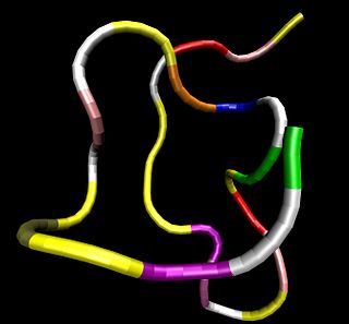
Human vasopressin, also called antidiuretic hormone (ADH), arginine vasopressin (AVP) or argipressin, is a hormone synthesized from the AVP gene as a peptide prohormone in neurons in the hypothalamus, and is converted to AVP. It then travels down the axon terminating in the posterior pituitary, and is released from vesicles into the circulation in response to extracellular fluid hypertonicity (hyperosmolality). AVP has two primary functions. First, it increases the amount of solute-free water reabsorbed back into the circulation from the filtrate in the kidney tubules of the nephrons. Second, AVP constricts arterioles, which increases peripheral vascular resistance and raises arterial blood pressure.

<span class="mw-page-title-main">Peptide hormone</span> Hormone whose molecules are peptides

Peptide hormones are hormones whose molecules are peptides. Peptide hormones have shorter amino acid chain lengths than protein hormones. These hormones have an effect on the endocrine system of animals, including humans. Most hormones can be classified as either amino acid–based hormones or steroid hormones. The former are water-soluble and act on the surface of target cells via second messengers; the latter, being lipid-soluble, move through the plasma membranes of target cells to act within their nuclei.

<span class="mw-page-title-main">Atrial natriuretic peptide</span> Cardiac hormone which increases renal sodium excretion

Atrial Natriuretic Peptide (ANP) or atrial natriuretic factor (ANF) is a natriuretic peptide hormone secreted from the cardiac atria that in humans is encoded by the NPPA gene. Natriuretic peptides are a family of hormone/paracrine factors that are structurally related. The main function of ANP is causing a reduction in expanded extracellular fluid (ECF) volume by increasing renal sodium excretion. ANP is synthesized and secreted by cardiac muscle cells in the walls of the atria in the heart. These cells contain volume receptors which respond to increased stretching of the atrial wall due to increased atrial blood volume.

<span class="mw-page-title-main">Glucocorticoid</span> Class of corticosteroids

Glucocorticoids are a class of corticosteroids, which are a class of steroid hormones. Glucocorticoids are corticosteroids that bind to the glucocorticoid receptor that is present in almost every vertebrate animal cell. The name "glucocorticoid" is a portmanteau and is composed from its role in regulation of glucose metabolism, synthesis in the adrenal cortex, and its steroidal structure.

<span class="mw-page-title-main">Apelin</span> Mammalian protein found in Homo sapiens

Apelin is a peptide that in humans is encoded by the APLN gene. Apelin is one of two endogenous ligands for the G-protein-coupled APJ receptor that is expressed at the surface of some cell types. It is widely expressed in various organs such as the heart, lung, kidney, liver, adipose tissue, gastrointestinal tract, brain, adrenal glands, endothelium, and human plasma.

Adenosine A<sub>1</sub> receptor Cell surface receptor found in humans

The adenosine A1 receptor (A1AR) is one member of the adenosine receptor group of G protein-coupled receptors with adenosine as endogenous ligand.

<span class="mw-page-title-main">Kisspeptin</span> Mammalian protein

Kisspeptins are proteins encoded by the KISS1 gene in humans. Kisspeptins are ligands of the G-protein coupled receptor, GPR54. Kiss1 was originally identified as a human metastasis suppressor gene that has the ability to suppress melanoma and breast cancer metastasis. Kisspeptin-GPR54 signaling has an important role in initiating secretion of gonadotropin-releasing hormone (GnRH) at puberty, the extent of which is an area of ongoing research. Gonadotropin-releasing hormone is released from the hypothalamus to act on the anterior pituitary triggering the release of luteinizing hormone (LH), and follicle stimulating hormone (FSH). These gonadotropic hormones lead to sexual maturation and gametogenesis. Disrupting GPR54 signaling can cause hypogonadotrophic hypogonadism in rodents and humans. The Kiss1 gene is located on chromosome 1. It is transcribed in the brain, adrenal gland, and pancreas.

<span class="mw-page-title-main">Adrenomedullin</span>

Adrenomedullin is a vasodilator peptide hormone of uncertain significance in human health and disease. It was initially isolated in 1993 from a pheochromocytoma, a tumor of the adrenal medulla: hence the name.

Atrial volume receptors are low pressure baroreceptors that are found in the atria of the heart. They are myelinated vagal fibres in the endocardium found at the junction between atria and the vena cava/pulmonary vein.

An atrial natriuretic peptide receptor is a receptor for atrial natriuretic peptide.

<span class="mw-page-title-main">Urotensin-II</span> Chemical compound

Urotensin-II (U-II) is a peptide ligand that is the strongest known vasoconstrictor. Because of the involvement of the UII system in multiple biological systems such as the cardiovascular, nervous, endocrine, and renal, it represents a promising target for the development of new drugs.

<span class="mw-page-title-main">Urodilatin</span> Chemical compound

Urodilatin (URO) is a hormone that causes natriuresis by increasing renal blood flow. It is secreted in response to increased mean arterial pressure and increased blood volume from the cells of the distal tubule and collecting duct. It is important in oliguric patients as it lowers serum creatinine and increases urine output.

<span class="mw-page-title-main">Natriuretic peptide precursor C</span> Protein-coding gene in the species Homo sapiens

Natriuretic peptide precursor C, also known as NPPC, is a protein that in humans is encoded by the NPPC gene. The precursor NPPC protein is cleaved to the 22 amino acid peptide C-type natriuretic peptide (CNP).

<span class="mw-page-title-main">NPR1</span> Protein-coding gene in the species Homo sapiens

Natriuretic peptide receptor A/guanylate cyclase A , also known as NPR1, is an atrial natriuretic peptide receptor. In humans it is encoded by the NPR1 gene.

<span class="mw-page-title-main">NPR3</span> Protein-coding gene in humans

Natriuretic peptide receptor C/guanylate cyclase C , also known as NPR3, is an atrial natriuretic peptide receptor. In humans it is encoded by the NPR3 gene.

<span class="mw-page-title-main">RAPGEF4</span> Protein-coding gene in the species Homo sapiens

Rap guanine nucleotide exchange factor (GEF) 4 (RAPGEF4), also known as exchange protein directly activated by cAMP 2 (EPAC2) is a protein that in humans is encoded by the RAPGEF4 gene.

<span class="mw-page-title-main">CORIN</span> Mammalian protein found in Homo sapiens

Corin, also called atrial natriuretic peptide-converting enzyme, is a protein that in humans is encoded by the CORIN gene.

Adolfo José de Bold was an Argentinian-born Canadian cardiovascular researcher, best known for his discovery of atrial natriuretic peptide (ANP), a polypeptide hormone secreted by heart muscle cells. The hormone plays a role in regulating blood pressure, blood volume, and cardiovascular growth, and its discovery proved the endocrine function of the heart.

Cenderitide is a natriuretic peptide developed by the Mayo Clinic as a potential treatment for heart failure. Cenderitide is created by the fusion of the 15 amino acid C-terminus of the snake venom dendroaspis natriuretic peptide (DNP) with the full C-type natriuretic peptide (CNP) structure. This peptide chimera is a dual activator of the natriuretic peptide receptors NPR-A and NPR-B and therefore exhibits the natriuretic and diuretic properties of DNP, as well as the antiproliferative and antifibrotic properties of CNP.

<span class="mw-page-title-main">Brain natriuretic peptide 32</span> Hormone secreted in the heart

Brain natriuretic peptide 32 (BNP), also known as B-type natriuretic peptide, is a hormone secreted by cardiomyocytes in the heart ventricles in response to stretching caused by increased ventricular blood volume. BNP is one of the three natriuretic peptides, in addition to ANP and CNP.

References

  1. 1 2 3 4 5 6 7 8 9 10 11 12 13 14 15 16 17 18 19 Kuwahara, Koichiro (2021-11-01). "The natriuretic peptide system in heart failure: Diagnostic and therapeutic implications". Pharmacology & Therapeutics. 227: 107863. doi: 10.1016/j.pharmthera.2021.107863 . ISSN   0163-7258.
  2. 1 2 3 4 Sangaralingham, S Jeson; Kuhn, Michaela; Cannone, Valentina; Chen, Horng H; Burnett Jr, John C (25 August 2022). "Natriuretic peptide pathways in heart failure: further therapeutic possibilities". Cardiovascular Research. 118 (18): 3416–3433 via Oxford Academic.
  3. 1 2 3 4 5 6 7 8 9 10 11 12 13 14 15 Potter, Lincoln R.; Yoder, Andrea R.; Flora, Darcy R.; Antos, Laura K.; Dickey, Deborah M. (2009), Schmidt, Harald H. H. W.; Hofmann, Franz; Stasch, Johannes-Peter (eds.), "Natriuretic Peptides: Their Structures, Receptors, Physiologic Functions and Therapeutic Applications", cGMP: Generators, Effectors and Therapeutic Implications, Handbook of Experimental Pharmacology, vol. 191, no. 191, Berlin, Heidelberg: Springer, pp. 341–366, doi:10.1007/978-3-540-68964-5_15, ISBN   978-3-540-68964-5, PMC   4855512 , PMID   19089336 , retrieved 2023-04-18
  4. Pandit, Kaushik; Mukhopadhyay, Pradip; Ghosh, Sujoy; Chowdhury, Subhankar (October 15, 2011). "Natriuretic peptides: Diagnostic and therapeutic use". Indian Journal of Endocrinology and Metabolism. 15 (8): S345-53. doi: 10.4103/2230-8210.86978 . ISSN   2230-8210. PMC   3230091 . PMID   22145138.