Related Research Articles

The external jugular vein receives the greater part of the blood from the exterior of the cranium and the deep parts of the face, being formed by the junction of the posterior division of the retromandibular vein with the posterior auricular vein.

The ansa cervicalis is a loop formed by muscular branches of the cervical plexus formed by branches of cervical spinal nerves C1-C3. The ansa cervicalis has two roots - a superior root and an inferior root - that unite distally, forming a loop. It is situated within the carotid sheath.

The dural venous sinuses are venous sinuses (channels) found between the endosteal and meningeal layers of dura mater in the brain. They receive blood from the cerebral veins, and cerebrospinal fluid (CSF) from the subarachnoid space via arachnoid granulations. They mainly empty into the internal jugular vein. Cranial venous sinuses communicate with veins outside the skull through emissary veins. These communications help to keep the pressure of blood in the sinuses constant. The major dural venous sinuses included the superior sagittal sinus, inferior sagittal sinus, transverse sinus, straight sinus, sigmoid sinus and cavernous sinus. These sinuses play a crucial role in cerebral venous drainage. A dural venous sinus, in human anatomy, is any of the channels of a branching complex sinus network that lies between layers of the dura mater, the outermost covering of the brain, and functions to collect oxygen-depleted blood. Unlike veins, these sinuses possess no muscular coat.

The straight sinus, also known as tentorial sinus or the sinus rectus, is an area within the skull beneath the brain. It receives blood from the inferior sagittal sinus and the great cerebral vein, and drains into the confluence of sinuses.
The superior sagittal sinus, within the human head, is an unpaired area along the attached margin of the falx cerebri. It allows blood to drain from the lateral aspects of anterior cerebral hemispheres to the confluence of sinuses. Cerebrospinal fluid drains through arachnoid granulations into the superior sagittal sinus and is returned to venous circulation.

In human anatomy, the hepatic portal system or portal venous system is the system of veins comprising the portal vein and its tributaries. The other portal venous systems in the body are the renal portal system, and the hypophyseal portal system.
The frontal vein begins on the forehead in a venous plexus which communicates with the frontal branches of the superficial temporal vein. The veins converge to form a single trunk, which runs downward near the middle line of the forehead parallel with the vein of the opposite side. The two veins are joined, at the root of the nose, by a transverse branch, called the nasal arch, which receives some small veins from the dorsum of the nose. At the root of the nose the veins diverge, and, each at the medial angle of the orbit, joins the supraorbital vein, to form the angular vein. Occasionally the frontal veins join to form a single trunk, which bifurcates at the root of the nose into the two angular veins.

The internal cerebral veins are two veins included in the group of deep cerebral veins that drain the deep parts of the hemispheres; each internal cerebral vein is formed near the interventricular foramina by the union of the superior thalamostriate vein and the superior choroid vein.

The cerebellar veins are veins which drain the cerebellum. They consist of the superior cerebellar veins and the inferior cerebellar veins. The superior cerebellar veins drain to the straight sinus and the internal cerebral veins. The inferior cerebellar veins drain to the transverse sinus, the superior petrosal sinus, and the occipital sinus.

The intercostal arteries are a group of arteries passing within an intercostal space. There are 9 anterior and 11 posterior intercostal arteries on each side of the body. The anterior intercostal arteries are branches of the internal thoracic artery and its terminal branch - the musculophrenic artery. The posterior intercostal arteries are branches of the supreme intercostal artery and thoracic aorta.

The hypogastric nerves are the continuation of the superior hypogastric plexus that descend into the pelvis anterior the sacrum and become the inferior hypogastric plexuses on either side of pelvic organs. The hypogastric nerves serve as a pathway for autonomic fibers to communicate between the lower abdomen and pelvis.
The esophageal veins drain blood from the esophagus to the azygos vein, in the thorax, and to the inferior thyroid vein in the neck. It also drains, although with less significance, to the hemiazygos vein, posterior intercostal vein and bronchial veins.

The efferent vessels of the tracheobronchial lymph nodes ascend upon the trachea and unite with efferents of the internal mammary and anterior mediastinal glands to form the right and left bronchomediastinal trunks.

Anterior spinal veins are veins that receive blood from the anterior spinal cord.
The anterior intercostal veins are the veins which drain the anterior intercostal space.

Lamina affixa is a layer of epithelium growing on the surface of the thalamus and forming the floor of the central part of lateral ventricle, on whose medial margin is attached the choroid plexus of the lateral ventricle; it covers the superior thalamostriate vein and the superior choroid vein. The torn edge of this plexus is called the tela choroidea.
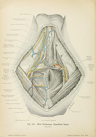
The vein of bulb of penis is a tributary of the internal pudendal vein.
The superficial dorsal veins of clitoris is a tributary of the external pudendal vein.
The posterior scrotal veins are veins of the scrotum. They accompany the posterior scrotal arteries. They drain into the vesical venous plexus. They help to drain blood from part of the scrotum.

The posterior labial veins are veins which drain to the vesical venous plexus.
References
![]() This article incorporates text in the public domain from the 20th edition of Gray's Anatomy (1918)
This article incorporates text in the public domain from the 20th edition of Gray's Anatomy (1918)