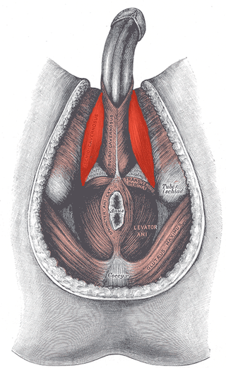
The ischiocavernosus muscle is a muscle just below the surface of the perineum, present in both men and women.

The bulbospongiosus muscles are one of the superficial muscles of the perineum. They have a slightly different origin, insertion and function in males and females. In males, these muscles cover the bulb of the penis, while in females, they cover the vestibular bulbs.

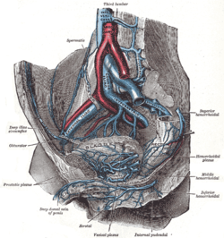
The internal pudendal artery is one of the three pudendal arteries. It branches off the internal iliac artery, and provides blood to the external genitalia.

In human anatomy, the inferior epigastric artery is an artery that arises from the external iliac artery. It is accompanied by the inferior epigastric vein; inferiorly, these two inferior epigastric vessels together travel within the lateral umbilical fold The inferior epigastric artery then traverses the arcuate line of rectus sheath to enter the rectus sheath, then anastomoses with the superior epigastric artery within the rectus sheath.

The sacrotuberous ligament is situated at the lower and back part of the pelvis. It is flat, and triangular in form; narrower in the middle than at the ends.

The inferior rectal artery is an artery that supplies blood to the lower third of the anal canal below the pectinate line.

The perineal nerve is a nerve of the pelvis. It arises from the pudendal nerve in the pudendal canal. It gives superficial branches to the skin, and a deep branch to muscles. It supplies the skin and muscles of the perineum. Its latency is tested with electrodes.

The ischioanal fossa is the fat-filled wedge-shaped space located lateral to the anal canal and inferior to the pelvic diaphragm. It is somewhat prismatic in shape, with its base directed to the surface of the perineum and its apex at the line of meeting of the obturator and anal fasciae.

The pudendal canal is an anatomical structure formed by the obturator fascia lining the lateral wall of the ischioanal fossa. The internal pudendal artery and veins, and pudendal nerve pass through the pudendal canal, and the perineal nerve arises within it.
The inferior rectal nerves usually branch from the pudendal nerve but occasionally arises directly from the sacral plexus; they cross the ischiorectal fossa along with the inferior rectal artery and veins, toward the anal canal and the lower end of the rectum, and is distributed to the sphincter ani externus and to the integument (skin) around the anus.

A corpus cavernosum penis (singular) is one of a pair of sponge-like regions of erectile tissue, which contain most of the blood in the penis during an erection.
The dorsal artery of the penis is an artery on the top surface of the penis. It is a branch of the internal pudendal artery. It runs forward on the dorsum of the penis to the glans, where it divides into two branches to the glans penis and the foreskin (prepuce).
The dorsal nerve of the penis is the deepest of three divisions of the pudendal nerve; it accompanies the internal pudendal artery along the ramus of the ischium; it then runs forward along the margin of the inferior ramus of the pubis, between the superior and inferior layers of the fascia of the urogenital diaphragm.

The submandibular triangle corresponds to the region of the neck immediately beneath the body of the mandible.

The inferior carotid triangle, is bounded, in front, by the median line of the neck from the hyoid bone to the sternum; behind, by the anterior margin of the sternocleidomastoid; above, by the superior belly of the omohyoid.

The urogenital triangle is the anterior part of the perineum. In female mammals, it contains the vagina and associated parts of the internal genitalia.
The dorsal nerve of the clitoris is a nerve in females that branches off the pudendal nerve to innervate the clitoris. The nerve is important for female sexual pleasure, and it may play a role in clitoral erections.
The posterior labial nerves are branches of the pudendal nerve. They supply the female labia majora.
The artery of bulb of vestibule is a branch of the internal pudendal artery. It supplies the vestibular bulbs and thus only exist in females.
The deep branch of the perineal nerve is a nerve of the perineum. It is a branch of the perineal nerve, from the pudendal nerve. It supplies the superficial transverse perineal muscle, bulbospongiosus muscle, ischiocavernosus muscle, the bulb of penis, levator ani, and the external anal sphincter.