Dendritic spike

Last updated
Figure A. shows the idealized phases of an action potential. Figure B. is a recording of an actual action potential N.B. Actual recordings of action potentials are often distorted compared to the schematic view because of variations in electrophysiological techniques used to make the recording. Action potential vert.png
Figure A. shows the idealized phases of an action potential. Figure B. is a recording of an actual action potential N.B. Actual recordings of action potentials are often distorted compared to the schematic view because of variations in electrophysiological techniques used to make the recording.

In neurophysiology, a dendritic spike refers to an action potential generated in the dendrite of a neuron. Dendrites are branched extensions of a neuron. They receive electrical signals emitted from projecting neurons and transfer these signals to the cell body, or soma. Dendritic signaling has traditionally been viewed as a passive mode of electrical signaling. Unlike its axon counterpart which can generate signals through action potentials, dendrites were believed to only have the ability to propagate electrical signals by physical means: changes in conductance, length, cross sectional area, etc. However, the existence of dendritic spikes was proposed and demonstrated by W. Alden Spencer, Eric Kandel, Rodolfo Llinás and coworkers in the 1960s [1] [2] and a large body of evidence now makes it clear that dendrites are active neuronal structures. Dendrites contain voltage-gated ion channels giving them the ability to generate action potentials. Dendritic spikes have been recorded in numerous types of neurons in the brain and are thought to have great implications in neuronal communication, memory, and learning. They are one of the major factors in long-term potentiation.

Contents

A dendritic spike is initiated in the same manner as that of an axonal action potential. Depolarization of the dendritic membrane causes sodium and potassium voltage-gated ion channels to open. The influx of sodium ions causes an increase in voltage. If the voltage increases past a certain threshold, the sodium current activates other voltage-gated sodium channels transmitting a current along the dendrite. Dendritic spikes can be generated through both sodium and calcium voltage-gated channels. Dendritic spikes usually transmit signals at a much slower rate than axonal action potentials. [3] Local voltage thresholds for dendritic spike initiation are usually higher than that of action potential initiation in the axon; therefore, spike initiation usually requires a strong input. [4]

Voltage-Gated Channels

Voltage-Gated Sodium Channel

Diagram of a voltage-sensitive sodium channel a-subunit. G - glycosylation, P - phosphorylation, S - ion selectivity, I - inactivation, positive (+) charges in S4 are important for transmembrane voltage sensing. Sodium-channel.svg
Diagram of a voltage-sensitive sodium channel α-subunit. G - glycosylation, P - phosphorylation, S - ion selectivity, I - inactivation, positive (+) charges in S4 are important for transmembrane voltage sensing.

Voltage-gated sodium channels are proteins found in the membrane of neurons. When electrically activated, they allow the movement of sodium ions across a plasma membrane. These channels are responsible for propagation of electrical signals in nerve cells. Voltage-gated sodium channels can be divided into two subunits: alpha and beta. A variety of alpha subunit voltage-gated sodium channels have been identified. Voltage-gated sodium channels found in mammals can be divided into three types: Nav1.x, Nav2.x, and Nav3.x. Nav1.x sodium channels are associated with the central nervous system. Nav1.1, Nav2.2, and Nav1.6 are three isoforms of the voltage-gated sodium channels that are present at high levels in the central nervous system of an adult rat brain. [5] These channels have been well documented in the axonal membrane of the central nervous system. Nav1.2 has been primarily identified in unmyelinated axons while high concentrations of Nav1.6 have been observed at nodes of Ranvier of axons. [6] Nav1.6 has been identified in the dendrites of hippocampal CA1 neurons that generate dendritic spikes; the density of Nav1.6 in these neurons is 35-80 times lower than in the initial segments of axons. [7]

Distribution of voltage-gated sodium channels along the dendritic membrane plays a crucial role in a dendrite's ability to propagate a signal. High dendritic membrane thresholds often make it harder for initiation of dendritic spikes. However, increased density of voltage-gated sodium channels may reduce the amplitude of a signal needed to initiate a spike. Clustering of voltage-gated sodium channels have been observed at the synapses of the globus pallidus neuron. [8] It has also been demonstrated through dendritic computational models that the threshold amplitude of a synaptic conductance needed to generate a dendritic spike is significantly less if the voltage-gated sodium channels are clustered at the synapse. [8] The same type of voltage-gated channels may differ in distribution between the soma and dendrite within the same neuron. There seems to be no general pattern of distribution for voltage-gated channels within dendrites. Different neuronal dendrites exhibit different density patterns which are subject to change during development and can be modulated by neurotransmitters. [4]

Voltage-Gated Calcium Channel

Like voltage-gated sodium channels, voltage-gated calcium channels are also integral membrane proteins found in the plasma membrane. Voltage-gated calcium channels generate action potentials by the same mechanisms as voltage-gated sodium channels. Various voltage-gated calcium channels have been identified in neurons. N- and P/Q-type voltage-gated calcium channels are the primary subtypes found to support synaptic transmission. [9] These channels are concentrated at nerve terminals. T-type and R-type voltage-gated calcium channels have been found in basal dendrites, and it is thought that the activation of these channels during action potential bursts lead to the generation of dendritic calcium spikes. [10] T-type and R-type channels are all part of the alpha 1 subunit class of calcium channels.

The various types of voltage-gated calcium channels result in two forms of voltage activation: low-voltage-activated (LVA) and high-voltage-activated (HVA) calcium currents. In deep cerebellar nuclei, calcium currents are not uniformly distributed along a dendrite. [11] The relative strength of LVA calcium currents are significantly more concentrated at the distal end of dendrites. The uneven distribution of LVA calcium currents suggests the important role of LVA calcium currents in dendritic integration at synaptic inputs. [11]

Voltage-Gated Potassium Channel

Potassium channel KcsA. 1r3j.png
Potassium channel KcsA.

Voltage-gated potassium channels are another set of voltage-gated channels that play a significant role in the initiation of dendritic spikes. Voltage-gated potassium channels, similar to voltage-gated sodium and calcium channels, facilitate the movement of cations across the plasma membrane. But unlike voltage-gated sodium and calcium channels, the voltage-gated potassium channel moves cations out of the cell thereby having an inhibitory effect on dendritic spike initiation.

The transient A-type voltage-gated potassium channel is a specific channel that plays a key role in dendritic spike initiation. The density of voltage-gated sodium and calcium channels is similar in both dendrites and axons; however, the dendritic membrane is far less excitable than the axonal membrane. [12] The difference in excitability can be attributed to the presence of these voltage-gated potassium channels. Voltage-gated potassium channels inhibit the ability of dendrites to generate action potentials and decrease the amplitude of dendritic spikes with increasing distance from the soma. The ability of voltage-gated potassium channels to modulate dendritic signaling may have significant effects on synaptic plasticity.

Spike Initiation

Action Potential

Action potentials initiated in the axon normally travel down the axon away from the soma. However, it is also possible for an action potential to travel in the opposite direction, invade the soma, and then travel down the dendrite as a dendritic spike. [13] This retrograde signal provides information to the synapse that the neuron has fired an output. [4] The efficacy of the signal varies among different neuronal types. For example, backward propagation of action potentials is very limited in cerebellar Purkinje cells [14] but is quite prevalent in interneurons of the medium ganglionic layer of the cerebellum-like lobe of some fish. [15]

Synaptic Input

Action potentials may be first generated at the dendrite if stimulated by strong synchronous synaptic inputs. [16] The ability of a dendrite to initiate an action potential is not only highly dependent on synaptic input but also on the number of voltage-gated channels and density of voltage-gated channels present in the membrane.

Spatial Summation

Hippocampal Pyramidal Cell Hippocampal-pyramidal-cell.png
Hippocampal Pyramidal Cell

Initiation of a dendritic spike through a single strong synaptic input does not guarantee that the spike will propagate reliably over long distances. [17] If multiple synapses are simultaneously activated, dendritic spikes may be formed through spatial summation. Spatial summation involves the addition of multiple input signals resulting in a larger signal and possibly a dendritic spike. Hippocampal CA1 neurons have been shown to produce reliable dendritic spike propagation through spatial summation of multiple synaptic inputs. In the hippocampus, the CA1 neurons contain two distinctive regions that receive excitatory synaptic inputs: the perforant path (PP) through the apical dendritic tuft (500-750 μm from soma) and the Schaffer-collateral (SC) through the basal and apical dendrites (250-500 μm from soma). [17] Studies show that individual stimulation of either the PP or SC was not sufficient enough to allow a dendritic spike to initiate an action potential. However, it was shown that when a dendritic spike occurred due to PP stimulations, the presence of a SC stimulation determined whether or not the signal would propagate to the soma. [17]

Spike Propagation

Backward Propagation

Dendritic spikes most commonly propagate backwards from the soma to distal dendritic branches[ citation needed ]. Backward propagation serves a number of functions in the neuron, and these functions vary based on the type of neuron. In general, backward propagation serves to communicate output information to the postsynaptic membrane. [4] In many neurotransmitter-releasing neurons, backward propagation of dendritic spikes signals the release of neurotransmitters. [18] For example, Mitral cells seem to serve both as projection neurons and as local interneurons. If the axonal output of a mitral cell is shut down by somatic inhibition, local dendritic action potentials cause the mitral cell to release neurotransmitters into the environment. [18] Backward propagation of dendritic spikes has been demonstrated in various neuronal types in the brain but has rarely been studied outside of the brain[ citation needed ]. Other than neurons in the brain, dendritic spikes have been observed in the neurons of the spinal cord[ citation needed ].

Forward Propagation

Forward propagation of dendritic spikes initiate due to synaptic activity, and refers to the transmission of the signal towards the soma. [17] The strength of synaptic stimulation required to generate a dendritic spike varies among neuronal types. Neurons which receive relatively few inputs cannot rely on spatial summation and therefore must rely on stronger synaptic inputs. Some relatively unbranched neurons, such as the globus pallidus neuron, bypass the need of strong synaptic input by increased concentrations of voltage-gated sodium channels at the synapse. [8] Other more branched neurons, such as pyramidal neurons, rely on spatial summation of multiple inputs to generate forward propagating dendritic spikes. Forward propagation is not well understood and much research is devoted to the subject[ citation needed ]. It is thought by most experts

that this phenomenon does not occur in neurons outside of the brain.

Spike-Timing-Dependent Plasticity

Schematic of a chemical synapse between an axon of one neuron and a dendrite of another. SynapseSchematic en.svg
Schematic of a chemical synapse between an axon of one neuron and a dendrite of another.

Spike-timing-dependent plasticity (STDP) refers to the functional changes in a neuron and its synapse due to time dependent action potentials. When an action potential reaches the pre-synaptic membrane it opens voltage-gated calcium channels causing an influx of calcium. The influx of calcium releases vesicles filled with neurotransmitters, usually glutamate, into the synaptic cleft. The neurotransmitters bind to receptors on the post-synaptic membrane opening ligand-gated channels causing the membrane to depolarize.

NMDA receptors are found throughout the post-synaptic membrane and act as a coincidence detector. The NMDA detects both glutamate released by pre-synaptic vesicles and depolarization of the post-synaptic membrane. The NMDA receptor exhibits voltage-dependent block by magnesium ions. Depolarization of the post-synaptic membrane (i.e. backward propagating dendritic spike) causes the magnesium ion to be removed from the channel, favoring channel opening. NMDA receptor activation thereby allows calcium influx. Neurons that “fire together wire together” refer to strengthening of synaptic connections through NMDA receptors when glutamate release is coincident with post-synaptic depolarization. [3] This form of wiring is known as long term potentiation. Synaptic connection can also be weakened when the activity of neurons is uncorrelated, also known as long term depression.

The dependence of post-synaptic depolarization in STDP indicates the importance of dendritic spikes. In general, post-synaptic depolarization occurs coincidentally with pre-synaptic activity when a backwards propagating signal reaches the post-synaptic membrane. Dendritic spikes allow backward propagating signals to reach and depolarize the post-synaptic membrane. The strengthening and weakening of synaptic connections is one proposed method of memory formation and learning.

Experimental Methods

Two-Photon Glutamate Uncaging

Two-photon glutamate uncaging, a type of photostimulation, has become the premier tool for studying dendritic spikes due to its high level of precision. [19]

Patch Clamp

The cell-attached patch clamp uses a micropipette attached to the cell membrane to allow recording from a single ion channel. Patch clamp.svg
The cell-attached patch clamp uses a micropipette attached to the cell membrane to allow recording from a single ion channel.

Patch clamp recording is used to measure electrical activity in neurons. The technique uses a one micrometer diameter open tip glass micropipette to suction the membrane of a cell. The pipette is filled with ionic solution, and a silver wire is placed in the solution to conduct and amplify electrical signals. The ion solution can be varied and drugs can be delivered through the micropipette to study the effects of current under various conditions. Receptor and voltage-gated channel antagonists are often applied (i.e. nickel used to block NMDA receptors) in order to study the effects of ion channels on dendritic spike initiation. [10] Current injection is often paired with patch clamp recordings in order to observe current modulation due to various experimental factors.

Extracellular Electrophysiology

Tetrode recording methods have also been shown to occasionally allow for observation of dendritic membrane potentials and dendritic action potentials. [20] Interestingly, the chronic recording paradigm that demonstrated this also showed that dendritic voltage properties exhibited egocentric spatial maps comparable to pyramidal neurons. This rare phenomenon may be due to a glial sheath [21] forming around the tetrode tips, creating a high impedance sea, similar to a gigaohm seal in patch recordings, that allows for such small and localized voltage measurement to be made.

Staining and Labeling

Staining and labeling techniques are often used in microscopy to help identify specific structures in a cell. Staining usually involves the use of dyes that are absorbed by various cell structures at different rates. Labeling involves the use of fluorescence to identify specific molecules. Fluorophores, fluorescent molecules, may be directly attached or attached to an antibody in order to detect a specific target. In the case of dendritic spikes, staining and labeling are used to identify and quantify the presence of certain voltage-gated channels. For example, rabbit polyclonal antibodies raised against synthetic peptide sequences have been used to identify the presence of Nav1.2, Nav1.3, and Nav1.6 sodium channels in dendrites of the globus pallidus neuron. [8]

Computational Modeling

Computational modeling of neurons, artificial neural networking, has become a very popular tool in investigating the properties of neuronal signaling. These models are based on biological neural networks. Computational modeling can be used to study single neurons, groups of neurons, or even networks of neurons. This field has generated much interest and serves as a tool for all branches of neuroscience research including dendritic spike initiation.

Related Research Articles

<span class="mw-page-title-main">Dendrite</span> Small projection on a neuron that receives signals

A dendrite or dendron is a branched protoplasmic extension of a nerve cell that propagates the electrochemical stimulation received from other neural cells to the cell body, or soma, of the neuron from which the dendrites project. Electrical stimulation is transmitted onto dendrites by upstream neurons via synapses which are located at various points throughout the dendritic tree.

<span class="mw-page-title-main">Neuron</span> Electrically excitable cell found in the nervous system of animals

Within a nervous system, a neuron, neurone, or nerve cell is an electrically excitable cell that fires electric signals called action potentials across a neural network. Neurons communicate with other cells via synapses, which are specialized connections that commonly use minute amounts of chemical neurotransmitters to pass the electric signal from the presynaptic neuron to the target cell through the synaptic gap.

<span class="mw-page-title-main">Chemical synapse</span> Biological junctions through which neurons signals can be sent

Chemical synapses are biological junctions through which neurons' signals can be sent to each other and to non-neuronal cells such as those in muscles or glands. Chemical synapses allow neurons to form circuits within the central nervous system. They are crucial to the biological computations that underlie perception and thought. They allow the nervous system to connect to and control other systems of the body.

<span class="mw-page-title-main">Action potential</span> Neuron communication by electric impulses

An action potential occurs when the membrane potential of a specific cell rapidly rises and falls. This depolarization then causes adjacent locations to similarly depolarize. Action potentials occur in several types of animal cells, called excitable cells, which include neurons, muscle cells, and in some plant cells. Certain endocrine cells such as pancreatic beta cells, and certain cells of the anterior pituitary gland are also excitable cells.

An inhibitory postsynaptic potential (IPSP) is a kind of synaptic potential that makes a postsynaptic neuron less likely to generate an action potential. IPSPs were first investigated in motorneurons by David P. C. Lloyd, John Eccles and Rodolfo Llinás in the 1950s and 1960s. The opposite of an inhibitory postsynaptic potential is an excitatory postsynaptic potential (EPSP), which is a synaptic potential that makes a postsynaptic neuron more likely to generate an action potential. IPSPs can take place at all chemical synapses, which use the secretion of neurotransmitters to create cell to cell signalling. Inhibitory presynaptic neurons release neurotransmitters that then bind to the postsynaptic receptors; this induces a change in the permeability of the postsynaptic neuronal membrane to particular ions. An electric current that changes the postsynaptic membrane potential to create a more negative postsynaptic potential is generated, i.e. the postsynaptic membrane potential becomes more negative than the resting membrane potential, and this is called hyperpolarisation. To generate an action potential, the postsynaptic membrane must depolarize—the membrane potential must reach a voltage threshold more positive than the resting membrane potential. Therefore, hyperpolarisation of the postsynaptic membrane makes it less likely for depolarisation to sufficiently occur to generate an action potential in the postsynaptic neurone.

<span class="mw-page-title-main">Depolarization</span> Change in a cells electric charge distribution

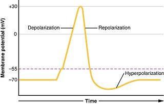
In biology, depolarization or hypopolarization is a change within a cell, during which the cell undergoes a shift in electric charge distribution, resulting in less negative charge inside the cell compared to the outside. Depolarization is essential to the function of many cells, communication between cells, and the overall physiology of an organism.

<span class="mw-page-title-main">Graded potential</span> Changes in membrane potential varying in size

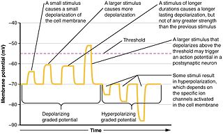
Graded potentials are changes in membrane potential that vary according to the size of the stimulus, as opposed to being all-or-none. They include diverse potentials such as receptor potentials, electrotonic potentials, subthreshold membrane potential oscillations, slow-wave potential, pacemaker potentials, and synaptic potentials. The magnitude of a graded potential is determined by the strength of the stimulus. They arise from the summation of the individual actions of ligand-gated ion channel proteins, and decrease over time and space. They do not typically involve voltage-gated sodium and potassium channels, but rather can be produced by neurotransmitters that are released at synapses which activate ligand-gated ion channels. They occur at the postsynaptic dendrite in response to presynaptic neuron firing and release of neurotransmitter, or may occur in skeletal, smooth, or cardiac muscle in response to nerve input. These impulses are incremental and may be excitatory or inhibitory.

<span class="mw-page-title-main">Excitatory synapse</span> Sort of synapse

An excitatory synapse is a synapse in which an action potential in a presynaptic neuron increases the probability of an action potential occurring in a postsynaptic cell. Neurons form networks through which nerve impulses travels, each neuron often making numerous connections with other cells of neurons. These electrical signals may be excitatory or inhibitory, and, if the total of excitatory influences exceeds that of the inhibitory influences, the neuron will generate a new action potential at its axon hillock, thus transmitting the information to yet another cell.

<span class="mw-page-title-main">Pyramidal cell</span> Projection neurons in the cerebral cortex and hippocampus

Pyramidal cells, or pyramidal neurons, are a type of multipolar neuron found in areas of the brain including the cerebral cortex, the hippocampus, and the amygdala. Pyramidal cells are the primary excitation units of the mammalian prefrontal cortex and the corticospinal tract. Pyramidal neurons are also one of two cell types where the characteristic sign, Negri bodies, are found in post-mortem rabies infection. Pyramidal neurons were first discovered and studied by Santiago Ramón y Cajal. Since then, studies on pyramidal neurons have focused on topics ranging from neuroplasticity to cognition.

<span class="mw-page-title-main">Axon hillock</span> Part of the neuronal cell soma from which the axon originates

The axon hillock is a specialized part of the cell body of a neuron that connects to the axon. It can be identified using light microscopy from its appearance and location in a neuron and from its sparse distribution of Nissl substance.

<span class="mw-page-title-main">Node of Ranvier</span> Gaps between myelin sheaths on the axon of a neuron

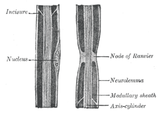
In neuroscience and anatomy, nodes of Ranvier, also known as myelin-sheath gaps, occur along a myelinated axon where the axolemma is exposed to the extracellular space. Nodes of Ranvier are uninsulated and highly enriched in ion channels, allowing them to participate in the exchange of ions required to regenerate the action potential. Nerve conduction in myelinated axons is referred to as saltatory conduction due to the manner in which the action potential seems to "jump" from one node to the next along the axon. This results in faster conduction of the action potential.

<span class="mw-page-title-main">End-plate potential</span>

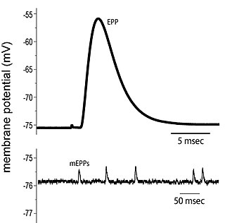
End plate potentials (EPPs) are the voltages which cause depolarization of skeletal muscle fibers caused by neurotransmitters binding to the postsynaptic membrane in the neuromuscular junction. They are called "end plates" because the postsynaptic terminals of muscle fibers have a large, saucer-like appearance. When an action potential reaches the axon terminal of a motor neuron, vesicles carrying neurotransmitters are exocytosed and the contents are released into the neuromuscular junction. These neurotransmitters bind to receptors on the postsynaptic membrane and lead to its depolarization. In the absence of an action potential, acetylcholine vesicles spontaneously leak into the neuromuscular junction and cause very small depolarizations in the postsynaptic membrane. This small response (~0.4mV) is called a miniature end plate potential (MEPP) and is generated by one acetylcholine-containing vesicle. It represents the smallest possible depolarization which can be induced in a muscle.

<span class="mw-page-title-main">Electrotonic potential</span>

In physiology, electrotonus refers to the passive spread of charge inside a neuron and between cardiac muscle cells or smooth muscle cells. Passive means that voltage-dependent changes in membrane conductance do not contribute. Neurons and other excitable cells produce two types of electrical potential:

Molecular neuroscience is a branch of neuroscience that observes concepts in molecular biology applied to the nervous systems of animals. The scope of this subject covers topics such as molecular neuroanatomy, mechanisms of molecular signaling in the nervous system, the effects of genetics and epigenetics on neuronal development, and the molecular basis for neuroplasticity and neurodegenerative diseases. As with molecular biology, molecular neuroscience is a relatively new field that is considerably dynamic.

An apical dendrite is a dendrite that emerges from the apex of a pyramidal cell. Apical dendrites are one of two primary categories of dendrites, and they distinguish the pyramidal cells from spiny stellate cells in the cortices. Pyramidal cells are found in the prefrontal cortex, the hippocampus, the entorhinal cortex, the olfactory cortex, and other areas. Dendrite arbors formed by apical dendrites are the means by which synaptic inputs into a cell are integrated. The apical dendrites in these regions contribute significantly to memory, learning, and sensory associations by modulating the excitatory and inhibitory signals received by the pyramidal cells.

<span class="mw-page-title-main">Synaptic potential</span> Potential difference across the postsynaptic membrane

Synaptic potential refers to the potential difference across the postsynaptic membrane that results from the action of neurotransmitters at a neuronal synapse. In other words, it is the “incoming” signal that a neuron receives. There are two forms of synaptic potential: excitatory and inhibitory. The type of potential produced depends on both the postsynaptic receptor, more specifically the changes in conductance of ion channels in the post synaptic membrane, and the nature of the released neurotransmitter. Excitatory post-synaptic potentials (EPSPs) depolarize the membrane and move the potential closer to the threshold for an action potential to be generated. Inhibitory postsynaptic potentials (IPSPs) hyperpolarize the membrane and move the potential farther away from the threshold, decreasing the likelihood of an action potential occurring. The Excitatory Post Synaptic potential is most likely going to be carried out by the neurotransmitters glutamate and acetylcholine, while the Inhibitory post synaptic potential will most likely be carried out by the neurotransmitters gamma-aminobutyric acid (GABA) and glycine. In order to depolarize a neuron enough to cause an action potential, there must be enough EPSPs to both depolarize the postsynaptic membrane from its resting membrane potential to its threshold and counterbalance the concurrent IPSPs that hyperpolarize the membrane. As an example, consider a neuron with a resting membrane potential of -70 mV (millivolts) and a threshold of -50 mV. It will need to be raised 20 mV in order to pass the threshold and fire an action potential. The neuron will account for all the many incoming excitatory and inhibitory signals via summative neural integration, and if the result is an increase of 20 mV or more, an action potential will occur.

Neural backpropagation is the phenomenon in which, after the action potential of a neuron creates a voltage spike down the axon, another impulse is generated from the soma and propagates towards the apical portions of the dendritic arbor or dendrites. In addition to active backpropagation of the action potential, there is also passive electrotonic spread. While there is ample evidence to prove the existence of backpropagating action potentials, the function of such action potentials and the extent to which they invade the most distal dendrites remain highly controversial.

<span class="mw-page-title-main">Summation (neurophysiology)</span>

Summation, which includes both spatial summation and temporal summation, is the process that determines whether or not an action potential will be generated by the combined effects of excitatory and inhibitory signals, both from multiple simultaneous inputs, and from repeated inputs. Depending on the sum total of many individual inputs, summation may or may not reach the threshold voltage to trigger an action potential.

Cellular neuroscience is a branch of neuroscience concerned with the study of neurons at a cellular level. This includes morphology and physiological properties of single neurons. Several techniques such as intracellular recording, patch-clamp, and voltage-clamp technique, pharmacology, confocal imaging, molecular biology, two photon laser scanning microscopy and Ca2+ imaging have been used to study activity at the cellular level. Cellular neuroscience examines the various types of neurons, the functions of different neurons, the influence of neurons upon each other, and how neurons work together.

<span class="mw-page-title-main">Nonsynaptic plasticity</span> Form of neuroplasticity

Nonsynaptic plasticity is a form of neuroplasticity that involves modification of ion channel function in the axon, dendrites, and cell body that results in specific changes in the integration of excitatory postsynaptic potentials and inhibitory postsynaptic potentials. Nonsynaptic plasticity is a modification of the intrinsic excitability of the neuron. It interacts with synaptic plasticity, but it is considered a separate entity from synaptic plasticity. Intrinsic modification of the electrical properties of neurons plays a role in many aspects of plasticity from homeostatic plasticity to learning and memory itself. Nonsynaptic plasticity affects synaptic integration, subthreshold propagation, spike generation, and other fundamental mechanisms of neurons at the cellular level. These individual neuronal alterations can result in changes in higher brain function, especially learning and memory. However, as an emerging field in neuroscience, much of the knowledge about nonsynaptic plasticity is uncertain and still requires further investigation to better define its role in brain function and behavior.

References

  1. Spencer, W. A.; Kandel, E. R. (1961). "Electrophysiology of Hippocampal Neurons: Iv. Fast Prepotentials". Journal of Neurophysiology. 24 (3): 272–285. doi:10.1152/jn.1961.24.3.272. ISSN   0022-3077. PMID   25286477.
  2. Llinás, R.; Nicholson, C.; Freeman, J. A.; Hillman, D. E. (1968-06-07). "Dendritic spikes and their inhibition in alligator Purkinje cells". Science. 160 (3832): 1132–1135. Bibcode:1968Sci...160.1132L. doi:10.1126/science.160.3832.1132. ISSN   0036-8075. PMID   5647436. S2CID   27657014.
  3. 1 2 Kampa BM, Letzkus JJ, Stuart GJ. 2007. Dendritic mechanisms controlling spike-timing-dependent synaptic plasticity. Trends in Neurosciences 30:456-63 doi : 10.1016/j.tins.2007.06.010
  4. 1 2 3 4 Häusser M, Spruston N, Stuart GJ. 2000. Diversity and dynamics of dendritic signaling. Science 290:739-744 doi : 10.1126/science.290.5492.739
  5. Goldin AL. 1999. Diversity of mammalian voltage-gated sodium channels. Annals New York Academy of Sciences 868:38-50 doi : 10.1111/j.1749-6632.1999.tb11272.x
  6. Caldwell JH, Schaller KL, Lasher RS, et al. 2000. Sodium channel Nav1.6 is localized at nodes of ranvier, dendrites, and synapses. Proceedings of the National Academy of Sciences 97.10:5616-5620
  7. Lorincz A, Nusser Z (2010). "Molecular identity of dendritic voltage-gated sodium channels". Science. 328 (5980): 906–9. Bibcode:2010Sci...328..906L. doi:10.1126/science.1187958. PMC   3546315 . PMID   20466935.
  8. 1 2 3 4 Hanson JE, Smith Y, Jaeger D. 2004. Sodium channels and dendritic spike initiation at excitatory synapses in globus pallidus neurons. Journal of Neuroscience 24:329-40
  9. Dolphin AC. 2006. A Short history of voltage-gated calcium channels. British Journal of Pharmacology 147:S56-S62
  10. 1 2 Kampa BM, Letzkus JJ, Stuart GJ. 2006. Requirement of dendritic calcium spikes for induction of spike-timing-dependent synaptic plasticity. Journal of Physiology 574.1:283-290
  11. 1 2 Gauck V, Thomann M, Jaeger D, et al. 2001. Spatial distribution of low- and high-voltage-activated calcium currents in neurons of the deep cerebellar nuclei. Journal of Neuroscience 21:1-4
  12. Hoffman DA, Magee JC, Colbert CM, et al. K+ channel regulation of signal propagation in dendrites of hippocampal pyramidal neuron. Nature 387:869-875
  13. Ma J, Lowe G. 2004. Action potential backpropagation and multiglomerular signaling in the rat vomeronasal system. Journal of Neuroscience 24(42):9341-9352
  14. Llinas R, Sugimori M. 1980. Electrophysiological properties of in vitro purkinje cell dendrites in mammalian cerebellar slices. Journal of Physiology 305:197-213
  15. Gomez L, Kanneworff M, Budelli R, Grant K. 2005. Dendritic spike back propagation in the electrosensory lobe of Gnathonemus petersii. Journal of Experimental Biology 208:141-55
  16. Golding NL, Spruston N. 1998. Dendritic sodium spikes are variable triggers of axonal action potentials in hippocampal CA1 pyramidal neurons. Neuron 21:1189-1200
  17. 1 2 3 4 Jarsky T, Roxin A, Kath WL, Spruston N. 2005. Conditional dendritic spike propagation following distal synaptic activation of hippocampal CA1 pyramidal neurons. Nature Neuroscience 8:1667-76
  18. 1 2 Chen WR, Shen GY, Shepherd G, et al. 2002. Multiple modes of action potential initiation and propagation in mitral cell primary dendrite. Journal of Neurophysiology 88:2755-2764
  19. Judkewitz, Benjamin; Roth, Arnd; Häusser, Michael (2006-04-20). "Dendritic Enlightenment: Using Patterned Two-Photon Uncaging to Reveal the Secrets of the Brain's Smallest Dendrites". Neuron. 50 (2): 180–183. doi: 10.1016/j.neuron.2006.04.011 . ISSN   0896-6273. PMID   16630828.
  20. Moore, Jason J.; Ravassard, Pascal M.; Ho, David; Acharya, Lavanya; Kees, Ashley L.; Vuong, Cliff; Mehta, Mayank R. (2017-03-24). "Dynamics of cortical dendritic membrane potential and spikes in freely behaving rats". Science. 355 (6331): eaaj1497. doi:10.1126/science.aaj1497. ISSN   1095-9203. PMID   28280248. S2CID   33117933.
  21. Polikov, Vadim S.; Tresco, Patrick A.; Reichert, William M. (2005-10-15). "Response of brain tissue to chronically implanted neural electrodes". Journal of Neuroscience Methods. 148 (1): 1–18. doi:10.1016/j.jneumeth.2005.08.015. ISSN   0165-0270. PMID   16198003. S2CID   11248506.