Last updated Don Wayne Fawcett (March 14, 1917 - May 7, 2009)
Don Wayne Fawcett (March 14, 1917 - May 7, 2009) was a pioneer of electron microscopy and one of its greatest practitioners for studying the organization of cells and tissues. His greatest achievement was his description of the structure of spermatozoa and the male reproductive system.
Don Fawcett was born in 1917 on a farm in Iowa, where his father and grandfather had raised purebred sheep and cattle until his father's poor health forced the family to leave the farm and move to Boston, where Fawcett's father managed a successful wool business. Fawcett attended high school at the Boston Latin School. Upon graduation, he matriculated at Harvard College in 1934, followed by Harvard Medical School in 1938, where he connected with anatomy professor George B. Wislocki. "I stole as much time as I could from my course work to do independent research on projects that included studies on the vascular bundles of aquatic mammals, and the amedullary bones[1] of the Florida manatee", he recalled.
While in college, Fawcett illustrated a book on athletic bandaging that had been written by the football team's physician. This may have been his only contribution as an illustrator; his later and more widely popular books were illustrated by Sylvia Collard Keen. In the summers, Fawcett worked on Bailey Island off the coast of Maine, where he embalmed and prepared small sharks for sale to colleges for comparative anatomy courses. This early interest in anatomy served him well when he trained in surgery at Harvard. Fawcett's predominant memory of his clinical training was being on duty in the emergency ward the night of the infamous Cocoanut Grove Nightclub disaster of 1942, which claimed 492 lives in one of the deadliest fires in American history. "We had been having a quiet evening when, without advance notice, we received 115 seriously burned patients within an hour and a half. Mobilizing all of the off-duty staff that I could reach, I continued on duty for 30 hours doing all I could to relieve the pain and dress the burns of the victims." He received his M.D. in 1942.
Fawcett served as a battalion surgeon and captain in the European Theater of World War II, but, before shipping out, he married Dorothy Secrest Fawcett, his wife for 68 years, who survived him. After the war, Fawcett chose a career in research instead of surgery. He returned to Harvard, where he served as an instructor.
Career
Fawcett perfected the art and technology of microscopy in the early days of cell biology, which emerged as a modern field after the electron microscope became more widely available in the 1940s. He would routinely cut thin sections at home, before venturing in the early morning hours to his lab at Harvard, where he viewed them with the microscope that would do so much to reveal the secrets of form and function. He kept a microtome at home, and it was rumored that he also had a private collection of diamond knives to help achieve his unparalleled results.
Tom Pollard, a student at Harvard during Fawcett's chairmanship, recalls how Fawcett used his skills in the darkroom to produce spectacular prints of electron micrographs to illustrate the key features of each image. He may have been most renowned as the first person to describe and depict in detail human spermatozoa, and he published extensively on the anatomy of male reproductive cellular anatomy.
Fawcett described the early days of electron microscopy as: "For morphologists the decade from 1950 to 1960 held the same anticipation and excitement that attends the opening of a new continent for exploration. The electron microscope revealed marvelous order and functional design in the organization of every tissue and organ that was examined and added significantly to our understanding of our own structure (...)". Fawcett published a collection of his fine-structure micrographs in The Cell, a classic cell biology text that features all the major cell structures,[2]
In 1955 Fawcett assumed the position of chair of the Department of Anatomy at Cornell Medical School in New York City and established an electron microscope laboratory. He had become what today might be characterized as the quintessential descriptive scientist; later generations of cell biologists and biochemists would build on his seminal work. After four years at Cornell, Fawcett again returned to Harvard as Hersey Professor of Anatomy and Chairman of the Department. The endowment for his chairmanship had been established by Ezekiel Hersey in 1770 with a gift of 1,000 pounds, and, Fawcett recalled, "My salary reflected the size of that endowment."
In 1976 Fawcett resigned the chairmanship and became Senior Associate Dean for Preclinical Science, a position for which, by his own description, he was ill-suited. After spending some time annually in Africa as examiner in the School of Veterinary Medicine of the University of Nairobi, Kenya, where he indulged his profound love of and talent for animal and nature photography, Fawcett left Boston in 1985 to take the position of senior research scientist and director of electron microscopy at the International Laboratory for Research on Animal Diseases in Nairobi. There, he worked in parasitology in a well-equipped laboratory financed by the World Bank and other international agencies. Its mission was to find methods of controlling two parasitic diseases, theileriosis (also known as East Coast Fever) and trypanosomiasis, which together killed hundreds of thousands of cattle annually in East and Central Africa. Fawcett relished the freedom from administrative duties that he enjoyed there. With just a small German microscope and all the accessories he needed, Fawcett could devote all his energy to studying what he considered an interesting new field. He found that he was able to "add significantly to what was then known about the parasites and their arachnid and dipteranvectors."
Fawcett is the author of The Cell, a classic cell biology text,[2] and was the author of several editions of Bloom and Fawcett: A Textbook of Histology, the definitive histology textbook to generations of students.[5] He published over 200 papers on the ultrastructure of cells and organelles.
Personal life
When Fawcett retired from his work in Africa, he and Dorothy established a new home in Montana in 1988, because the rural environment suited them and they wanted to be close to family.
Despite his leadership roles, Fawcett was uncommonly solitary in his work and private in his personal life.
Similarly, in later years, though Fawcett was devoted to his and Dorothy's four children (Robert, Mary, Dona, and Joseph) his colleagues cannot recall ever meeting them. A contributing factor may have been that Fawcett suffered profoundly from migraine headaches, which he did not reveal to his colleagues and for which he delayed seeking treatment for many decades—a choice that may have caused him to withdraw for hours of silent endurance—until he finally agreed to seek medical attention.
Fawcett died at his home in Missoula, Montana on May 7, 2009, at the age of 92.
Legacy
His greatest professional legacy, according to Harvard colleague Dan Goodenough, may have been his talent for identifying and recruiting young talent, including Susumu Ito, Betty Hay (who succeeded him as Chair of Anatomy at Harvard and later as President of the ASCB), and Jean-Paul Revel, also to become an ASCB president. Fawcett recruited dozens of postdoctoral fellows, many of whom also went on to become leaders of cell biology. He "guided" his protégés by procuring for them modest start-up funds, finding them a lab or bench, and wishing them well. They were left completely free to pursue their own research questions alone or through other relationships they may have developed.
Goodenough describes him as "fair, generous, and austere," recounting how Fawcett encouraged his early interest in black-and-white photography at a time when Goodenough was a graduate student and couldn't even dream of purchasing good equipment. Fawcett unhesitatingly lent him his fine cameras, lenses, and darkroom equipment. Yet, Fawcett never failed to greet a young faculty member to Harvard with the admonition that they had not a prayer of being asked to stay on the senior faculty.
Tom Pollard, the chair of Cell Biology and Anatomy at Johns Hopkins for many years and now at Yale, says that Fawcett "changed my life." Pollard describes how, after accepting a neurology residency at University of California, San Francisco, Fawcett, who had attended a talk that Pollard had given as a medical student, called him and asked him to consider basic science instead. Pollard rerouted back to Harvard, where Fawcett gave him a princely start-up fund of US$500 and left him to follow his curiosity.
An electron microscope is a microscope that uses a beam of electrons as a source of illumination. They use electron optics that are analogous to the glass lenses of an optical light microscope to control the electron beam, for instance focusing them to produce magnified images or electron diffraction patterns. As the wavelength of an electron can be up to 100,000 times smaller than that of visible light, electron microscopes have a much higher resolution of about 0.1 nm, which compares to about 200 nm for light microscopes. Electron microscope may refer to:
Histology, also known as microscopic anatomy or microanatomy, is the branch of biology that studies the microscopic anatomy of biological tissues. Histology is the microscopic counterpart to gross anatomy, which looks at larger structures visible without a microscope. Although one may divide microscopic anatomy into organology, the study of organs, histology, the study of tissues, and cytology, the study of cells, modern usage places all of these topics under the field of histology. In medicine, histopathology is the branch of histology that includes the microscopic identification and study of diseased tissue. In the field of paleontology, the term paleohistology refers to the histology of fossil organisms.
A microscope is a laboratory instrument used to examine objects that are too small to be seen by the naked eye. Microscopy is the science of investigating small objects and structures using a microscope. Microscopic means being invisible to the eye unless aided by a microscope.
George Emil Palade was a Romanian-American cell biologist. Described as "the most influential cell biologist ever", in 1974 he was awarded the Nobel Prize in Physiology and Medicine along with Albert Claude and Christian de Duve. The prize was granted for his innovations in electron microscopy and cell fractionation which together laid the foundations of modern molecular cell biology, the most notable discovery being the ribosomes of the endoplasmic reticulum – which he first described in 1955.
The microscopic scale is the scale of objects and events smaller than those that can easily be seen by the naked eye, requiring a lens or microscope to see them clearly. In physics, the microscopic scale is sometimes regarded as the scale between the macroscopic scale and the quantum scale. Microscopic units and measurements are used to classify and describe very small objects. One common microscopic length scale unit is the micrometre, which is one millionth of a metre.
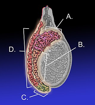
The epididymis is an elongated tubular structure attached to the posterior side of each one of the two male reproductive glands, the testicles. It is a single, narrow, tightly coiled tube in adult humans, 6 to 7 centimetres in length; uncoiled the tube would be approximately 6 m long. It connects the testicle to the vas deferens in the male reproductive system. The epididymis serves as an interconnection between the multiple efferent ducts at the rear of a testicle (proximally), and the vas deferens (distally). Its primary function is the storage, maturation and transport of sperm cells.
A micrograph or photomicrograph is a photograph or digital image taken through a microscope or similar device to show a magnified image of an object. This is opposed to a macrograph or photomacrograph, an image which is also taken on a microscope but is only slightly magnified, usually less than 10 times. Micrography is the practice or art of using microscopes to make photographs.
Hugh Esmor Huxley MBE FRS was a British molecular biologist who made important discoveries in the physiology of muscle. He was a graduate in physics from Christ's College, Cambridge. However, his education was interrupted for five years by the Second World War, during which he served in the Royal Air Force. His contribution to development of radar earned him an MBE.
Peter Gerald Satir is an American microbiologist who has spent his career studying the basis of motion by studying the cilium. He is a native of New York, graduated from the Bronx High School of Science in 1952, received his PhD from the Rockefeller University in 1961 and worked at the Department of Anatomy and Structural Biology at the Albert Einstein College of Medicine.
Keith Roberts Porter was a Canadian-American cell biologist. He created pioneering biology techniques and research using electron microscopy of cells. Porter also contributed to the development of other experimental methods for cell culture and nuclear transplantation. He was also responsible for naming the endoplasmic reticulum, conducting work on the 9 + 2 microtubule structure in the axoneme of cilia, and coining the term "microtrabecular lattice." In collaborations with other scientists, he contributed to the understanding of cellular structures and concepts such as compartmentalization, flagella, centrioles, fibrin, collagen, T-tubules and sarcoplasmic reticulum. He also introduced microtome cutting.
The American Society for Cell Biology (ASCB) is a professional society that was founded in 1960.
John E. Heuser is an American Professor of Biophysics in the department of Cell Biology and Physiology at the Washington University School of Medicine as well as a Professor at the Institute for Integrated Cell-Material Sciences (iCeMS) at Kyoto University.
Sanford Louis "Sandy" Palay was an American scientist and educator.
Clara Franzini-Armstrong is an Italian-born American electron microscopist, and Professor Emeritus of Cell and Developmental Biology at University of Pennsylvania.
June Dalziel Almeida was a Scottish virologist, a pioneer in virus imaging and identification. Her skills in electron microscopy earned her an international reputation.
Elizabeth Dexter "Betty" Hay was an American cell and developmental biologist. She was best known for her research in limb regeneration, the role of the extracellular matrix (ECM) in cell differentiation, and epithelial-mesenchymal transitions (EMT). Hay led many research teams in discovering new findings in these related fields, which led her to obtain several high honors and awards for her work. Hay primarily worked with amphibians during her years of limb regeneration work and then moved onto avian epithelia for research on the ECM and EMT. Hay was thrilled by the introduction of transmission electron microscopy (TEM) during her lifetime, which aided her in many of her findings throughout her career. Moreover, Hay was a huge advocate of women in science during her lifetime.
The American Microscopical Society (AMS) is a society of biologists dedicated to promoting the use of microscopy.
Joachim Frank ; born September 12, 1940) is a German-American biophysicist at Columbia University and a Nobel laureate. He is regarded as the founder of single-particle cryo-electron microscopy (cryo-EM), for which he shared the Nobel Prize in Chemistry in 2017 with Jacques Dubochet and Richard Henderson. He also made significant contributions to structure and function of the ribosome from bacteria and eukaryotes.
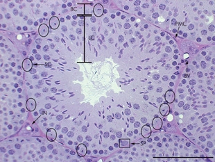
A peritubular myoid (PTM) cell is one of the smooth muscle cells which surround the seminiferous tubules in the testis. These cells are present in all mammals but their organization and abundance varies between species. The exact role of PTM cells is still somewhat uncertain and further work into this is needed. However, a number of functions of these cells have been established. They are contractile cells which contain actin filaments and are primarily involved in transport of spermatozoa through the tubules. They provide structural integrity to the tubules through their involvement in laying down the basement membrane. This has also been shown to affect Sertoli cell function and PTM cells also communicate with Sertoli cells through the secretion of growth factors and ECM components. Studies have shown PTM cells to be critical in achieving normal spermatogenesis. Overall, PTM cells have a role in both maintaining the structure of the tubules and regulating spermatogenesis through cellular interaction.
George Bunker Chapman was a professor and a pioneer in research of cell biology and ultrastructure using transmission-light and transmission electron microscopy. He was the first person to see the interior structure of four bacterium species in electron micrographs he produced, described in his Ph.D. dissertation completed in 1953. As a professor, he changed the lives of hundreds of students, colleagues, and others through his mentorship.
This page is based on this Wikipedia article Text is available under the CC BY-SA 4.0 license; additional terms may apply. Images, videos and audio are available under their respective licenses.