Double-strand break repair model

Last updated
Various pathways for double-strand break repair. NHEJ in this article refers to cNHEJ. MMEJ in this article refers to a-EJ. Double-strand break repair pathway models.png
Various pathways for double-strand break repair. NHEJ in this article refers to cNHEJ. MMEJ in this article refers to a-EJ.

A double-strand break repair model refers to the various models of pathways that cells undertake to repair double strand-breaks (DSB). DSB repair is an important cellular process, as the accumulation of unrepaired DSB could lead to chromosomal rearrangements, tumorigenesis or even cell death. [1] In human cells, there are two main DSB repair mechanisms: Homologous recombination (HR) and non-homologous end joining (NHEJ). HR relies on undamaged template DNA as reference to repair the DSB, resulting in the restoration of the original sequence. [2] NHEJ modifies and ligates the damaged ends regardless of homology. [2] In terms of DSB repair pathway choice, most mammalian cells appear to favor NHEJ rather than HR. This is because the employment of HR may lead to gene deletion or amplification in cells which contains repetitive sequences. [1] In terms of repair models in the cell cycle, HR is only possible during the S and G2 phases, while NHEJ can occur throughout whole process. [3] These repair pathways are all regulated by the overarching DNA damage response mechanism. [4] Besides HR and NHEJ, there are also other repair models which exists in cells. Some are categorized under HR, such as synthesis-dependent strain annealing, break-induced replication, and single-strand annealing; while others are an entirely alternate repair model, namely, the pathway microhomology-mediated end joining (MMEJ). [5]

Contents

Causes

DSB can occur naturally due to the presence of reactive species generated by metabolism, and various external factors (e.g. ionizing radiation or chemotherapeutic drugs). [1]

In mammalian cells, there are numerous cellular processes that induce DSB. Firstly, DNA topological strain from topoisomerase during normal cell growth can cause the majority a cell’s DSB. [6] Secondly, cellular processes such as meiosis and the maturation of antibodies can cause nuclease-induced DSB. [7] Thirdly, the cleavage of different DNA structures such as reversed or blocked DNA replication forks, R-loops and DNA interstrand crosslinks can also cause DSB. [7]

Different models

Homologous recombination

Homologous recombination involves the exchange of DNA materials between homologous chromosomes. There are multiple pathways of HR to repair DSBs, which includes double-strand break repair (DSBR), synthesis-dependent strand annealing (SDSA), break-induced replication (BIR), and single-strand annealing (SSA). [8]

The regulation of HR in mammalian cells involves key HR proteins such as BRCA1 and BRCA2. [9] And as mentioned, since HR can lead to aggressive chromosomal rearrangement, loss of genetic information that could contribute to cell death, it explains why HR is strictly regulated. [8]

Double-strand break repair

Three possible sub-pathways for a double-strand break to repair via homologous recombination: Gene conversion, BIR and SDSA. The gene conversion is referring to the double-strand break repair model. The other sub-pathway is the synthesis-dependent strain annealing. SSA is the fourth sub-pathway and it is not shown in this diagram. Double-strand break repair models that act via homologous recombination.png
Three possible sub-pathways for a double-strand break to repair via homologous recombination: Gene conversion, BIR and SDSA. The gene conversion is referring to the double-strand break repair model. The other sub-pathway is the synthesis-dependent strain annealing. SSA is the fourth sub-pathway and it is not shown in this diagram.

HR repairs DSB by copying intact and homologous DNA molecules. The blunt ends of the DSB are processed into ssDNA with 3’ extensions, which allows RAD51 recombinase (eukaryotic homologue of prokaryotic RecA) to bind to it to form a nucleoprotein filament. [3] [10] The function of the filament is to locate the template DNA and form a joint heteroduplex molecule. Other proteins such as RP-A protein and RAD52 also coordinate in the heteroduplex formation, [10] the RP-A protein has to be removed for the RAD51 to form the filament, [11] whereas the RAD52 is a key HR mediator. [3] Afterwards, the 3’ ssDNA invades the template DNA, and displaces a DNA strand to form a D-loop. DNA polymerase and other accessory factors follows by replacing the missing DNA via DNA synthesis. Ligase then attaches the DNA strand break, [10] resulting in the formation of 2 Holliday junctions. The recombined DNA strands then undergoes resolution by cleavage. The orientation of the cleavage determines whether the resolution results in either cross-over or noncross-over products. [12] Lastly, the strands finally separate and revert to its original form.

, the main pathway for resolution relies on the BTR (BLM helicase-TopoisomeraseIIIα-RMI1-RM2) complex, where it induces the resolution of the 2 Holliday junctions, but this pathway favors the noncross-over cleavage. [12]

Synthesis-dependent strain annealing

Synthesis-dependent strain annealing is the most preferred repair mechanism in somatic cells. [3] The pathway of SDSA is similar to DSBR until just after the D-loop formation. Instead of forming Holliday junctions after DNA synthesis, the nascent strand dissociates via RETL1 helicase and anneals back to the other end of the resected strand. [3] [9] [13] This explains why SDSA results in a non-crossover pathway. [3] The remaining gap is filled in and the nick is attached by the ligase. [9]

Break-induced replication

Although there is little research in regards of break-induced replication, it is known that it is a one-ended recombination mechanism, where only of the one ends of a DSB will be involved in strand invasion. [14] This means that unlike DSBR, BIR does not link back to the second DSB end after the strand invasion and replication. [14]

Single-strand annealing

Single-strand annealing involves homologous/repeated sequences flanking a DSB. [7] The process starts with the key end resection factor CtlP, which mediates the end resection of DSBs, resulting in the formation of a 3' ssDNA extension. Meditated by RAD52, the flanking homologous sequences are annealed, and forms a synapse intermediate. [7] Then, the nonhomologous 3’ extension is removed by the ERCC1-XPF complex through endonucleolytic cleavage, with RAD52 increasing the efficiency of the ERCC1-XPF complex activity. [7] It is only after the removal of 3’ ssDNA, where the polymerase will fill the missing gaps and the ligase to ligate the strands. [7] Since SSA results in the deletion of repetitive sequences, this could potentially lead to error-prone repair. [3]

Single-strand annealing differs from SDSA and DSBR in numerous ways. For instance, the 3’ extension after the end resection in SSA anneals to the repeated/homologous sequences of the other end, whereas in other pathways the strand invasion to another homologous DNA template. [15] Moreover, SSA does not require RAD51, because it does not involve strand invasion, but rather the annealing of homologous sequences. [3]

Non-homologous end joining

Non-homologous end joining (NHEJ) is one of the major pathways in DSB repair besides HR. [16] The basic concept of NHEJ involves three steps. First, the ends of a DSB is captured by a group of enzymes. The enzymes then form a bridge which connects the DSB ends together, and is lastly followed by religation of the DNA strands. [17] To initiate whole process, the Ku70/80 protein complex binds to the damaged ends of the DSB strands. This forms a preliminary scaffold which allows the recruitment of various NHEJ factors, such as the DNA-dependent protein kinase catalytic subunit (DNA-PKcs), DNA Ligase IV and X-ray cross complementing protein 4 (XRCC4) to form a bridge and bring both ends of the damaged DNA strands together. [18] [19] [20] [21] This is then followed by the processing of any non-ligatable DNA termini by a group of proteins including Artemis, PNKP, APLF and Ku, before the XRCC4 and DNA Ligase IV ligate the bridged DNA. [17] [22]

Microhomology-mediated end joining

Microhomology-mediated end joining (MMEJ), also known as alt-non-homologous end joining, is another pathway to repair DSBs. The process of MMEJ can be summarized in five steps: the 5' to 3' cutting of DNA ends, annealing of microhomology, removing heterologous flaps, and ligation and synthesis of gap filling DNA. [5] It was found that the selection between MMEJ and NHEJ is mainly dependent on Ku levels and the concurrent cell cycle. [23]

The regulation of double-strand break repair pathways

DNA damage response

DNA damage response (DDR) is the overarching mechanism which mediates the cell's detection and response to DNA damage. This includes the process of detecting DSB within the cell, and the subsequent triggering and regulation of DSB repair pathways. Upstream detections of DNA damage via DDR will lead to the activation of downstream responses such as senescence, cell apoptosis, halting transcription and activating DNA repair mechanisms. [4] Proteins such as the proteins ATM, ATR and DNA-dependent protein kinase (DNA-PK) are vital for the process of detection of DSB in DDR, and these proteins are recruited to the DSB site in the DNA. [24] In particular, ATM has been identified as the protein kinase in charge of the global meditation of cellular responses to DSB, which includes various DSB repair pathways. [24] Following the recruitment of the aforementioned proteins to DNA damage sites, they will in turn trigger cellular responses and repair pathways to mitigate and repair the damage caused. [4] In short, these vital upstream proteins and downstream repair pathways altogether forms the DDR, which plays a vital role in DSB repair pathways regulation.

Fanconi anemia complex in one DNA damage response pathway

Recombinational repair of DNA double-strand damage.jpg

The image in this section illustrates molecular steps in a DNA damage response pathway in which a Fanconi anemia complex is activated during repair of a double-strand break. ATM (ATM) is also a protein kinase that is recruited and activated by DNA double-strand breaks. DNA double-strand damages activate the Fanconi anemia core complex (FANCA/B/C/E/F/G/L/M). [25] The FA core complex monoubiquitinates the downstream targets FANCD2 and FANCI. [26] ATM activates (phosphorylates) CHEK2 and FANCD2 [27] CHEK2 phosphorylates BRCA1. [28] Ubiquinated FANCD2 complexes with BRCA1 and RAD51. [29] The PALB2 protein acts as a hub, [30] bringing together BRCA1, BRCA2 and RAD51 at the site of a DNA double-strand break, and also binds to RAD51C, a member of the RAD51 paralog complex RAD51B-RAD51C-RAD51D-XRCC2 (BCDX2). The BCDX2 complex is responsible for RAD51 recruitment or stabilization at damage sites. [31] RAD51 plays a major role in homologous recombinational repair of DNA during double strand break repair. In this process, an ATP dependent DNA strand exchange takes place in which a template strand invades base-paired strands of homologous DNA molecules. RAD51 is involved in the search for homology and strand pairing stages of the process.

Double-strand break repair pathway choice

As cells have developed various DSB repair models, it is said that specific pathways are favoured for their ability to repair DSB depending on the cellular context. [32] These conditions include the type of DSB involved, the species of cells involved, and the stage of the cell cycle. [33]

In various types of DSB

Cells have evolved a multitude of DSB repair pathways in response to the various types of DSB. [33] Hence, various pathways are favoured in different situations. For instance, frank DSB, which are DSB induced by substances like as ionizing radiation, and nucleases, can be repaired by both HR and NHEJ. On the other hand, DSB due to replication fork collapse mainly favours HR. [33] [34]

In higher eukaryotes and yeast cells

It is said that the favoured pathway in a particular situations is also largely dependent on the species of the cell, the cell type, and cell cycle phases; and are all modulated and triggered by different upstream regulatory proteins. [33] As compared to higher eukaryotes, yeast cells have adopted HR as the main repair pathway for DSB. [35] Imprecise NHEJ, the primary pathway for NHEJ to repair "dirty" ends due to IR, was found to be inefficent at repairing DSB in yeast cells. It was hypothesized that this inefficiency as compared to mammalian cells is due to the lack of three vital NHEJ proteins, including DNA-PKcs, BRCA1, and Artemis. [33] Contrary to yests, higher eukaryotes has a much higher frequency and efficiency at adopting NHEJ pathways. [36] Research hypothesize that this is due to the higher eukaryote's larger genome size, as it means that more NHEJ related proteins are encoded for NHEJ repair pathways; and a larger genome implies a challenging obstacle to find a homologous template for HR. [33]

In cell cycle

HR and NHEJ pathways are favoured in various phases of cell cycles for a multitude of factors. As S and G2 phases of the cell cycle generate more chromatids, the increased availability of template access for HR results in the up-regulation of the pathway. [37] This rise is further increased due to the activation of CDK1 and the increase of RAD51 and RAD52 levels during G1 phase. [33] [38] Despite this, NHEJ not is inactive during the HR up-regulation. In fact, NHEJ was shown to be active throughout all stages of the cell cycle, and is favoured in G1 phase during low resection action intervals. [39] [40] This suggests the competition between HR and NHEJ for DSB repair in cells. [38] It should be noted, however, that there is a shift of favour from NHEJ to HR when the cell cycle is progressing from G1 to S/G2 phases in eukaryotic cells. [38]

During meiosis

In diploid eukaryotic organisms, the events of meiosis can be viewed as occurring in three steps. (1) Haploid gametes undergo syngamy/fertilisation with the result that chromosome sets of different parental origin come together to share the same nucleus. (2) Homologous chromosomes originating from different cells (i.e. non-sister chromosomes) align in pairs and undergo recombination involving double-strand break repair. (3) Two successive cell divisions (without duplication of chromosomes) result in haploid gametes that can then repeat the meiotic cycle. During step (2), damages in DNA of the germline can be removed by double-strand break repair. [41] In particular, double-strand breaks in one duplex DNA molecule can be accurately repaired using information from a homologous intact DNA molecule by the process of homologous recombination. [41]

Defective DSB repair

Although there is no universal model to explain disease etiology caused by DNA repair deficiency, it is said that the accumulation of unrepaired DNA damage may lead to various diseases, including various metabolic syndromes and types of cancers. [42] [43] Some examples of diseases caused by defects of DSB repair mechanisms are listed below:

See also

Related Research Articles

<span class="mw-page-title-main">Chromosomal crossover</span> Cellular process

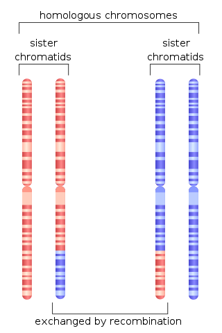
Chromosomal crossover, or crossing over, is the exchange of genetic material during sexual reproduction between two homologous chromosomes' non-sister chromatids that results in recombinant chromosomes. It is one of the final phases of genetic recombination, which occurs in the pachytene stage of prophase I of meiosis during a process called synapsis. Synapsis begins before the synaptonemal complex develops and is not completed until near the end of prophase I. Crossover usually occurs when matching regions on matching chromosomes break and then reconnect to the other chromosome.

<span class="mw-page-title-main">DNA repair</span> Cellular mechanism

DNA repair is a collection of processes by which a cell identifies and corrects damage to the DNA molecules that encodes its genome. In human cells, both normal metabolic activities and environmental factors such as radiation can cause DNA damage, resulting in tens of thousands of individual molecular lesions per cell per day. Many of these lesions cause structural damage to the DNA molecule and can alter or eliminate the cell's ability to transcribe the gene that the affected DNA encodes. Other lesions induce potentially harmful mutations in the cell's genome, which affect the survival of its daughter cells after it undergoes mitosis. As a consequence, the DNA repair process is constantly active as it responds to damage in the DNA structure. When normal repair processes fail, and when cellular apoptosis does not occur, irreparable DNA damage may occur. This can eventually lead to malignant tumors, or cancer as per the two-hit hypothesis.

RecQ helicase is a family of helicase enzymes initially found in Escherichia coli that has been shown to be important in genome maintenance. They function through catalyzing the reaction ATP + H2O → ADP + P and thus driving the unwinding of paired DNA and translocating in the 3' to 5' direction. These enzymes can also drive the reaction NTP + H2O → NDP + P to drive the unwinding of either DNA or RNA.

<span class="mw-page-title-main">Non-homologous end joining</span> Pathway that repairs double-strand breaks in DNA

Non-homologous end joining (NHEJ) is a pathway that repairs double-strand breaks in DNA. It is called "non-homologous" because the break ends are directly ligated without the need for a homologous template, in contrast to homology directed repair (HDR), which requires a homologous sequence to guide repair. NHEJ is active in both non-dividing and proliferating cells, while HDR is not readily accessible in non-dividing cells. The term "non-homologous end joining" was coined in 1996 by Moore and Haber.

<span class="mw-page-title-main">Heteroduplex</span>

A heteroduplex is a double-stranded (duplex) molecule of nucleic acid originated through the genetic recombination of single complementary strands derived from different sources, such as from different homologous chromosomes or even from different organisms.

<span class="mw-page-title-main">Homologous recombination</span> Genetic recombination between identical or highly similar strands of genetic material

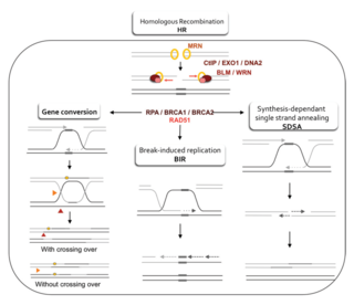
Homologous recombination is a type of genetic recombination in which genetic information is exchanged between two similar or identical molecules of double-stranded or single-stranded nucleic acids.

<span class="mw-page-title-main">RAD51</span>

DNA repair protein RAD51 homolog 1 is a protein encoded by the gene RAD51. The enzyme encoded by this gene is a member of the RAD51 protein family which assists in repair of DNA double strand breaks. RAD51 family members are homologous to the bacterial RecA, Archaeal RadA and yeast Rad51. The protein is highly conserved in most eukaryotes, from yeast to humans.

<span class="mw-page-title-main">Nibrin</span> Protein-coding gene in the species Homo sapiens

Nibrin, also known as NBN or NBS1, is a protein which in humans is encoded by the NBN gene.

<span class="mw-page-title-main">PARP1</span> Mammalian protein found in Homo sapiens

Poly [ADP-ribose] polymerase 1 (PARP-1) also known as NAD+ ADP-ribosyltransferase 1 or poly[ADP-ribose] synthase 1 is an enzyme that in humans is encoded by the PARP1 gene. It is the most abundant of the PARP family of enzymes, accounting for 90% of the NAD+ used by the family. PARP1 is mostly present in cell nucleus, but cytosolic fraction of this protein was also reported.

<span class="mw-page-title-main">TP53BP1</span> Protein-coding gene in the species Homo sapiens

Tumor suppressor p53-binding protein 1 also known as p53-binding protein 1 or 53BP1 is a protein that in humans is encoded by the TP53BP1 gene.

<span class="mw-page-title-main">RAD52</span> Protein-coding gene in the species Homo sapiens

RAD52 homolog , also known as RAD52, is a protein which in humans is encoded by the RAD52 gene.

<span class="mw-page-title-main">RAD54L</span> Protein-coding gene in the species Homo sapiens

DNA repair and recombination protein RAD54-like is a protein that in humans is encoded by the RAD54L gene.

<span class="mw-page-title-main">FANCL</span> Protein-coding gene in the species Homo sapiens

E3 ubiquitin-protein ligase FANCL is an enzyme that in humans is encoded by the FANCL gene.

<span class="mw-page-title-main">BRE (gene)</span> Protein-coding gene in the species Homo sapiens

BRCA1-A complex subunit BRE is a protein that in humans is encoded by the BRE gene.

<span class="mw-page-title-main">Homology directed repair</span>

Homology-directed repair (HDR) is a mechanism in cells to repair double-strand DNA lesions. The most common form of HDR is homologous recombination. The HDR mechanism can only be used by the cell when there is a homologous piece of DNA present in the nucleus, mostly in G2 and S phase of the cell cycle. Other examples of homology-directed repair include single-strand annealing and breakage-induced replication. When the homologous DNA is absent, another process called non-homologous end joining (NHEJ) takes place instead.

Microhomology-mediated end joining (MMEJ), also known as alternative nonhomologous end-joining (Alt-NHEJ) is one of the pathways for repairing double-strand breaks in DNA. As reviewed by McVey and Lee, the foremost distinguishing property of MMEJ is the use of microhomologous sequences during the alignment of broken ends before joining, thereby resulting in deletions flanking the original break. MMEJ is frequently associated with chromosome abnormalities such as deletions, translocations, inversions and other complex rearrangements.

<span class="mw-page-title-main">Synthesis-dependent strand annealing</span>

Synthesis-dependent strand annealing (SDSA) is a major mechanism of homology-directed repair of DNA double-strand breaks (DSBs). Although many of the features of SDSA were first suggested in 1976, the double-Holliday junction model proposed in 1983 was favored by many researchers. In 1994, studies of double-strand gap repair in Drosophila were found to be incompatible with the double-Holliday junction model, leading researchers to propose a model they called synthesis-dependent strand annealing. Subsequent studies of meiotic recombination in S. cerevisiae found that non-crossover products appear earlier than double-Holliday junctions or crossover products, challenging the previous notion that both crossover and non-crossover products are produced by double-Holliday junctions and leading the authors to propose that non-crossover products are generated through SDSA.

Lumír Krejčí is a Czech biochemist. His research is focused on regulatory processes involved in maintaining genome integrity. Currently, as Associate Professor in biochemistry, he leads the laboratory of recombination and DNA repair (LORD) at the Department of Biology, Faculty of Medicine, at Masaryk University in Brno.

Telomeres, the caps on the ends of eukaryotic chromosomes, play critical roles in cellular aging and cancer. An important facet to how telomeres function in these roles is their involvement in cell cycle regulation.

<span class="mw-page-title-main">DNA end resection</span> Biochemical process

DNA end resection, also called 5′–3′ degradation, is a biochemical process where the blunt end of a section of double-stranded DNA (dsDNA) is modified by cutting away some nucleotides from the 5' end to produce a 3' single-stranded sequence. The presence of a section of single-stranded DNA (ssDNA) allows the broken end of the DNA to line up accurately with a matching sequence, so that it can be accurately repaired.

References

  1. 1 2 3 Featherstone C, Jackson SP (October 1999). "DNA double-strand break repair". Current Biology. 9 (20): R759–R761. Bibcode:1999CBio....9.R759F. doi: 10.1016/S0960-9822(00)80005-6 . PMID   10531043. S2CID   1941783.
  2. 1 2 Mao Z, Bozzella M, Seluanov A, Gorbunova V (October 2008). "Comparison of nonhomologous end joining and homologous recombination in human cells". DNA Repair. 7 (10): 1765–1771. doi:10.1016/j.dnarep.2008.06.018. PMC   2695993 . PMID   18675941.
  3. 1 2 3 4 5 6 7 8 Scully R, Panday A, Elango R, Willis NA (November 2019). "DNA double-strand break repair-pathway choice in somatic mammalian cells". Nature Reviews. Molecular Cell Biology. 20 (11): 698–714. doi:10.1038/s41580-019-0152-0. PMC   7315405 . PMID   31263220.
  4. 1 2 3 Zhou BB, Elledge SJ (November 2000). "The DNA damage response: putting checkpoints in perspective". Nature. 408 (6811): 433–439. Bibcode:2000Natur.408..433Z. doi:10.1038/35044005. PMID   11100718. S2CID   4419141.
  5. 1 2 Sfeir A, Symington LS (November 2015). "Microhomology-Mediated End Joining: A Back-up Survival Mechanism or Dedicated Pathway?". Trends in Biochemical Sciences. 40 (11): 701–714. doi:10.1016/j.tibs.2015.08.006. PMC   4638128 . PMID   26439531.
  6. Tan XY, Huen MS (October 2020). "Perfecting DNA double-strand break repair on transcribed chromatin". Essays in Biochemistry. 64 (5): 705–719. doi:10.1042/EBC20190094. PMID   32309851. S2CID   216030185.
  7. 1 2 3 4 5 6 Bhargava R, Onyango DO, Stark JM (September 2016). "Regulation of Single-Strand Annealing and its Role in Genome Maintenance". Trends in Genetics. 32 (9): 566–575. doi:10.1016/j.tig.2016.06.007. PMC   4992407 . PMID   27450436.
  8. 1 2 Sebesta M, Krejci L (2016). "Mechanism of Homologous Recombination". In Hanaoka F, Sugasawa K (eds.). DNA Replication, Recombination, and Repair. Tokyo: Springer Japan. pp. 73–109. doi:10.1007/978-4-431-55873-6_4. ISBN   978-4-431-55873-6 . Retrieved 2021-03-31.{{cite book}}: |work= ignored (help)
  9. 1 2 3 Do AT, Brooks JT, Le Neveu MK, LaRocque JR (March 2014). "Double-strand break repair assays determine pathway choice and structure of gene conversion events in Drosophila melanogaster". G3. 4 (3): 425–432. doi:10.1534/g3.113.010074. PMC   3962482 . PMID   24368780.
  10. 1 2 3 Modesti M, Kanaar R (2001-04-26). "Homologous recombination: from model organisms to human disease". Genome Biology. 2 (5): REVIEWS1014. doi: 10.1186/gb-2001-2-5-reviews1014 . PMC   138934 . PMID   11387040.
  11. Wright WD, Shah SS, Heyer WD (July 2018). "Homologous recombination and the repair of DNA double-strand breaks". The Journal of Biological Chemistry. 293 (27): 10524–10535. doi: 10.1074/jbc.TM118.000372 . PMC   6036207 . PMID   29599286.
  12. 1 2 Shah Punatar R, Martin MJ, Wyatt HD, Chan YW, West SC (January 2017). "Resolution of single and double Holliday junction recombination intermediates by GEN1". Proceedings of the National Academy of Sciences of the United States of America. 114 (3): 443–450. Bibcode:2017PNAS..114..443S. doi: 10.1073/pnas.1619790114 . PMC   5255610 . PMID   28049850.
  13. Currall BB, Chiang C, Talkowski ME, Morton CC (June 2013). "Mechanisms for Structural Variation in the Human Genome". Current Genetic Medicine Reports. 1 (2): 81–90. doi:10.1007/s40142-013-0012-8. PMC   3665418 . PMID   23730541.
  14. 1 2 Kraus E, Leung WY, Haber JE (July 2001). "Break-induced replication: a review and an example in budding yeast". Proceedings of the National Academy of Sciences of the United States of America. 98 (15): 8255–8262. Bibcode:2001PNAS...98.8255K. doi: 10.1073/pnas.151008198 . PMC   37429 . PMID   11459961.
  15. Lok BH, Powell SN (December 2012). "Molecular pathways: understanding the role of Rad52 in homologous recombination for therapeutic advancement". Clinical Cancer Research. 18 (23): 6400–6406. doi:10.1158/1078-0432.CCR-11-3150. PMC   3513650 . PMID   23071261.
  16. Radhakrishnan SK, Jette N, Lees-Miller SP (May 2014). "Non-homologous end joining: emerging themes and unanswered questions". DNA Repair. Recent Developments in Non-Homologous End Joining. 17: 2–8. doi:10.1016/j.dnarep.2014.01.009. PMC   4084493 . PMID   24582502.
  17. 1 2 Weterings E, Chen DJ (January 2008). "The endless tale of non-homologous end-joining". Cell Research. 18 (1): 114–124. doi: 10.1038/cr.2008.3 . PMID   18166980. S2CID   2090745.
  18. Uematsu N, Weterings E, Yano K, Morotomi-Yano K, Jakob B, Taucher-Scholz G, et al. (April 2007). "Autophosphorylation of DNA-PKCS regulates its dynamics at DNA double-strand breaks". The Journal of Cell Biology. 177 (2): 219–229. doi:10.1083/jcb.200608077. PMC   2064131 . PMID   17438073.
  19. Mari PO, Florea BI, Persengiev SP, Verkaik NS, Brüggenwirth HT, Modesti M, et al. (December 2006). "Dynamic assembly of end-joining complexes requires interaction between Ku70/80 and XRCC4". Proceedings of the National Academy of Sciences of the United States of America. 103 (49): 18597–18602. Bibcode:2006PNAS..10318597M. doi: 10.1073/pnas.0609061103 . PMC   1693708 . PMID   17124166.
  20. Costantini S, Woodbine L, Andreoli L, Jeggo PA, Vindigni A (June 2007). "Interaction of the Ku heterodimer with the DNA ligase IV/Xrcc4 complex and its regulation by DNA-PK". DNA Repair. 6 (6): 712–722. doi:10.1016/j.dnarep.2006.12.007. PMID   17241822.
  21. Nick McElhinny SA, Snowden CM, McCarville J, Ramsden DA (May 2000). "Ku recruits the XRCC4-ligase IV complex to DNA ends". Molecular and Cellular Biology. 20 (9): 2996–3003. doi:10.1128/MCB.20.9.2996-3003.2000. PMC   85565 . PMID   10757784.
  22. Davis AJ, Chen DJ (June 2013). "DNA double strand break repair via non-homologous end-joining". Translational Cancer Research. 2 (3): 130–143. doi:10.3978/j.issn.2218-676X.2013.04.02. PMC   3758668 . PMID   24000320.
  23. Truong LN, Li Y, Shi LZ, Hwang PY, He J, Wang H, et al. (May 2013). "Microhomology-mediated End Joining and Homologous Recombination share the initial end resection step to repair DNA double-strand breaks in mammalian cells". Proceedings of the National Academy of Sciences of the United States of America. 110 (19): 7720–7725. Bibcode:2013PNAS..110.7720T. doi: 10.1073/pnas.1213431110 . PMC   3651503 . PMID   23610439.
  24. 1 2 Blackford AN, Jackson SP (June 2017). "ATM, ATR, and DNA-PK: The Trinity at the Heart of the DNA Damage Response". Molecular Cell. 66 (6): 801–817. doi: 10.1016/j.molcel.2017.05.015 . PMID   28622525. S2CID   206997898.
  25. D'Andrea AD (2010). "Susceptibility pathways in Fanconi's anemia and breast cancer". N. Engl. J. Med. 362 (20): 1909–19. doi:10.1056/NEJMra0809889. PMC   3069698 . PMID   20484397.
  26. Sobeck A, Stone S, Landais I, de Graaf B, Hoatlin ME (2009). "The Fanconi anemia protein FANCM is controlled by FANCD2 and the ATR/ATM pathways". J. Biol. Chem. 284 (38): 25560–8. doi: 10.1074/jbc.M109.007690 . PMC   2757957 . PMID   19633289.
  27. Castillo P, Bogliolo M, Surralles J (2011). "Coordinated action of the Fanconi anemia and ataxia telangiectasia pathways in response to oxidative damage". DNA Repair (Amst.). 10 (5): 518–25. doi:10.1016/j.dnarep.2011.02.007. PMID   21466974.
  28. Stolz A, Ertych N, Bastians H (2011). "Tumor suppressor CHK2: regulator of DNA damage response and mediator of chromosomal stability". Clin. Cancer Res. 17 (3): 401–5. doi: 10.1158/1078-0432.CCR-10-1215 . PMID   21088254.
  29. Taniguchi T, Garcia-Higuera I, Andreassen PR, Gregory RC, Grompe M, D'Andrea AD (2002). "S-phase-specific interaction of the Fanconi anemia protein, FANCD2, with BRCA1 and RAD51". Blood. 100 (7): 2414–20. doi:10.1182/blood-2002-01-0278. PMID   12239151.
  30. Park JY, Zhang F, Andreassen PR (2014). "PALB2: the hub of a network of tumor suppressors involved in DNA damage responses". Biochimica et Biophysica Acta (BBA) - Reviews on Cancer. 1846 (1): 263–75. doi:10.1016/j.bbcan.2014.06.003. PMC   4183126 . PMID   24998779.
  31. Chun J, Buechelmaier ES, Powell SN (2013). "Rad51 paralog complexes BCDX2 and CX3 act at different stages in the BRCA1-BRCA2-dependent homologous recombination pathway". Mol. Cell. Biol. 33 (2): 387–95. doi:10.1128/MCB.00465-12. PMC   3554112 . PMID   23149936.
  32. Chapman JR, Taylor MR, Boulton SJ (August 2012). "Playing the end game: DNA double-strand break repair pathway choice". Molecular Cell. 47 (4): 497–510. doi: 10.1016/j.molcel.2012.07.029 . PMID   22920291.
  33. 1 2 3 4 5 6 7 Shrivastav M, De Haro LP, Nickoloff JA (January 2008). "Regulation of DNA double-strand break repair pathway choice". Cell Research. 18 (1): 134–147. doi: 10.1038/cr.2007.111 . PMID   18157161. S2CID   20992607.
  34. Rothstein R, Michel B, Gangloff S (January 2000). "Replication fork pausing and recombination or "gimme a break"". Genes & Development. 14 (1): 1–10. doi: 10.1101/gad.14.1.1 . PMID   10640269. S2CID   40751623.
  35. Sugawara N, Haber JE (February 1992). "Characterization of double-strand break-induced recombination: homology requirements and single-stranded DNA formation". Molecular and Cellular Biology. 12 (2): 563–575. doi:10.1128/MCB.12.2.563. PMC   364230 . PMID   1732731.
  36. Lin Y, Lukacsovich T, Waldman AS (December 1999). "Multiple pathways for repair of DNA double-strand breaks in mammalian chromosomes". Molecular and Cellular Biology. 19 (12): 8353–8360. doi:10.1128/MCB.19.12.8353. PMC   84924 . PMID   10567560.
  37. Dronkert ML, Beverloo HB, Johnson RD, Hoeijmakers JH, Jasin M, Kanaar R (May 2000). "Mouse RAD54 affects DNA double-strand break repair and sister chromatid exchange". Molecular and Cellular Biology. 20 (9): 3147–3156. doi:10.1128/MCB.20.9.3147-3156.2000. PMC   85609 . PMID   10757799.
  38. 1 2 3 Chen F, Nastasi A, Shen Z, Brenneman M, Crissman H, Chen DJ (September 1997). "Cell cycle-dependent protein expression of mammalian homologs of yeast DNA double-strand break repair genes Rad51 and Rad52". Mutation Research. 384 (3): 205–211. doi:10.1016/S0921-8777(97)00020-7. PMID   9330616.
  39. Aylon Y, Liefshitz B, Kupiec M (December 2004). "The CDK regulates repair of double-strand breaks by homologous recombination during the cell cycle". The EMBO Journal. 23 (24): 4868–4875. doi:10.1038/sj.emboj.7600469. PMC   535085 . PMID   15549137.
  40. Ira G, Pellicioli A, Balijja A, Wang X, Fiorani S, Carotenuto W, et al. (October 2004). "DNA end resection, homologous recombination and DNA damage checkpoint activation require CDK1". Nature. 431 (7011): 1011–1017. Bibcode:2004Natur.431.1011I. doi:10.1038/nature02964. PMC   4493751 . PMID   15496928.
  41. 1 2 Bernstein, H.; Hopf, F.A.; Michod, R.E. (1987). "The Molecular Basis of the Evolution of Sex". Molecular Genetics of Development. Advances in Genetics. Vol. 24. pp. 323–370. doi:10.1016/s0065-2660(08)60012-7. ISBN   978-0-12-017624-3. PMID   3324702.
  42. Tiwari V, Wilson DM (August 2019). "DNA Damage and Associated DNA Repair Defects in Disease and Premature Aging". American Journal of Human Genetics. 105 (2): 237–257. doi:10.1016/j.ajhg.2019.06.005. PMC   6693886 . PMID   31374202.
  43. Mercer JR, Cheng KK, Figg N, Gorenne I, Mahmoudi M, Griffin J, et al. (October 2010). "DNA damage links mitochondrial dysfunction to atherosclerosis and the metabolic syndrome". Circulation Research. 107 (8): 1021–1031. doi:10.1161/CIRCRESAHA.110.218966. PMC   2982998 . PMID   20705925.
  44. 1 2 Katsuki Y, Takata M (October 2016). "Defects in homologous recombination repair behind the human diseases: FA and HBOC". Endocrine-Related Cancer. 23 (10): T19–T37. doi: 10.1530/ERC-16-0221 . PMID   27550963.
  45. Gröschel S, Hübschmann D, Raimondi F, Horak P, Warsow G, Fröhlich M, et al. (April 2019). "Defective homologous recombination DNA repair as therapeutic target in advanced chordoma". Nature Communications. 10 (1): 1635. Bibcode:2019NatCo..10.1635G. doi:10.1038/s41467-019-09633-9. PMC   6456501 . PMID   30967556.
  46. Koh KH, Kang HJ, Li LS, Kim NG, You KT, Yang E, et al. (September 2005). "Impaired nonhomologous end-joining in mismatch repair-deficient colon carcinomas". Laboratory Investigation; A Journal of Technical Methods and Pathology. 85 (9): 1130–1138. doi: 10.1038/labinvest.3700315 . PMID   16025146. S2CID   21197331.