Enthesis

Last updated
Enthesis
Joint.svg
Typical joint
Identifiers
TH H3.03.00.0.00034
Anatomical terminology

The enthesis (plural entheses) is the connective tissue between tendon or ligament and bone. [1]

Contents

There are two types of entheses: Fibrous entheses and fibrocartilaginous entheses. [2] [3]

In a fibrous enthesis, the collagenous tendon or ligament directly attaches to the bone.

In a fibrocartilaginous enthesis, the interface presents a gradient that crosses four transition zones: [4]

  1. Tendinous area displaying longitudinally oriented fibroblasts and a parallel arrangement of collagen fibres
  2. Fibrocartilaginous region of variable thickness where the structure of the cells changes to chondrocytes
  3. Abrupt transition from cartilaginous to calcified fibrocartilage—often called 'tidemark' or 'blue line'
  4. Bone

Clinical significance

A disease of the entheses is known as an enthesopathy or enthesitis . [5]

Enthetic degeneration is characteristic of spondyloarthropathy and other pathologies.

The enthesis is the primary site of disease in ankylosing spondylitis.

Society and culture

Bioarchaeology

Entheses are widely recorded in the field of bioarchaeology where the presence of anomalies at these sites, called entheseal changes, has been used to infer repetitive loading to study the division of labour in past populations. [6] Several different recording methods have been proposed to record the variety of changes seen at these sites. [7] [8] [9] [10] [11] [12] [13] [14] [15] However, research has shown that, whichever recording method is used, entheseal changes occur more frequently in older individuals. [16] [8] [17] [18] [19] Research demonstrates that diseases, such as ankylosing spondylitis and calcific tendinitis, [20] also have to be taken into consideration. Experimental studies have demonstrated how loading history (physical activity) can increase the relative size of muscle attachment sites. [21] [22] [23] [24]

History

"Enthesis" is rooted in the Ancient Greek word, "ἔνθεσις" or "énthesis," meaning “putting in," or "insertion." This refers to the role of the enthesis as the site of attachment of bones with tendons or ligaments. Relatedly, in muscle terminology, the insertion is the site of attachment at the end with predominant movement or action (opposite of the origin). Thus the words (enthesis and insertion [of muscle]) are proximal in the semantic field, but insertion in reference to muscle can refer to any relevant aspect of the site (i.e., the attachment per se, the bone, the tendon, or the entire area), whereas enthesis refers to the attachment per se and to ligamentous attachments as well as tendinous ones.

See also

Related Research Articles

<span class="mw-page-title-main">Coccyx</span> Bone of the pelvis

The coccyx, commonly referred to as the tailbone, is the final segment of the vertebral column in all apes, and analogous structures in certain other mammals such as horses. In tailless primates since Nacholapithecus, the coccyx is the remnant of a vestigial tail. In animals with bony tails, it is known as tailhead or dock, in bird anatomy as tailfan. It comprises three to five separate or fused coccygeal vertebrae below the sacrum, attached to the sacrum by a fibrocartilaginous joint, the sacrococcygeal symphysis, which permits limited movement between the sacrum and the coccyx.

<span class="mw-page-title-main">Coracoid process</span> Small hook-like structure on the lateral edge of the superior anterior portion of the scapula

The coracoid process is a small hook-like structure on the lateral edge of the superior anterior portion of the scapula. Pointing laterally forward, it, together with the acromion, serves to stabilize the shoulder joint. It is palpable in the deltopectoral groove between the deltoid and pectoralis major muscles.

<span class="mw-page-title-main">Shoulder</span> Part of the body

The human shoulder is made up of three bones: the clavicle (collarbone), the scapula, and the humerus as well as associated muscles, ligaments and tendons.

<span class="mw-page-title-main">Tennis elbow</span> Condition in which the outer part of the elbow becomes sore and tender

Tennis elbow, also known as lateral epicondylitis or enthesopathy of the extensor carpi radialis origin, is an enthesopathy of the origin of the extensor carpi radialis brevis on the lateral epicondyle. The outer part of the elbow becomes painful and tender. The pain may also extend into the back of the forearm. Onset of symptoms is generally gradual although they can seem sudden and be misinterpreted as an injury. Golfer's elbow is a similar condition that affects the inside of the elbow.

The term bioarchaeology has been attributed to British archaeologist Grahame Clark who, in 1972, defined it as the study of animal and human bones from archaeological sites. Redefined in 1977 by Jane Buikstra, bioarchaeology in the United States now refers to the scientific study of human remains from archaeological sites, a discipline known in other countries as osteoarchaeology, osteology or palaeo-osteology. Compared to bioarchaeology, osteoarchaeology is the scientific study that solely focus on the human skeleton. The human skeleton is used to tell us about health, lifestyle, diet, mortality and physique of the past. Furthermore, palaeo-osteology is simple the study of ancient bones.

The biceps femoris is a muscle of the thigh located to the posterior, or back. As its name implies, it consists of two heads; the long head is considered part of the hamstring muscle group, while the short head is sometimes excluded from this characterization, as it only causes knee flexion and is activated by a separate nerve.

<span class="mw-page-title-main">Enthesopathy</span> Medical condition

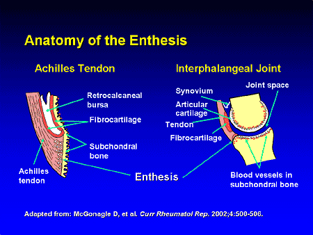
An enthesopathy refers to a disorder involving the attachment of a tendon or ligament to a bone. This site of attachment is known as the enthesis . If the condition is known to be inflammatory, it can more precisely be called an enthesitis.

<span class="mw-page-title-main">Flexor retinaculum of the hand</span> Thickened fascia over the carpal tunnel

The flexor retinaculum is a fibrous band on the palmar side of the hand near the wrist. It arches over the carpal bones of the hands, covering them and forming the carpal tunnel.

<span class="mw-page-title-main">SLAP tear</span> Medical condition

A SLAP tear or SLAP lesion is an injury to the superior glenoid labrum that initiates in the back of the labrum and stretches toward the front into the attachment point of the long head of the biceps tendon. SLAP is an acronym for "Superior Labrum Anterior and Posterior". SLAP lesions are commonly seen in overhead throwing athletes but middle-aged labor workers can also be affected, and they can be caused by chronic overuse or an acute stretch injury of the shoulder.

<span class="mw-page-title-main">Glenoid fossa</span> Part of the shoulder

The glenoid fossa of the scapula or the glenoid cavity is a bone part of the shoulder. The word glenoid is pronounced or and is from Greek: gléne, "socket", reflecting the shoulder joint's ball-and-socket form. It is a shallow, pyriform articular surface, which is located on the lateral angle of the scapula. It is directed laterally and forward and articulates with the head of the humerus; it is broader below than above and its vertical diameter is the longest.

<span class="mw-page-title-main">Enthesitis</span> Medical condition

Enthesitis is inflammation of the entheses, the sites where tendons or ligaments insert into the bone. It is an enthesopathy, a pathologic condition of the entheses. Early clinical manifestations are an aching sensation akin to "working out too much", and it gets better with activity. It is worse in the morning. The muscle insertion hurts very focally as it joins into the bone, but there is little to no pain at all with passive motion. There are some cases of isolated, primary enthesitis which are very poorly studied and understood. It is known to be associated with other autoimmune diseases, like spondyloarthropathies and psoriasis. A common autoimmune enthesitis is at the heel, where the Achilles tendon attaches to the calcaneus.

The endurance running hypothesis is a series of conjectures which presume humans evolved anatomical and physiological adaptations to run long distances and, more strongly, that "running is the only known behavior that would account for the different body plans in Homo as opposed to apes or australopithecines".

<span class="mw-page-title-main">Wezmeh</span> Cave and archaeological site in Iran

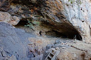
The Wezmeh Cave is an archaeological site near Islamabad Gharb, western Iran, around 470 km (290 mi) southwest of the capital Tehran. The site was discovered in 1999 and excavated in 2001 by a team of Iranian archaeologists under the leadership of Dr. Kamyar Abdi. Wezmeh cave was re-excavated by a team under direction of Fereidoun Biglari in 2019.

<span class="mw-page-title-main">Taforalt</span> Cave and archaeological site in Morocco

Taforalt, or Grotte des Pigeons, is a cave in the province of Berkane, Aït Iznasen region, Morocco, possibly the oldest cemetery in North Africa. It contained at least 34 Iberomaurusian adolescent and adult human skeletons, as well as younger ones, from the Upper Palaeolithic between 15,100 and 14,000 calendar years ago. There is archaeological evidence for Iberomaurusian occupation at the site between 23,200 and 12,600 calendar years ago, as well as evidence for Aterian occupation as old as 85,000 years.

<span class="mw-page-title-main">Medieval bioarchaeology</span> Study of human remains recovered from medieval archaeological sites

Medieval Bioarchaeology is the study of human remains recovered from medieval archaeological sites. Bioarchaeology aims to understand populations through the analysis of human skeletal remains and this application of bioarchaeology specifically aims to understand medieval populations. There is an interest in the Medieval Period when it comes to bioarchaeology, because of how differently people lived back then as opposed to now, in regards to not only their everyday life, but during times of war and famine as well. The biology and behavior of those that lived in the Medieval Period can be analyzed by understanding their health and lifestyle choices.

<span class="mw-page-title-main">Linear enamel hypoplasia</span>

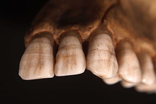
Linear enamel hypoplasia is a failure of the tooth enamel to develop correctly during growth, leaving bands of reduced enamel on a tooth surface. It is the most common type of enamel hypoplasia reported in clinical and archaeological samples, with other types including plane-form enamel hypoplasia and pitting enamel hypoplasia.

Mortuary archaeology is the study of human remains in their archaeological context. This is a known sub-field of bioarchaeology, which is a field that focuses on gathering important information based on the skeleton of an individual. Bioarchaeology stems from the practice of human osteology which is the anatomical study of skeletal remains. Mortuary archaeology, as well as the overarching field it resides in, aims to generate an understanding of disease, migration, health, nutrition, gender, status, and kinship among past populations. Ultimately, these topics help to produce a picture of the daily lives of past individuals. Mortuary archaeologists draw upon the humanities, as well as social and hard sciences to have a full understanding of the individual.

In the skeleton of humans and other animals, a tubercle, tuberosity or apophysis is a protrusion or eminence that serves as an attachment for skeletal muscles. The muscles attach by tendons, where the enthesis is the connective tissue between the tendon and bone. A tuberosity is generally a larger tubercle.

Mary Lewis is Professor of Bioarchaeology at the University of Reading. After completing a PhD in bioarchaeology at the University of Bradford in 1999, Lewis went on to lecture at Bournemouth University (2000–2004) before moving to the University of Reading in 2004. She conducted the first osteological study of a body which has been hanged, drawn, and quartered. Lewis has held editorial roles with the International Journal of Osteoarchaeology, International Journal of Paleopathology, and the American Journal of Biological Anthropology.

<span class="mw-page-title-main">Near Eastern bioarchaeology</span> Archaeological sub-discipline

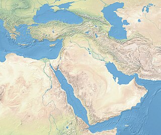
Near Eastern bioarchaeology covers the study of human skeletal remains from archaeological sites in Cyprus, Egypt, Levantine coast, Jordan, Turkey, Iran, Saudi Arabia, Qatar, Kuwait, Bahrain, United Arab Emirates, Oman, and Yemen.

References

  1. "enthesis". Medcyclopaedia. GE. Archived from the original on 2012-02-05.
  2. Thomopoulos S, Birman V, Genin G, eds. (2012). Structural Interfaces and Attachments in Biology. New York: Springer. ISBN   9781461433163.
  3. Rothrauff BB, Tuan RS (January 2014). "Cellular therapy in bone-tendon interface regeneration". Organogenesis. 10 (1): 13–28. doi:10.4161/org.27404. PMC   4049890 . PMID   24326955.
  4. Genin GM, Thomopoulos S (May 2017). "The tendon-to-bone attachment: Unification through disarray". Nature Materials. 16 (6): 607–608. Bibcode:2017NatMa..16..607G. doi:10.1038/nmat4906. PMC   5575797 . PMID   28541313.
  5. Benjamin M, Toumi H, Ralphs JR, Bydder G, Best TM, Milz S (April 2006). "Where tendons and ligaments meet bone: attachment sites ('entheses') in relation to exercise and/or mechanical load". Journal of Anatomy. 208 (4): 471–490. doi:10.1111/j.1469-7580.2006.00540.x. PMC   2100202 . PMID   16637873.
  6. Jurmain R, Cardoso FA, Henderson C, Villotte S (2011-01-01). Grauer AL (ed.). A Companion to Paleopathology. Wiley-Blackwell. pp. 531–552. doi:10.1002/9781444345940.ch29. ISBN   9781444345940.
  7. Hawkey DE, Merbs CF (1995-12-01). "Activity-induced musculoskeletal stress markers (MSM) and subsistence strategy changes among ancient Hudson Bay Eskimos". International Journal of Osteoarchaeology. 5 (4): 324–338. doi:10.1002/oa.1390050403. ISSN   1099-1212.
  8. 1 2 Henderson CY, Mariotti V, Pany-Kucera D, Villotte S, Wilczak C (2013-03-01). "Recording Specific Entheseal Changes of Fibrocartilaginous Entheses: Initial Tests Using the Coimbra Method". International Journal of Osteoarchaeology. 23 (2): 152–162. doi:10.1002/oa.2287. hdl: 10316/44423 . ISSN   1099-1212. S2CID   145571511.
  9. Henderson CY, Mariotti V, Pany-Kucera D, Villotte S, Wilczak C (2016-09-01). "The New 'Coimbra Method': A Biologically Appropriate Method for Recording Specific Features of Fibrocartilaginous Entheseal Changes" (PDF). International Journal of Osteoarchaeology. 26 (5): 925–932. doi:10.1002/oa.2477. hdl: 10316/44421 . ISSN   1099-1212.
  10. Mariotti V, Facchini F, Belcastro MG (June 2004). "Enthesopathies--proposal of a standardized scoring method and applications". Collegium Antropologicum. 28 (1): 145–159. PMID   15636072.
  11. Mariotti V, Facchini F, Giovanna Belcastro M (March 2007). "The study of entheses: proposal of a standardised scoring method for twenty-three entheses of the postcranial skeleton". Collegium Antropologicum. 31 (1): 291–313. PMID   17598416.
  12. Villotte S. "Practical protocol for scoring the appearance of some fibrocartilaginous entheses on the human skeleton".
  13. Villotte S, Castex D, Couallier V, Dutour O, Knüsel CJ, Henry-Gambier D (June 2010). "Enthesopathies as occupational stress markers: evidence from the upper limb". American Journal of Physical Anthropology. 142 (2): 224–234. doi:10.1002/ajpa.21217. PMID   20034011.
  14. Villotte S, Assis S, Cardoso FA, Henderson CY, Mariotti V, Milella M, et al. (June 2016). "In search of consensus: Terminology for entheseal changes (EC)" (PDF). International Journal of Paleopathology. 13: 49–55. doi:10.1016/j.ijpp.2016.01.003. PMID   29539508. S2CID   3902457.
  15. Karakostis, Fotios Alexandros; Lorenzo, Carlos (2016). "Morphometric patterns among the 3D surface areas of human hand entheses". American Journal of Biological Anthropology. 160 (4): 694–707. doi:10.1002/ajpa.22999. PMID   27166777.
  16. Cardoso FA, Henderson C (2013-03-01). "The Categorisation of Occupation in Identified Skeletal Collections: A Source of Bias?". International Journal of Osteoarchaeology. 23 (2): 186–196. doi:10.1002/oa.2285. hdl: 10316/21142 . ISSN   1099-1212.
  17. Michopoulou E, Nikita E, Valakos ED (December 2015). "Evaluating the efficiency of different recording protocols for entheseal changes in regards to expressing activity patterns using archival data and cross-sectional geometric properties". American Journal of Physical Anthropology. 158 (4): 557–568. doi: 10.1002/ajpa.22822 . PMID   26239396.
  18. Milella M, Giovanna Belcastro M, Zollikofer CP, Mariotti V (July 2012). "The effect of age, sex, and physical activity on entheseal morphology in a contemporary Italian skeletal collection". American Journal of Physical Anthropology. 148 (3): 379–388. doi:10.1002/ajpa.22060. PMID   22460619.
  19. Henderson CY, Mariotti V, Santos F, Villotte S, Wilczak CA (2017-06-20). "The new Coimbra method for recording entheseal changes and the effect of age-at-death". BMSAP. 29 (3–4): 140–149. doi:10.1007/s13219-017-0185-x. hdl: 10316/44430 . ISSN   0037-8984. S2CID   29420179.
  20. Henderson CY (March 2013). "Do diseases cause entheseal changes at fibrous entheses?". International Journal of Paleopathology. 3 (1): 64–69. doi:10.1016/j.ijpp.2013.03.007. hdl: 10316/44415 . PMID   29539362. S2CID   3916560.
  21. Karakostis FA, Jeffery N, Harvati K (November 2019). "Experimental proof that multivariate patterns among muscle attachments (entheses) can reflect repetitive muscle use". Scientific Reports. 9 (1): 16577. Bibcode:2019NatSR...916577K. doi:10.1038/s41598-019-53021-8. PMC   6851080 . PMID   31719626.
  22. Karakostis FA, Wallace IJ, Konow N, Harvati K (December 2019). "Experimental evidence that physical activity affects the multivariate associations among muscle attachments (entheses)". The Journal of Experimental Biology. 222 (Pt 23): jeb213058. doi:10.1242/jeb.213058. PMC   6918778 . PMID   31712353.
  23. Castro AA, Karakostis FA, Copes LE, McClendon HE, Trivedi AP, Schwartz NE, Garland T (February 2022). "Effects of selective breeding for voluntary exercise, chronic exercise, and their interaction on muscle attachment site morphology in house mice". Journal of Anatomy. 240 (2): 279–295. doi:10.1111/joa.13547. PMC  8742976. PMID   34519035.
  24. Karakostis, Fotios Alexandros; Wallace, Ian J. (2023-01-31). "Climbing influences entheseal morphology in the humerus of mice: An experimental application of the VERA methodology". American Journal of Biological Anthropology: ajpa.24700. doi: 10.1002/ajpa.24700 . ISSN   2692-7691. S2CID   256486347.

Further reading