
A bone is a rigid organ that constitutes part of the skeleton in most vertebrate animals. Bones protect the various other organs of the body, produce red and white blood cells, store minerals, provide structure and support for the body, and enable mobility. Bones come in a variety of shapes and sizes and have complex internal and external structures. They are lightweight yet strong and hard and serve multiple functions.

Bone healing, or fracture healing, is a proliferative physiological process in which the body facilitates the repair of a bone fracture.

An endotherm is an organism that maintains its body at a metabolically favorable temperature, largely by the use of heat released by its internal bodily functions instead of relying almost purely on ambient heat. Such internally generated heat is mainly an incidental product of the animal's routine metabolism, but under conditions of excessive cold or low activity an endotherm might apply special mechanisms adapted specifically to heat production. Examples include special-function muscular exertion such as shivering, and uncoupled oxidative metabolism, such as within brown adipose tissue.
Osteoblasts are cells with a single nucleus that synthesize bone. However, in the process of bone formation, osteoblasts function in groups of connected cells. Individual cells cannot make bone. A group of organized osteoblasts together with the bone made by a unit of cells is usually called the osteon.

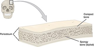
A trabecula is a small, often microscopic, tissue element in the form of a small beam, strut or rod that supports or anchors a framework of parts within a body or organ. A trabecula generally has a mechanical function, and is usually composed of dense collagenous tissue. It can be composed of other material such as muscle and bone. In the heart, muscles form trabeculae carneae and septomarginal trabeculae. Cancellous bone is formed from groupings of trabeculated bone tissue.

An osteocyte, an oblate shaped type of bone cell with dendritic processes, is the most commonly found cell in mature bone. It can live as long as the organism itself. The adult human body has about 42 billion of them. Osteocytes do not divide and have an average half life of 25 years. They are derived from osteoprogenitor cells, some of which differentiate into active osteoblasts. Osteoblasts/osteocytes develop in mesenchyme.

Intramembranous ossification is one of the two essential processes during fetal development of the gnathostome skeletal system by which rudimentary bone tissue is created. Intramembranous ossification is also an essential process during the natural healing of bone fractures and the rudimentary formation of bones of the head.
In osteology, the osteon or haversian system is the fundamental functional unit of much compact bone. Osteons are roughly cylindrical structures that are typically between 0.25 mm and 0.35 mm in diameter. Their length is often hard to define, but estimates vary from several millimeters to around 1 centimeter. They are present in many bones of most mammals and some bird, reptile, and amphibian species.

Thrinaxodon is an extinct genus of cynodonts, including the species T. liorhinus which lived in what are now South Africa and Antarctica during the Early Triassic. Thrinaxodon lived just after the Permian–Triassic mass extinction event, its survival during the extinction may have been due to its burrowing habits.

Transcranial Doppler (TCD) and transcranial color Doppler (TCCD) are types of Doppler ultrasonography that measure the velocity of blood flow through the brain's blood vessels by measuring the echoes of ultrasound waves moving transcranially. These modes of medical imaging conduct a spectral analysis of the acoustic signals they receive and can therefore be classified as methods of active acoustocerebrography. They are used as tests to help diagnose emboli, stenosis, vasospasm from a subarachnoid hemorrhage, and other problems. These relatively quick and inexpensive tests are growing in popularity. The tests are effective for detecting sickle cell disease, ischemic cerebrovascular disease, subarachnoid hemorrhage, arteriovenous malformations, and cerebral circulatory arrest. The tests are possibly useful for perioperative monitoring and meningeal infection. The equipment used for these tests is becoming increasingly portable, making it possible for a clinician to travel to a hospital, to a doctor's office, or to a nursing home for both inpatient and outpatient studies. The tests are often used in conjunction with other tests such as MRI, MRA, carotid duplex ultrasound and CT scans. The tests are also used for research in cognitive neuroscience.

The mastoid part of the temporal bone is the posterior (back) part of the temporal bone, one of the bones of the skull. Its rough surface gives attachment to various muscles and it has openings for blood vessels. From its borders, the mastoid part articulates with two other bones.
Angioblasts are embryonic cells from which the endothelium of blood vessels arises. They are derived from embryonic mesoderm. Blood vessels first make their appearance in several scattered vascular areas that are developed simultaneously between the endoderm and the mesoderm of the yolk-sac, i. e., outside the body of the embryo. Here a new type of cell, the angioblast, is differentiated from the mesoderm.
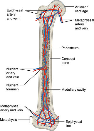
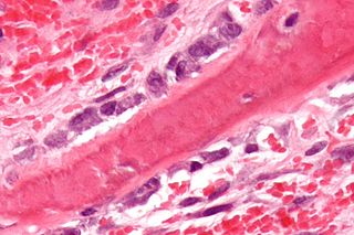
The nutrient artery, usually accompanied by one or two nutrient veins, enters the bone through the nutrient foramen, runs obliquely through the cortex, sends branches upward and downward to the bone marrow, which ramify in the endosteum–the vascular membrane lining the medullary cavity–and give twigs to the adjoining canals. Nutrient arteries are the most apparent blood vessels of the bones.
The physiology of dinosaurs has historically been a controversial subject, particularly their thermoregulation. Recently, many new lines of evidence have been brought to bear on dinosaur physiology generally, including not only metabolic systems and thermoregulation, but on respiratory and cardiovascular systems as well.
Flat bones are bones whose principal function is either extensive protection or the provision of broad surfaces for muscular attachment. These bones are expanded into broad, flat plates, as in the cranium (skull), the ilium, ischium, and pubis (pelvis), sternum and the rib cage. The flat bones are: the occipital, parietal, frontal, nasal, lacrimal, vomer, sternum, ribs, and scapulae.

Volkmann's canals, also known as perforating holes or channels, are anatomic arrangements in cortical bones that allow blood vessels to enter the bones from periosteum. They interconnect the haversian canals with each other and the periosteum. They usually run at obtuse angles to the haversian canals and contain anastomosing vessels between haversian capillaries. They were named after German physiologist Alfred Volkmann (1800-1878).

Theriognathus is an extinct genus of therocephalian therapsid belonging to the family Whaitsiidae, known from fossils from South Africa, Zambia, and Tanzania. Theriognathus has been dated as existing during the Late Permian. Although Theriognathus means mammal jaw, the lower jaw is actually made up of several bones as seen in modern reptiles, in contrast to mammals. Theriognathus displayed many different reptilian and mammalian characteristics. For example, Theriognathus had canine teeth like mammals, and a secondary palate, multiple bones in the mandible, and a typical reptilian jaw joint, all characteristics of reptiles. It is speculated that Theriognathus was either carnivorous or omnivorous based on its teeth, and was suited to hunting small prey in undergrowth. This synapsid adopted a sleek profile of a mammalian predator, with a narrow snout and around 1 meter long. Theriognathus is represented by 56 specimens in the fossil record.
A fibrin scaffold is a network of protein that holds together and supports a variety of living tissues. It is produced naturally by the body after injury, but also can be engineered as a tissue substitute to speed healing. The scaffold consists of naturally occurring biomaterials composed of a cross-linked fibrin network and has a broad use in biomedical applications.

Orthopedic surgery is the branch of surgery concerned with conditions involving the musculoskeletal system. Orthopedic surgeons use both surgical and nonsurgical means to treat musculoskeletal injuries, sports injuries, degenerative diseases, infections, bone tumours, and congenital limb deformities. Trauma surgery and traumatology is a sub-specialty dealing with the operative management of fractures, major trauma and the multiply-injured patient.
Hard tissue, refers to "normal" calcified tissue, is the tissue which is mineralized and has a firm intercellular matrix. The hard tissues of humans are bone, tooth enamel, dentin, and cementum. The term is in contrast to soft tissue.


